online | memorias.ioc.fiocruz.br Culex quinquefasciatus Say, 1823 (Diptera:
Culici-dae) is an anthropophilic mosquito (Forattini et al. 2000) that has a widespread distribution in tropical and sub-tropical regions. It is found abundantly in the southern parts of the United States, Central and South America, the tropics of Africa, the Middle and Far East, southern Asia, New Guinea and Australia. The wide distribution of this mosquito both in the northern and southern hemi-spheres exposes it to a variety of climates and conditions that challenge its survival. On a global level, this species has been associated with the transmission of philaria, as well as Wuchereria bancrofti, Dirofilaria immitis, West Nile virus and other viruses causing Saint Louis enceph-alitis and Venezuelan equine encephenceph-alitis (Rivas et al. 1997, Goddard et al. 2002).
The first arthropod cell line was established from tissues of Antheraea eucalypti moth pupae in Australia by Grace (1962). The study of insect cell cultures has progressed in recent years and more than 500 cell lines from different insect species, mainly corresponding to the Diptera, Lepidoptera, Hemiptera, Homoptera and Orthoptera orders, have been described (Lynn 2001). These cell lines have become an important tool for use in a wide range of studies, including research on intra-cellular parasites and viral infections, in vitro pathogen
propagation, the development of vaccines, pest manage-ment, the production of recombinant proteins and mo-lecular studies (Sudeep et al. 2005, Hoshino et al. 2009). In Colombia, Rey et al. (2000), Bello et al. (2001) and Zapata et al. (2005) generated primary cell cultures and cell lines from different species of mosquitoes and sand flies that were capable of continuous growth. These cells were characterised morphologically, cytogenetically, biochemically and/or molecularly and, in some cases, were assessed for susceptibility to infection with arbo-viruses and parasites.
Mosquito cell lines of importance to public health are considered important tools in basic and applied bio-medical studies. Although there are a significant number of cell lines that have been established from different mosquito species, they do not address all of the needs of various basic and applied fields of research where cells are required. In addition, mosquito cell cultures, even when derived from the same species but from different tissues, are not necessarily interchangeable or useful for the same purposes.
Hsu et al. (1970) established a cell line that was derived from the ovarian tissues of Cx. quinquefasciatus. These cells were characterised morphologically and cytogeneti-cally and showed notable particularities in their pattern of growth and cell shape. In addition, Hsu (1971) studied the susceptibility of the ovarian cells to infection with nine arboviruses and showed that all of the viruses tested, ex-cept for Eastern equine encephalitis virus and Sindbis, replicated in Cx. quinquefasciatus cell cultures to various degrees. The present paper describes, for the first time, the establishment and analysis (morphological, cytogenetic, biochemical and molecular) of a new cell line derived from the embryonic tissues of Cx. quinquefasciatus.
Financial support: Rosario University, NIH (Colombia) + Corresponding author: nidya.segura@urosario.edu.co Received 13 May 2011
Accepted 29 November 2011
Establishment and characterisation of a new cell line derived from
Culex quinquefasciatus (Diptera: Culicidae)
Nidya A Segura1/+, Erika Santamaría², Olga L Cabrera², Felio Bello1
1Laboratorio de Entomología Médica y Forense, Universidad del Rosario, Calle 63D 24-31, Bogotá, Colombia
²Grupo de Entomología, Instituto Nacional de Salud, Bogotá, Colombia
Insect cell cultures are an important biotechnological tool for basic and applied studies. The objective of this work was to establish and characterise a new cell line from Culex quinquefasciatus embryonic tissues. Embryonated eggs were taken as a source of tissue to make explants that were seeded in L-15, Grace’s, Grace’s/L-15, MM/VP12, Schneider’s and DMEM culture media with a pH range from 6.7-6.9 and incubated at 28ºC. The morphological, cytogenetic, biochemical and molecular characteristics of the cell cultures were examined by observing the cell shapes, obtaining the karyotypes, using a cellulose-acetate electrophoretic system and performing random ampli-fied polymorphic DNA-polymerase chain reaction analysis, respectively. The Grace’s/L-15 medium provided the optimal nutritional conditions for cell adhesion and proliferation. Approximately 40-60 days following the explant procedure, a confluent monolayer was formed. Cellular morphology in the primary cultures and the subcultures was heterogeneous, but in the monolayer the epithelioid morphology type predominated. A karyotype with a dip-loid number of six chromosomes (2n = 6) was observed. Isoenzymatic and molecular patterns of the mosquito cell cultures matched those obtained from the immature and adult forms of the same species. Eighteen subcultures were generated. These cell cultures potentially constitute a useful tool for use in biomedical applications.
MATERIALS AND METHODS
Obtaining biological material - Embryonated eggs from Cx. quinquefasciatus were taken from a colony that was maintained in the insectary of the Entomology Group, National Health Institute, in March 2009. The colony was established from adults collected in an urban area of Bogotá located at 4º36’43”N and 74º04’07”W at 2,600 meters above sea level, which is the geographical centre of Colombia.
Primary culture initiation - For each explant of em-bryonated tissues, eight-10 rafts, composed of approxi-mately 600 eggs, were placed in a Petri dish. After an incubation period of 12-20 h, the eggs were refrigerated for 24 h and then incubated at 28ºC for 6 h following the protocol of Oelofsen et al. (1990). Subsequently, the eggs were manually separated using a soft bristle paint-brush inside a laminar flow chamber. The eggs were then placed into a 50 mL centrifuge tube that was previ-ously rinsed with a 95% solution of ethanol and rinsed two times for 1 min with the same solution. During this period, the tube was stirred repeatedly. The ethanol solu-tion was then removed, 0.5% sodium hypochlorite was added and the tube was continuously stirred for 5 min. The eggs were washed three times with sterile distilled water and then rinsed with the growth medium to be used. One millilitre of the egg mass was placed into a 2 mL Ten Brock homogeniser, where the eggs were dis-rupted mechanically. The resultant solution was placed in a 25 cm2 plastic tissue culture flask containing 10 mL
of the growth medium. To assess the best culture me-dium, the seeding of embryonic tissues was carried out separately in the following culture media: L-15 (Gibco), Grace’s (Gibco), Grace’s/L-15, MM/VP12 (Varma & Pudney 1969), Schneider’s (Sigma) and DMEM (Gibco). All media were supplemented with 20% foetal bovine serum (FBS) and a mixture of penicillin (100 units/mL), streptomycin (100 units/mL) and antimycotics (2.5 μg/ mL Amphotericin B) and the pH of the media was ad-justed to between 6.7-6.9. The culture flasks, each con-taining both the selected growth medium and the cells, were incubated at 28ºC without CO2.
Subcultures - The first successful subculture was obtained in March 2010 and since that time 18 passages have been performed to date. The disruption of the con-fluent monolayers was done mechanically with the help of a rubber policeman. After the cells were resuspended, they were mixed by vigorous pipetting and the cell solu-tion was transferred to a new flask containing 5 mL of fresh medium. The first five subcultures were passaged at a ratio of 1:1 for each subculture with an average duration of 30 days between each passage. From the sixth-12th pas-sages, subcultures were carried out in a passage split ratio of 1:1 at 15 day intervals. Thereafter, the split ratio was increased gradually to a 1:3 ratio every eight days.
Morphological characteristics - Cell shapes were de-termined by daily observation using an inverted micro-scope with a microphotographic system (Leica DMLI) at 100-400X magnification. The most representative cells were photographed.
Cytogenetic characteristics - Samples of subcultures (passage 15) were used to obtain metaphasic chromo-somes. Colchicine (0.6 µg/mL) was added to the cul-tures for a period of 3 h. The cell suspension was then removed and centrifuged at 800 g for 10 min. The super-natant was discarded and 0.56% KCl was added to the pellet. The mixture was stirred by flushing with a Pas-teur pipette and left for 30 min. The mixture was then centrifuged again and Carnoy’s fixative (methanol and acetic acid, 3:1) was added for 15 min. After three suc-cessive washes with Carnoy’s, 1 mL of the cell suspen-sion was dropped onto clean and degreased slides. The dried preparation was stained with 2% Giemsa’s stain. The best metaphase arrays from the slides were selected and microphotographed.
Analyses of isozyme patterns - The isoenzymatic phe-notypes of the following four enzymes were examined: malic dehydrogenase, glucose-6-phosphate dehydroge-nase, phosphoglucose isomerase and phosphoglucose mutase. Isoenzymes were separated using electrophoresis on cellulose acetate according to the procedure described by Brown and Knudson (1980). Cell samples were run simultaneously with Cx. quinquefasciatus larvae and pu-pae extracts from the same colony. The four isozymatic patterns from the cell cultures were compared with a cell line from Lutzomyia longipalpis (Lulo) (Rey et al. 2000) and two isozymatic patterns were compared with a cell line from Lutzomyia spinicrassa (Zapata et al. 2005). The relative electrophoretic mobility (REM) was calculated using the formula REM = e/a × 100, where “e” corre-sponds to the distance run in mm for each enzyme in the Cx. quinquefasciatus sample and “a” corresponds to the distance run in mm for each enzyme in the Lulo sample. Migration was measured from the edge of the well, where the sample had been applied to the corresponding band’s midpoint (Zapata et al. 2005).
Molecular characterisation [random amplified polymorphic DNA (RAPD)-polymerase chain reaction (PCR)] - DNA was extracted from confluent monolay-ers of the Cx. quinquefasciatus and Lulo cell lines using the method of Landry et al.(1993). DNA was extracted from adults of the species under study following the pro-cedure proposed by Coen et al. (1982).
The PCR technique was standardised to minimise the potential effects of the variable substances in the reaction mixture on the amplification process. The re-action mixture had the following composition: 2.5 µL of Buffer A (10 x), 1 µL of dNTPs (10 mM), 1.25 µL of MgCl2 (50 mM), primer 1 (10 µM), 0.2 µL of Taq DNA polymerase (1 U/µL), 5 µL of DNA (10 ng/µL) and 14.05 µL of nuclease-free water (final volume of 25 µL per sample). Four primers, synthesised by Invitro-genTM, were selected. The sequence for each of the
step) with an additional 5 min at 72ºC for the last cycle (Williams et al. 1990, Kawai & Mitsuhashi 1997).
The reaction products were electrophoresed in a 1.4% agarose gel at 35 mA for 120 min. The agarose plate was then stained in 0.5 µg/mL ethidium bromide in TAE buffer (40 mM Tris, 20 mM acetic acid, 1 mM Na2EDTA) and photographed under ultraviolet light. In-dividual bands were scored as present or absent in the amplification profile of each sample (Williams et al. 1990, Stevens & Wall 1997).
Band patterns were compared using the similarity coefficient (SAB) of Ney and Li (1979), which is repre-sented by the following formula: SAB = 2NAB / (NA + NB), where NA and NB correspond to the total number of bands shown by individual A and individual B, respec-tively, and NAB is the number of shared bands.
Cryopreservation - Semi-confluent culture monolay- ers were disrupted and the detached cells were adjusted to 5 x 10⁶/mL in freezing medium (50% Grace’s/L15,
40% FBS and 10% dimethyl sulfoxide). The cell suspen-dimethyl sulfoxide). The cell suspen-. The cell suspen-sion was dispensed into sterile cryotubes and refriger-ated at 5ºC for 20 min, then frozen at -70ºC overnight prior to permanent storage in liquid nitrogen.
RESULTS
Primary culture initiation - Obtaining primary cultures from embryonic tissues was successful us-ing Grace’s/L15 medium. There was no cell growth in Grace’s, L-15, Schneider’s, MM, VP12, MM/VP12 or DMEM media. The best results at the initiation of the cell cultures were achieved using eggs that had been in-cubated for 16-20 h followed by refrigeration (4ºC) for up to 24 h and then additionally incubated for 4-6 h at 28ºC. Cell growth during the primary culture was slow and in the first weeks of adhesion and cell growth some tissue fragments with pulsating movement were observed and continued to be observed in some cases for more than three weeks. The presence of vesicles was also observed (Fig. 1A), many of which were in suspension and some of which adhered to the cells in the monolayer. Observa-tion through an inverted microscope revealed that the vesicles were formed by a monolayer of epithelioid cells surrounding an empty space. With increased time in cul-ture, the vesicles became greatly increased in size and number. After rupturing, these vesicles were an impor-tant source of cell release that contributed to cell pro-liferation in the primary cell culture and hence, to the formation of the confluent monolayer.
A confluent monolayer was achieved after 40-60 days of incubation (Fig. 1B), at which time the cells were growing and adhering firmly to the flask surface. Eigh-teen serial subcultures were performed and the initial subcultures demonstrated a similar slow growth rate with low cell proliferation as the primary cultures. How-ever, after the sixth passage, cell division increased in the cultures. At this time, the cells were subcultured at a ratio of 1:3 once per week. The viability of frozen cells was shown at five months after freezing.
Morphological characteristics - Cx. quinquefas-ciatus cell cultures in the initial growth stages showed heterogeneous cell morphology, demonstrating
spheri-cal, elongated, irregular and, occasionally, giant shapes. However, the predominant morphological type was the epithelioid type, which was observed in the confluent monolayer (Fig. 2), as well as in the subcultures.
Cytogenetic characteristics - The metaphase arrays obtained from primary cultures and subcultures of Cx. quinquefasciatus exhibited a diploid chromosomal num-ber (2n = 6) (Fig. 3). The classification of the chromo-somes was performed according to the size in ascending order and the position of the centromeres. Thus, pairs 1 and 2 were classified as metacentric, whereas pair 3 was classified as sub-metacentric.
Fig. 1A: vesicles in suspension during the initiation process of the pri-mary cell cultures from embryonic tissues of Culex quinquefasciatus; B: confluent monolayer Cx. quinquefasciatus formed at 60 days of embryonic tissues explanted. Bar = 200 µm.
Fig. 3: diploid chromosomes from Culex quinquefasciatus cells in cul-ture. Bar = 25 µm.
Isoenzymatic profiles - The four isoenzymatic systems used to characterise the Cx. quinquefasciatus cell cultures were used to establish the identity of the new cell line by comparing its isoenzymatic band patterns with those from mosquito larvae and pupae, as band patterns exhib-ited by the Lulo and Lu. spinicrassa cell lines were differ-ent from each other and from the new cell line. The four systems analysed (ME, G-6-PDH, PGI and PGM) showed one band in both the cell line and the Cx. quinquefasciatus larvae and pupae. However, the Lulo cell line also showed a zymogram with one band in each of the isoenzymatic systems evaluated, but with different relative mobility values, whereas the cell line from Lu. spinicrassa was heterozygotic for the PGM system (Table I).
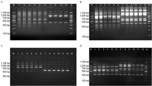
RAPD-PCR analysis - With the A2 primer, seven DNA fragments were recorded for the Lulo cell line and for the cell cultures and adults from Cx. quinquefascia-tus, the number of DNA fragments was 13 (Fig. 4A). With the A10 primer, a profile of 17 DNA fragments was obtained from cell cultures and adults from Cx. quin-quefasciatus, whereas 13 fragments were obtained from the Lulo cell line (Fig. 4B). Using the A20 primer, 11 DNA fragments were obtained from Lulo and six were obtained from the adult mosquito and cell culture sam-ples (Fig. 4C). Finally, with the EO7 primer, the results showed eight DNA fragments for the Cx. quinquefas-ciatus species (cell cultures and adults), nine for the Lu. spinicrassa cell line and six for the Lulo cell line (Fig. 4D). Generally, the DNA fragments obtained covered a range from 300-1,100 bp.
The SAB values between DNA bands of the cell cul-tures and adults of Cx. quinquefasciatus were identical. In contrast, the values obtained from Cx. quinquefas-ciatus and the Lulo cell line and, in one case (primer E07), from the Lu. spinicrassa cell line, were all differ-ent (Table II).
DISCUSSION
One of the important variables in the initiation of cell cultures is the timing of egg embryogenesis used in the tissue explants. In the present study, an optimal incuba-tion time of between 16-20 h was determined to achieve successful cell cultures, which corresponds to approxi-mately 2/3 of the total time of embryo formation before the egg hatches. A similar finding has been published in previous studies on the establishment of cell lines de-rived from mosquitoes (Pant & Dandha 1980, Bello et al. 1997). In addition to the above-mentioned incubation period, the embryonated eggs that produced the best re-sults at the initiation of the cell cultures were those that received an additional 4-6 h of incubation at 28ºC. It was also observed that the eggs retained their viability for up to 24 h when they were refrigerated at 4ºC and after leaving them at room temperature for 30 min and then incubating them under the additional conditions previ-ously indicated, the eggs produced even better results in establishing the primary cell cultures. Similar results using this same technique were reported by Oelofsen et al. (1990), who suggested that it was possibly the tem-perature changes that stimulated cell division.
Cell growth in the primary cell cultures until the for-mation of the confluent monolayer was relatively slow, which is in agreement with previous studies (Rey et al. 2000, Bello et al. 2001, Sudeep et al. 2009). The pattern of cell division and growth depended initially on the em-bryonic tissue fragments that adhered firmly to the flask surface 12 h after explantation and, at later times, the cells began to migrate and proliferate from these tissues.
Additionally, during the initial culturing stages, the pulsating movement of the cell cultures suggested the ac-tivity of muscle tissue, which is dependent on contractile proteins. Fibre connections could also be observed origi-nating from several fragments, which is perhaps why the
cells were able to extend and cover more area. These re-sults are in line with observations by Hsu et al. (1970) in ovarian cell cultures of Cx. quinquefasciatus and those by Duce and Usherwood (1986) in cell cultures of Lo-custa migratoria and Schistocerca gregaria (Orthoptera: Acrididae). Nevertheless, in the present study, actively growing cells later detached from around the fragments to form localised cell colonies that achieved better exten-sion and were therefore better able to proliferate.
Another important characteristic of the cell growth pattern was the occurrence of vesicles surrounded by cells that facilitated the formation of the confluent mono-layer. The occurrence of vesicles in the cell cultures of insects and other arthropods is very common and has been demonstrated in several studies (Charpentier et al. 1995, Rey et al. 2000, Silva et al. 2008).
The culture medium in which the cells were able to adapt, grow and proliferate consisted of a mixture of equal parts of Grace’s and L-15 media, indicating that this mixture provided the necessary and sufficient com-ponents to initiate the primary cell cultures of Cx. quin-quefasciatus. However, the growth of the first five sub-cultures was slow and they were similar to the primary cultures in their pattern of growth and division, with an interval of 30 days between subcultures. In addition, particles that could possibly represent metabolite by-products in response to either an excess or a deficiency of certain substances were observed within the cultured cells and in the medium. However, these particles were not observed subsequent to the sixth subculture.
The cell morphology at the initiation of the primary cell cultures was heterogeneous, but an epithelioid mor-phology became the predominant cell shape later in the confluent cell monolayer and in the subcultures. This re-sult is likely explained by the variety of tissues present in the initial mixed culture that then further divide and de-velop predominantly into one of the two cell shapes (epi-thelioid and fibroblastoid) that are commonly observed in insect cells. In this work, the epithelioid type was pre-dominant, which is in agreement with many other stud-ies on the initiation and establishment of mosquito cell cultures (Bello et al. 2001, Athawale et al. 2002, Zapata et al. 2005, Sudeep et al. 2009).
Regarding the karyological analysis, the number of chromosomes observed matched previous reports on other cell lines of the Culicidae family, including spe-cies from the Culex genus (Hsu et al. 1970, Charpentier et al. 1995, Athawale et al. 2002, Sudeep et al. 2009). According to Kitzmiller (1976), the number (2n = 6) and morphology of the Culicidae chromosomes are highly conserved among the whole family. Additionally, the absence of polyploidy demonstrates the integrity of this cell line and indicates that there has not been a trans-formation like that which occurred in a cell line from the neonate larvae of Aedes aegypti, which showed that greater than 75% of the cells contained a diploid number of chromosomes (Sudeep et al. 2009).
The isoenzymatic profiles of the Cx. quinquefasciatus cell line matched identically the profiles of the samples from the immature forms of the same species, showing
TABLE II
Similarity coefficients for random amplified polymorphic DNA-polymerase chain reaction profiles using four different primers
Primer
Culex quinquefasciatus cell line vs.
C. quinquefasciatus adults
C. quinquefasciatus cell line vs.
Lutzomyia longipalpis cell line
C. quinquefasciatus cell line vs.
Lutzomyia spinicrassa cell line
A2 1 0.3
-A10 1 0.2
-A20 1 0.35
-E07 1 0.28 0.35
TABLE I
Relative electrophoretic mobility for the four isoenzymes used in the study
Systems
Lutzomyia longipalpis Lutzomyia spinicrassa Culex quinquefasciatus
Cells Cells Larvae Pupa Cells
Malic dehydrogenase 100 - 111 111 111
Glucose-6-phosphate dehydrogenase 100 - 108 108 108
Phosphoglucose isomerase 100 133 166 166 166
in all cases the same mobility patterns, which is indica-tive of a common origin. In contrast, a comparison of the isoenzymatic profiles of the four systems evaluated with the profiles obtained from the Lulo and Lu. spinicrassa cell lines revealed large differences, indicating diverse origins and ruling out cross contamination among the cell lines used in our laboratory. This methodology has commonly been used to characterise and authenticate different cell lines from insects (Rey et al. 2000, Lynn & Ferkovich 2004, Zapata et al. 2005, Hoshino et al. 2009).
The identity of the new cell line was also determined using RAPD profiles. Differences were not observed between the Cx. quinquefasciatus cell line and the adult samples using the four primers described above, thereby confirming the identity of the cell line as being derived from Cx. quinquefasciatus. This result suggests that the cultured cells did not incur losses of genetic material since they had been established and that their molecular com-position, according to the markers assessed, reflected the low allelic diversity of the colonised Cx. quinquefascia-tus strain (Léry et al. 2003). In contrast, when comparing the RAPD profiles of the new cell line with the Lulo cell line and the line derived from Lu. spinicrassa, a SAB lower than 0.3 was shown, corresponding to homology of some bands (2 in most cases) and indicating that the cell lines came from different insect species. The four primers used were able to differentiate and confirm the identity of the original sources of the lines from Cx. quinquefasciatus, Lu. longipalpis and Lu. spinicrassa, as well as determine that there was no cross contamination. The efficiency of this technique has been previously reported and it has been shown to differentiate related species, but the tech-nique has not been used to identify clones of cell lines. In studies using Lu. longipalpis and Lu. spinicrassa, the E07 primer was able to differentiate between two species of the same genus, therefore making it an effective tool to authenticate cell lines that are taxonomically close (McIn-tosh et al. 1996, Kawai & Mitsuhash 1997).
A new cell line derived from the embryonic tissues of Cx. quinquefasciatus was generated and character-ised in this study. This cell line will be tested in future studies to assess its usefulness in drug development against arbovirus infections and to identify the molecu-lar mechanism of action of such inhibitors. In addition, it is important to have a larger panel of mosquito cell lines, as they serve as efficient tools to advance studies in multiple areas of biology and biomedicine, including immunology, endocrinology, toxicology, biochemistry, parasitology and evolutionary biology.
ACKNOWLEDGEMENTS
To Marco Fidel Suarez, for collecting the adults that began the colony of Cx. quinquefasciatus.
REFERENCES
Athawale SS, Sudeep AB, Barde PV, Jadi R, Mishra AC, Mourya D 2002. A new cell line from the embryonic tissues of Culex tri-taeniorhynchus and its susceptibility to certain flaviviruses. Acta virologica 46: 237-240.
Bello FJ, Brochero H, Boshell J, Olano V, Rey G 1997. Establishment and characterization of a cell line from mosquito Anopheles albi-manus (Diptera: Culicidae). Mem Inst Oswaldo Cruz 92: 123-128.
Bello FJ, Rodríguez JA, Escovar J, Olano VA, Morales A, González M, Rey G 2001. A new continuous cell line from the mosquito Psoro-phora confinnis (Diptera: Culicidae) and its susceptibility to infec-tions with some arboviruses. Mem Inst Oswaldo Cruz96: 865-873.
Brown JE, Knudson DK 1980. Characterization of invertebrate cells lines. III. Isozyme analyses employing cellulose-acetate electro-phoresis. In vitro14: 255-260.
Charpentier G, Belloncik S, Ducros G, Fontenille D, Tian L, Quiot M 1995. Establishment and characterization of three cell lines from Aedes triseriatus (Diptera: Culicidae). J Med Entomol32: 793-800.
Coen JE, Strachan T, Dover G 1982. Dynamics of concerted evolution of ribosomal DNA and histone gene families in the Melanogaster
species subgroup of Drosophila. J Molec Biol158: 17-35.
Duce JA, Usherwood PN 1986. Primary cultures of muscle from em-bryonic locusta (Locusta migratoria, Schistocerca gregaria): de-velopmental, electrophysiological and patch-clams studies. J Exp Biol12: 307-323.
Forattini O, Kakitani I, La Corte Dos Santos R, Kobayashi K, Ueno H, Fernández Z 2000. Potencial sinantrópico de mosquitos Kerte-sia e Culex (Diptera: Culicidae) no sudeste do Brasil. Rev Saude Publica34: 565-569.
Goddard LB, Roth AR, Reisen WK, Scott TW 2002. Vector com-petence of California mosquitoes for West Nile virus. Em Infect Dis 8: 1385-1391.
Grace TDC 1962. Establishment of four strains of cells from insect tissues grown in vitro. Nature 195: 788-789.
Hoshino K, Hirose M, Iwabuchi K 2009. A new insect cell line from the longicorn beetle Plagionotus christophi (Coleoptera: Ceram-bycidae). In Vitro Cell Dev Biol Anim45: 19-22.
Hsu 1971. Growth of arboviruses in arthropod cell cultures: compara-tive studies. I. Preliminary observations on growth of arboviruses in a newly established line of mosquito cell (Culex quinquefascia-tus). Curr Top Microbiol Immunol55: 140-148.
Hsu SH, Mao WH, Cross JH 1970. Establishment of a line of cells derived from ovarian tissue of Culex quinquefasciatus Say. J Med Entomol7: 703-707.
Kawai Y, Mitsuhashi J 1997. An insect cell line discrimination meth-od by RAPD-PCR. In Vitro Cell Dev Biol Anim33: 512-515.
Kitzmiller JB 1976. Genetics, cytogenetics and evolution of mosqui-toes. Adv Genet18: 416-433.
Landry BS, Dextraze L, Boivin G 1993. Random amplified polymor-phic DNA markers for DNA fingerprinting and genetic variabil-ity assessment of minute parasitic wasp species (Hymenoptera: Mymariadae and Trichogrammatidae) used in biological control programs of phytophagous insects. Genome36: 580-587.
Léry X, LaRue B, Cossette J, Charpentier G 2003. Characterization and authentication of insect cell lines using RAPD markers. In-sect Biochem Mol Biol33: 1035-1041.
Lynn D 2001. Novel techniques to establish new insect cell lines. In vitro Cell Dev Biol Animal37: 319-321.
Lynn DE, Ferkovich FM 2004. New cell lines from Ephestia kue-hniella: characterization and susceptibility to baculoviruses.
J Insect Sci4: 9.
McIntosh AH, Grasela JJ, Matteri RL 1996. Identification of insect cell lines by DNA amplication fingerprinting (DAF). Insect Mol Biol5: 187-195.
Oelofsen M, Gericke A, Smith M, Van der Linde TC 1990. Establish-ment and characterization of a cell line from the mosquito Culex
(Culex) theileri (Diptera: Culicidae) and its susceptibility to in-fection with arboviruses. J Med Entomol27: 939-944.
Pant U, Dandha V 1980. Establishment of a cell line from Culex bitae-niorhynchus. J Tissue Cult Methods 6: 61-63.
Rey GJ, Ferro C, Bello FJ 2000. Establishment and characteriza-tion of a new continuous cell line from Lutzomyia longipalpis
(Diptera: Psychodidae) and its susceptibility to infections with arboviruses and Leishmania chagasi. Mem Inst Oswaldo Cruz 95: 103-110.
Rivas F, Diaz L, Cárdenas V, Daza E, Bruzón L, Alcalá A, De la Hoz O, Caceres FM, Aristizabal G, Martinez JW, Revelo D, De la Hoz F, Boshell J, Camacho T, Calderon L, Olano VA, Villarreal LI, Roselli D, Alvarez G, Ludwig G, Tsai T 1997. Epidemic Venezu-elan equine encephalitis in La Guajira, Colombia 1995. J Infect Dis175: 828-832.
Silva LM, Lages CP, Venuto T, Lima RM, Diniz MV, Valentim CL, Baba EH, Pimenta PF, Fortes-Dias CL 2008. Primary culture of venom glands from the Brazilian armed spider, Phoneutria ni-griventer (Araneae, Ctenidae). Toxicon 51: 428-434.
Stevens J, Wall R 1997. Genetic variation in populations of the Blow-flies Lucilia cuprina and Lucilia sericata (Diptera: Calliphori-dae). Random amplified polymorphic ADN analysis and mito-chondrial ADN sequences. Biochem Syst Ecol25: 81-97.
Sudeep AB, Mourya DT, Mishra AC 2005. Insect cell culture in re-search: Indian scenario. Indian J Med Res121: 725-738.
Sudeep AB, Parashar D, Jadi R, Basu A, Mokashi C, Arankalle V, Mishra A 2009. Establishment and characterization of a new Aedes aegypti (L.) (Diptera: Culicidae) cell line with special emphasis on virus susceptibility. In vitro Cell Dev Biol Animal45: 491-495.
Varma MGR, Pudney M 1969. The growth and serial passage of cell lines from Aedes aegypti (L) larvae in different media. J Med Entomol6: 432-439.
Williams JG, Kubelik AR, Livak KJ, Rafalski JA, Tingey SV 1990. DNA polymorphisms amplified by arbitrary primers are useful as genetic markers. Nucleic Acids Res 18: 6531-6535.
Zapata AC, Cárdenas E, Bello F 2005. Characterization of cell cul-tures derived from Lutzomyia spinicrassa (Diptera: Psychodidae) and their susceptibility to infection with Leishmania (Viannia)