495
A systematic revision of
Tatia
(Siluriformes: Auchenipteridae: Centromochlinae)
Luisa Maria Sarmento-Soares
1,2and Ronaldo Fernando Martins-Pinheiro
2The auchenipterid catfish genus Tatia is revised. Twelve species are recognized including three described as new. Tatia is diagnosed by the hyomandibula elongated anterodorsally, the anal-fin base of adult males reduced in length, and the caudal peduncle laterally compressed and deep with a middorsal keel. Tatia aulopygia occurs in the Madeira river drainage and is distinguished by the reduced cranial fontanel in adults and male modified anal fin with middle rays reduced in length. Tatia boemia, known from the upper Uruguay river drainage, is distinguished by its unique color pattern with dark chromatophores on the sides of body. Tatia brunnea from river basins in Suriname and French Guiana and the Negro river drainage, Amazon basin, is recognized by its wide head and mouth and by the male modified anal fin with sharply pointed tip. Tatia dunni, from the upper Amazon basin, is recognized by its narrow head, long postcleithral process in some specimens, and body coloration with irregular blotches or stripes. Tatia galaxias, endemic to the Orinoco river basin, is distinguished by its large eye and short snout.Tatia gyrina, distributed in the upper and central Amazon basin and in northern Suriname, has a uniquely reduced mesethmoid, slightly protruding lower jaw, second nuchal plate with slightly concave lateral borders, third nuchal plate reduced, small prevomer, low number of ribs, low number of vertebrae and sexual dimorphism regarding intumescent male genital papilla. Tatia intermedia, recorded from central and lower Amazon basin, Tocantins river, and coastal drainages in Guyana, Suriname, French Guiana, and eastern Pará State, Brazil, is distinguished by the short postcleithral process, small eye and long snout. Tatia neivai, from the upper Paraná river, Paraguay river and upper Paraíba do Sul river basin, is distinguished by its unique vertebral count and caudal-fin coloration consisting of transverse dark bars. Tatia strigata, from central Amazon basin and Negro river, is distinguished by its horizontally striped color pattern and the modified male anal fin with middle rays reduced in length. Tatia caxiuanensis, a new species described from the Curuá river, lower Amazon basin, is recognized by its wide cranial fontanel and distinctive anal fin in mature males. Tatia meesi, a new species described from the Essequibo river basin, Guyana, is distinguished from congeners by the cranial fontanel with two separate openings and thin nasal bone. Tatia nigra, a new species described from the central Amazon basin, is distinguished by its short postcleithral process, low number of vertebrae, and dark color pattern. All twelve species of Tatia are described or redescribed and a key to species is provided. O gênero Tatia de auquenipterídeos é revisado. Doze espécies são reconhecidas incluindo três descritas como novas. Tatia é reconhecido pelo hiomandibular fortemente fendido anterodorsalmente, base da nadadeira anal de machos maduros reduzida em tamanho, e pelo pedúnculo caudal lateralmente comprimido e alto com uma quilha médio-dorsal. Tatia aulopygia ocorre na drenagem do rio Madeira e é distinguida pela fontanela craniana reduzida em adultos e pela nadadeira anal em machos maduros fendida, pela redução em tamanho dos raios medianos. Tatia boemia, conhecida da drenagem do alto rio Uruguai, é reconhecida por seu padrão de colorido único com cromatóforos escuros pelas laterais do corpo. Tatia brunnea, de bacias hidrográficas no Suriname e Guiana Francesa e ainda da drenagem do rio Negro na Amazônia, é reconhecida pela ampla largura da cabeça e boca e pela nadadeira anal modificada em machos com extremidade pontiaguda. Tatia dunni, do alto Amazonas, é reconhecida pela cabeça estreita, pelo processo pós-cleitral longo em alguns espécimens, e pela coloração do corpo com manchas irregulares ou faixas claras. Tatia galaxias, endêmica da bacia do rio Orinoco, é distinguida pelos grandes olhos e focinho curto. Tatia gyrina, com ocorrência pelo alto e médio Amazonas, e pelos rios do norte do Suriname, possui mesetmóide reduzido, mandíbula levemente prognata, segunda placa nucal com bordo lateral estreito, terceira placa nucal reduzida, pré-vomer pequeno, reduzido número de costelas, pequeno número de vértebras e dimorfismo sexual onde a papila genital masculina é entumescida. Tatia
1Laboratório de Ecologia de Peixes, sala 525, Pavilhão Haroldo Lisboa, Departamento de Ecologia, Instituto de Biologia Roberto Alcântara Gomes, Universidade Estadual do Rio de Janeiro. Av. São Francisco Xavier, 524; Maracanã, 20550-013 Rio de Janeiro, RJ, Brazil. [email protected]
intermedia é registrada para o médio e baixo rio Amazonas, rio Tocantins e ainda para drenagens costeiras na Guiana, Suriname, Guiana Francesa e leste do Pará no Brasil. É distinguida pelo processo do pós-cleitro curto, olhos pequenos e focinho longo. Tatia neivai, do alto rio Paraná, rio Paraguai e alto rio Paraíba do Sul, é distinguida pela contagem vertebral exclusiva e pela coloração da nadadeira caudal com barras transversais escuras. Tatia strigata, distribuída pelo médio Amazonas e rio Negro é reconhecida pelo padrão de colorido com listras horizontais irregulares e nadadeira anal modificada fendida em machos, com raios medianos reduzidos em tamanho. Tatia caxiuanensis, nova espécie, descrita para o rio Curuá no baixo Amazonas, é reconhecida pela fontanela craniana ampla e pela nadadeira anal de machos maduros distinta. Tatia meesi, nova espécie, descrita para o rio Essequibo, Guiana, é diferenciada de seus congêneres pela fontanela craniana com duas aberturas separadas e pelo osso nasal afilado. Tatia nigra, nova espécie, descrita para o médio Amazonas, é distinguida pelo processo pós-cleitral curto, pelo número reduzido de vértebras e pelo padrão de colorido escurecido. Todas as doze espécies de Tatia são redescritas ou descritas e uma chave de identificação é fornecida.
Key words: South America, Freshwater, Catfish, Taxonomy, Centromochlus.
Introduction
The subfamily Centromochlinae comprises small to me-dium size auchenipterid catfishes that share derived anal-fin morphology in males (Ferraris, 1988; Soares-Porto, 1998). Four genera,Centromochlus,Tatia,Glanidium, and Gelanoglanis, comprising 31 species, are presently recognized as valid in the subfamily (Ferraris, 2007).
Tatia comprises a group of relatively small (20-150 mm standard length), nocturnal auchenipterids [or centromochlins] that feed on small fruits and invertebrates (H. A. Britski, M. Goulding, pers. comm.). They are endemic to South America east of the Andes and are present in most of major drainages, including the Orinoco, Amazon, Paraná-Paraguay, and Uruguay, as well as northern coastal rivers from the Essequibo to Amapá State and Marajó Island, Brazil. Tatia is absent in the São Francisco river basin and small riverine basins along the Brazilian east coast. Tatia is the most speciose genus in the subfamily, with 12 species recognized here (Table 1). Tatia species occur in lentic sections of igarapés, rivers, and lakes, where they remain hidden in submerged trunks or rocky crevices during the day and emerge only at night to forage (Lowe-McConnell, 1964, 1975, 1987; Soares-Porto, 1995). At night, individuals of Tatia usually remain close to the surface near river banks or in the middle of the river, swimming in a very peculiar way (fast and erratic), and capturing fallen insects (F.C.T. Lima, pers. comm.). Some species occur in black water rivers, but none are restricted to these environments. Many members of the genus are beautifully colored and, consequently, very attractive to the ornamental fish trade. Tatia species, however, are not well suited for captivity because of their apparent intolerance for low oxygen conditions. Most do not survive transport to Europe and North America (Sands, 1984).
The generic name Tatia was proposed by Miranda Ribeiro (1911: 360) for Charles Tate Regan of the British Museum of Natural History, London, in honor of his many contributions to the knowledge of the South American freshwater fishes. The group was erected to include two species previously ascribed to Centromochlus:Tatia intermedia (Steindachner) andT. aulopygia (Kner), based on the presence of a genital papilla over the anterior anal-fin rays (Miranda Ribeiro,
1911:353). After Miranda Ribeiro’s (1911) description the name Tatia was mentioned only in catalogues (e.g. Gosline, 1945) and practically forgotten. Subsequent authors such as Eigen-mann & Allen (1942) and Fowler (1945a) described new spe-cies in the genus Centromochlus, without mentioning Tatia. Boeseman (1953) referred to Tatia, but described a new spe-cies as Centromochlus creutzbergi. Britski (1972) considered Tatia to be a junior synonym of Glanidium Lütken due to similarities of sexually dimorphic features. In his study of auchenipterids primarily from Suriname Mees (1974) re-describedTatia and expanded the genus to 14 species, six of which he described as new: T. brunnea, T. concolor, T. galaxias, T. punctata, T. reticulata and T.simplex. Four addi-tional species of Tatia have been described subsequently (Mees, 1988; Royero, 1992; Soares-Porto, 1995; Koch & Reis, 1996).
Regarding interrelationships among auchenipterid gen-era, Ferraris (1988) and Curran (1989) presented independent phylogenetic hypotheses based on cladistic analysis. Ferraris (1988) recognized a monophyletic basal clade within auchenipterids “Centromochlidae” and removed some spe-cies previously assigned to Tatia to two undescribed genera, restricting the nominate genus to: T. aulopygia, T. intermedia, T. dunni, T. galaxias, and one undescribed species. Curran (1989) questioned the monophyly of the speciose genus Tatia. The author considered Tatia and Centromochlus as only dis-tantly related; assigning Tatia as close to Auchenipterichthys. Soares-Porto (1998) stated Tatia sensu stricto as a mono-phyletic unit restricted to eight species: T. aulopygia, T. boemia, T. brunnea, T. creutzbergi, T. gyrina, T. intermedia, T. neivai, and T. strigata. Other species previously assigned toTatia were transferred to Centromochlus, on the basis of derived characters (Soares-Porto, 1996, 1998). Most recently Ferraris (2007) listed twelve valid species of Tatia: T. aulopygia, T. boemia, T. brunnea, T. creutzbergi, T. dunni, T. galaxias, T. gyrina, T. intermedia, T. musaica, T. neivai, T. simplex, and T. strigata.
Material and Methods
Osteological features were examined in cleared and stained (CS) specimens prepared according to the procedures of Tay-lor & van Dyke (1985). Prior to clearing and staining, speci-mens were dissected when possible to determine gut con-tents, sexual maturity of gonads, and record myological in-formation. Osteological data from some types or specimens poorly represented in ichthyological collections were ob-tained from radiographs (noted as “R” in the Material Exam-ined section). Nomenclature of osteological elements follows The Zebrafish Information Network (ZFIN). Muscle names follow Sarmento-Soares & Porto (2006). Drawings were ren-dered from digital photographs prefereably of cleared and stained specimens. Straight-line measurements were made with a digital caliper, and recorded in tenths of a millimeter.
Measurements included: standard length (SL, from snout tip to caudal-fin base); body depth (on nuchal shield, from origin of dorsal-fin spine to the belly); body width (widest distance between lateral surfaces of cleithra, taken between anterior-most margin of cleithral bone beneath pectoral-fin origin); caudal-peduncle depth (least distance between dor-sal and ventral surfaces of caudal peduncle); caudal-peduncle length (from base of posterior-most anal-fin ray to point coin-ciding with origin of lower unbranched caudal-fin ray); predorsal length (from snout tip to origin of dorsal fin); prea-nal length (from snout tip to aprea-nal-fin origin); prepelvic length (from snout tip to pelvic-fin origin); dorsal-fin origin to pec-toral-fin origin; dorsal-fin origin to pelvic-fin origin; pectoral-fin origin to pelvic-pectoral-fin origin; prepectoral length (from snout tip to pectoral-fin origin); dorsal-fin base (from origin of dor-sal-fin spine to posterior-most base of dordor-sal-fin insertion); adipose-fin base (from origin of adipose fin fold to its poste-rior-most base); anal-fin base (distance between genital open-ing and posterior-most base of anal-fin insertion, measured
under the same criteria for both males and females); dorsal-fin spine length (from base of spine to its distal tip); pectoral-fin spine length (from origin of spine to its distal tip, taken with the spine erected); postcleithral (humeral) process length (from anterodorsal margin of exposed process to posterior-most tip of process); first branched pelvic-fin ray (from base to tip of first branched pelvic-fin ray); longest anal-fin ray (from base to tip of first branched anal-fin ray); head length (HL, from snout tip to bony end of opercle); head width (be-tween dorsalmost extents of opercular openings); snout depth (distance in median sagittal plane between posterior nostril and midventral contour of body); interorbital distance (least distance between dorsalmost margins of bony orbit); left internarial width (between left anterior and left posterior nos-trils); anterior internarial distance (transverse distance be-tween anterior nostrils); posterior internarial distance (trans-verse distance between posterior nostrils); snout length (from snout tip to anteriormost margin of eye); maxillary-barbel length (from base to tip, with barbel retracted); outer mental-barbel length (from base to tip, with mental-barbel retracted); inner mental-barbel length (from base to tip, with barbel retracted); orbital diameter (greatest horizontal dimension of eyeball); mouth width (between corners of closed mouth).
Counts of fin rays and bony elements were obtained from alcohol-preserved and cleared and stained specimens. Verte-bral counts included all rib-bearing centra but did not include any of the anterior, complex centrum elements without ribs, following Ferraris & Fernandez (1987), and included the com-pound caudal centrum (PU1+U1) as the last element. Hemal spine counts accompany the numbers for correspondent ver-tebra, as shown in Fig. 1. Count of branchiostegal rays were done only in cleared and stained specimens. The direction of dorsal and pectoral-fin spine serrations are referred to as antrorse for those pointing away from base of spine and ret-rorse for those bent towards base of spine. Male modified
Nominal species Mees (1974) Ferraris (2007) Assignment herein Taxonomic status
Centromochlus aulopygius Kner, 1857 Tatia aulopygia Tatia aulopygia Tatia aulopygia Valid
Tatia boemia Koch & Reis, 1996 - Tatia boemia Tatia boemia Valid
Tatia brunnea Mees, 1974 Tatia brunnea Tatia brunnea Tatia brunnea Valid
Tatia caxiuanensis, new species - - Tatia caxiuanensis n.sp. Valid
Centromochlus dunni Fowler, 1945 Tatia intermedia Tatia dunni Tatia dunni Valid Tatia galaxias Mees, 1974 Tatia galaxias Tatia galaxias Tatia galaxias Valid Centromochlus gyrinus Eigenmann & Allen, 1942 Tatia gyrina Tatia gyrina Tatia gyrina Valid
Centromochlus intermedius Steindachner, 1877 Tatia intermedia Tatia intermedia Tatia intermedia Valid
Tatia meesi, new species - - Tatia meesi n.sp. Valid
Glanidium neivai Ihering, 1930 Tatia neivai Tatia neivai Tatia neivai Valid
Tatia nigra, new species - - Tatia nigra n.sp. Valid
Tatia strigata Soares-Porto, 1995 - Tatia strigata Tatia strigata Valid
Centromochlus altae Fowler, 1945 Tatia altae Centromochlus altae Centromochlus altae Valid
Tatia concolor Mees, 1974 Tatia concolor Centromochlus concolor Centromochlus concolor Valid
Centromochlus creutzbergi Boeseman,1953 Tatia creutzbergi Tatia creutzbergi Junior synonym of Tatia gyrina Junior synonim
Tatia musaica Royero, 1992 - Tatia musaica “Centromochlus” musaicus [incertae sedis in Centromochlinae]
Valid, incertae sedis
in Centromochlinae
Centromochlus perugiae Steindachner, 1882 Tatia perugiae Centromochlus perugiae Centromochlus perugiae Valid Tatia punctata Mees, 1974 Tatia punctata Centromochlus punctatus Centromochlus punctatus Valid
Tatia reticulate Mees, 1974 Tatia reticulata Centromochlus reticulatus Centromochlus reticulatus Valid
Centromochlus schultzi Rössel, 1962 Tatia schultzi Centromochlus schultzi Centromochlus schultzi Valid
Tatia simplex Mees, 1974 Tatia simplex Tatia simplex “Centromochlus” simplex
[incertae sedis in Centromochlinae] Valid,
incertae sedis
in Centromochlinae
anal-fin segmented rays (lepidotrichia) may have adjacent segments with curved border, forming denticulations herein refered to as antrorse or retrorse.
Institutional abbreviations follow Reis et al. (2003), with the exception of MBML for Museu de Biologia Professor Mello Leitão, Santa Teresa, Espírito Santo, Brazil and NUPELIA for Núcleo de Pesquisas em Limnologia, Ictiologia e Aquicultura/ Fundação Universidade Estadual de Maringá, Maringá, Paraná, Brazil.
Results
Tatia Miranda Ribeiro
Tatia Miranda Ribeiro, 1911: 360 [type species: Centromochlus intermediusSteindachner, 1877, by subsequent designa-tion by Jordan, 1920: 545. Gender: feminine].
Diagnosis.Tatia is distinguished among the Centromochlinae by three uniquely derived features. The hyomandibula is elon-gate anterodorsally, not contacting the narrow metapterygoid and, instead, connected only to the trapezoidal quadrate (Fig. 2); anal-fin base of adult males is reduced (anal-fin base length 3.3-8.0% SL); and caudal peduncle is compressed and deep (caudal-peduncle depth 10.1-18.6% SL), with a middorsal keel posterior to adipose fin.
A unique combination of restricted characters aids in dis-tinguishing Tatia: first (anteriormost) nuchal plate present (Fig. 3, n1); infraorbital 1 bone short, limited to anterior corner of orbit; eye moderately large, between 17.4-42.9% HL; max-illa shorter than or same length as autopalatine; retractor tentaculimuscle absent, functionally substituted by maxillo-mandibular ligament (Sarmento-Soares & Porto, 2006); adult males with modified anal fin (all species), and first unbranched anal-fin ray non-segmented or with segments fused (except inT. brunnea).
Generic description. Tatia comprises species with somewhat robust head; caudal peduncle much compressed laterally and deep; well-developed adipose eye lid; eye moderately large,
and latero-dorsally located. Following characters used to dis-tinguish species of Tatia (illustrations based on T. gyrina): Head: cranial fontanel always present (Fig. 3, fo); premaxilla transversely elongated, meeting its counterpart medially; autopalatine tubular, oriented obliquely to longitudinal axis of body; maxilla very small, shorter than or same length as autopalatine; three nuchal plates present (Fig. 3, n1, n2, n3). Epioccipital process small, connected to third nuchal plate by ligaments (Fig. 3, pe). Mullerian ramus with distal tip shaped as slightly curved disc, its concave face superficially adjoined to anterior wall of gasbladder. Prevomer usually with well developed arrow-shaped lateral processes or with short lat-eral process. Prevomerian teeth present in T. galaxias and T. intermedia.
Suspensorium (Fig. 2): Suspensorium bones in Tatia simi-lar among all species and distinctive among Centromochlinae. Hyomandibula narrow, elongate, projected anteriorly as short membranous lamina (Fig. 2, hy). Anterior laminar projection of hyomandibula sutured only to quadrate, through cartilage ventrally and deeply dentate suture dorsally. Metapterygoid conical, dorsally sharp, with little laminar extension (Fig. 2, mt), joined to quadrate via cartilage block only (T. gyrina, T. caxiuanensis, T. meesi, T. strigata) or cartilage plus dentate suture (T. aulopygia, T. boemia, T. brunnea, T. dunni, T. galaxias, T. intermedia, T. neivai, T. nigra). Suspensorium deeply notched anterodorsally, thus metapterygoid not con-tacting hyomandibula. Quadrate trapezoidal, with broad base (Fig. 2, qu), connected to preopercle, hyomandibula and metapterygoid; long preopercle (Fig. 2, po) ventral margins sutured to both quadrate and hyomandibula; suprapreopercle present as short or robust canal bone (Fig. 2, sp); opercle laminate and broadly subtriangular (Fig. 2, op).
Hyoid arch (Fig. 4): Urohyal narrow (Fig. 4, uh); short dorsal hypohyal (Fig. 4, dh) associated with comparatively large ventral hypohyal (Fig. 4, vh); anterior ceratohyal well developed (Fig. 4, ac), posterior ceratohyal smaller (Fig. 4, pc); branchiostegal ray articulated to hyoid arch; branchiostegal rays 5-7; 3-5 on anterior ceratohyal and two on posterior ceratohyal; posteriormost usually largest and flattened. No branchiostegal ray associated with interceratohyal cartilage between bones.
Pharyngeal (gill) arches (Fig. 5): Basibranchial 1, in con-tact with urohyal (Fig. 5, ab), anterior to basibranchial 2; basibranchial 2 forming osseous rod, broadest anteriorly (Fig. 5, bb2) and usually separated by gap from basibranchial 3; basibranchial 3 shorter (Fig. 5, bb3), also broadest anteriorly; basibranchial 4 large, flattened and completely cartilaginous (Fig. 5, bb4); basibranchial 2 bordered laterally by cartilagi-nous head of hypobranchial 1; basibranchial 3 between carti-laginous head of hypobranchial 2 and carticarti-laginous hypo-branchial 3; basihypo-branchial 4 bordered laterally by cartilagi-nous head of ceratobranchial 4 and caudally by cartilagicartilagi-nous head of ceratobranchial 5. Hypobranchial 1 mostly osseous, elongate and expanded laterally, subtriangular, with cartilagi-nous ends; hypobranchial 2 mostly cartilagicartilagi-nous, subtriangular, with medial osseous part; hypobranchial 3
com-Fig. 1. Schematic representation of partial axial skeleton of Tatia boemia MCP 12949, 59.9 mm SL. Left side lateral view. Abbre-viations:af, anal fin; dr, distal radials; hs15- hs18, hemal spines
15 and 18; pf, pelvic fin; pr, proximal radials; r8- r11, ribs 8 to
pletely cartilaginous, more trapezoidal (Fig. 5, hb 1-3); hypo-branchial 4 absent. Five ceratohypo-branchials present, mostly ossified, with cartilage on both ends (Fig. 5, cb 1-5). First ceratobranchial supporting single row of rakers (not illus-trated); fifth ceratobranchial expanded postero-medially to support lower pharyngeal toothplate with short conical teeth. Five epibranchials, all but fifth largely ossified except for car-tilaginous ends; epibranchials 1 and 2 rod-like, without rak-ers; epibranchial 3 with posterior uncinate process; epibranchial 4 broad, with laminar extension; epibranchial 5 cartilaginous, reduced, located in axil between cartilaginous ends of epibranchial 4 and ceratobranchial 4. Pharyngobran-chial 1 absent; pharyngobranPharyngobran-chial 2 short, cartilaginous, some-what rounded, placed between anteromedial cartilaginous tips of epibranchials 1 and 2; pharyngobranchial 3 elongate, ossi-fied, with expanded posterior border; pharyngobranchial 4 ossified, supporting upper pharyngeal tooth plate with short conical teeth.
Lateral line system: Infraorbital 1 short, with ventro-lat-eral process either clearly restricted to anterior border of eye or almost reaching ventral border of eye; infraorbitals thin and canalicular, forming incomplete or complete infraorbital series. Lateral line on body straight, inconspicuous, with os-sified canal bones just anteriorly.
Fins: Dorsal fin with major spine preceded by small dorsal locking spine and followed by 3-5 branched rays; pectoral fin with one spine plus 3-5 branched rays; pelvic fin with one unbranched plus 5 branched rays, margin rounded; adipose fin above anal-fin base; anal fin with 3 unbranched plus 6-8 branched rays; caudal fin with 8+9 principal rays, forked, lobes with rounded tips; dorsal caudal-fin lobe slightly elongate in males of some species.
Ribs and vertebrae: Pleural ribs 7-11; first rib thicker than others; ribs becoming progressively smaller posteriorly. Post-Weberian vertebrae 30-39.
Sexual dimorphism. In catfishes of subfamily
Centromochlinae the anal fin of sexually mature males is modi-fied into a sperm conductor with the anal-fin rays and proxi-mal radials directed posteriorly, aligned nearly parallel to ver-tebral axis (Ferraris, 1988; Soares-Porto, 1998). In mature males the anal fin is strongly modified such that it is supported by enlarged and joined proximal anal-fin radials (Ferraris, 1988; Soares-Porto, 1998) and by partially ossified distal radials. Anal-fin proximal radials are associated with the thickened and lengthened hemal spines of adjacent vertebrae. Modi-fied male anal fin has three developed unbranched rays. The first unbranched ray is shortest, about one-quarter length of second unbranched ray, and with segments fused (except in T. brunnea). Some species have the first unbranched anal-fin ray preceded immediately by a tegumentary keel. The second and third unbranched rays are thick. In the Centromochlinae, Tatia is the only genus with species that bear segments that curve outwards, forming antrorse denticulations along the anterior margin of the third unbranched anal fin ray in adult males.
Remarks. Two of the nominal species assigned to Tatia by Ferraris (2007) are not herein recognized as members of this genus. Both Tatia musaica Royero and Tatia simplex Mees lack the derived characters listed above for Tatia: thin, anterodorsally elongate hyomandibula (vs. broad in both C. musaicus and C. simplex); caudal peduncle laterally com-pressed (vs. ellipsoid); proportionally deeper caudal peduncle, depth 10.1-18.6% SL (vs. 7.9-9.6% SL in C. musaicus and 9.2-10.0% SL in C. simplex); first nuchal plate present (vs. ab-Fig. 2. Right suspensorium of Tatia gyrina, INPA 20971, 28.0 mm SL. Lateral view. Abbreviations: aa, angulo-articular; dn,
dentary; hy, hyomandibula; io, interopercle; mt,
metapterygoid;op, opercle; po, preopercle; qu, quadrate; sb,
subpreopercle;sp, suprapreopercle. Scale bar = 1.0 mm.
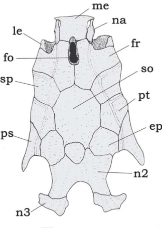
Fig. 3. Neurocranium of Tatia gyrina, INPA 20971, 28.0 mm SL. Dorsal view. Abbreviations: ep, epioccipital; fo, single
cra-nial fontanel, fr, frontal; le, lateral ethmoid; me, mesethmoid; na, nasal; n1, first nuchal plate, n2; second nuchal plate; n3,
third nuchal plate; pe, posterior epioccipital process; ps,
sent); maxilla shorter than or same length as autopalatine (vs. longer than autopalatine). These two species are incertae sedis in Centromochlinae (see Table 1), based in part on inter-relationships analysis by Soares-Porto (1996), and are herein provisionally allocated under Centromochlus.
The monophyly and interrelationships among species of Tatia were hypothesized by Soares-Porto (1998), who con-sidered the clade T. gyrina plus T. creutzbergi as sister to all otherTatia species.
Inseminating reproductive mode (sensuBurnset al., 1997) has been documented within the Auchenipteridae (von Ihering, 1937; Loir et al., 1989; Meisner et al., 2000). In the centromochlin catfishes, inseminating is supposed to take place (Loir et al.,
1989; Franke, 1990), but a reproductive behavior is not yet reported. During reproductive phase, the anal-fin rays change their position probably channeling for discharge of seminal ducts, acting as a sperm conductor during reproduction. Such a morphological change was observed in some males of Centromochlus romani and also in T. nigra. The sperm prob-ably runs along the spoon shaped anal-fin rays, and the fish may press the fin against the belly of the female, for inseminat-ing. Morphological evidences suggesting inseminating repro-ductive mode in Tatia are the intumescent female genital pa-pilla (as in T. gyrina and T. nigra) and the spoon-shaped later-ally curved male anal fin (observed in T. nigra). A detailed analy-sis of morphological changes of the reproductive tract needs to be accomplished by histological preparations, and is out of the scope of present contribution.
Distribution.Species of Tatia are broadly distributed in the regions east of the Andean Cordilleras (Fig. 6). Tatia ranges from the Guarapiche river, Orinoco basin, in Venezuela, to the Pelotas river, Uruguay drainage, in Brazil. The group is found in major continental river basins, such as the Amazon, Orinoco and Paraná-Paraguay; as well as in the Essequibo and small coastal drainages in French Guiana, Suriname and Amapá and Marajó island in northern Brazil. Although well distributed in cis-Andean South America, the group is not known from cer-tain drainage basins, including the São Francisco and small coastal river drainages of northeastern and far eastern Brazil. Tatia is found, however, in headwaters of Paraíba do Sul ba-sin, São Paulo State. The absence of Tatia from certain areas may be a function of inadequate collections from those river basins or may reflect actual disjunct distributions of the group as whole.
Species Account
Tatia aulopygia (Kner, 1857)
Figs. 7-9
Centromochlus aulopygius Kner, 1857: 432, Pl. 8 (fig. 26) [type locality: Guaporé river]. -Steindachner, 1876: 664-665 [no locality]. Eigenmann & Eigenmann, 1890: 270 [citation]. Pearson, 1937: 110 [Mamoré river drainage].
Tatia aulopygia. Miranda Ribeiro, 1911: 361 [generic desig-nation]. Gosline, 1945: 10 [listing]. Mees, 1974: 59-63 [notes and distribution, in part, Maciel, Guaporé river]. Sands, 1984: 38-39 [listing]. Lauzanne & Loubens, 1985: 112 [Mamoré river drainage]. Burgess, 1989:242 [listing]. Soares-Porto, 1995:204 [citation]. Soares-Porto, 1998: 331-350 [citation]. Lasso et al., 2001:97 [citation]. Ferraris, 2003:476 [checklist]. Ferraris, 2007: 77 [checklist]. Centromochlus intermedius. Fisher, 1917: 422 [in part, Maciel,
Guaporé river]. Pearson, 1937: 110 [Mamoré river drainage].
Diagnosis.Tatia aulopygia is distinguished from all other species of Tatia by having a short cranial fontanel, with the opening restricted to the frontals (Fig. 8); a genital papilla
Fig. 4. Right hyoid arch of Tatia gyrina, INPA 20971, 28.0 mm SL. Medial view. Abbreviations: ac, anterior ceratohyal; br,
branchiostegal rays; dh, dorsal hypohyal; ic, interceratohyal
cartilage;ih, interhyal; pc, posterior ceratohyal; vh, ventral
hypohyal;uh, urohyal. Scale bar = 1.0 mm.
Fig. 5. Pharyngeal arches of Tatia gyrina, INPA 20971, 28.0 mm SL. Dorsal view. Abbreviations: ab, anterior (first)
basibranchial;bb2, basibranchial 2; bb3, basibranchial 3; bb4,
basibranchial 4; cb1-5, ceratobranchials 1 to 5 (anterior to
posterior);eb1-4, epibranchials 1 to 4 (anterior to posterior); hb1-3, hypobranchials 1 to 3; pb3, pharingobranchial 3; pb4,
with thick flap of skin around the deferent duct in males (Fig. 9); and a notched anal fin in adult males (Fig. 9). The species also is distinguished from congeners by a combination of characteristics: nasal bone partially sutured to lateral margin of mesethmoid; anal fin with 7-8 branched rays; 10-11 ribs; 38-39 post-Weberian vertebrae. Additional features useful for distinguishing this species include: third nuchal plate well projected laterally with curved tip; and caudal-fin lobes of mature females similar in length, but mature males with slightly elongated upper lobe.
Description. Measured adult specimens 75.2-159.0 mm SL;
morphometric data presented in Table 2. Body deep, head slightly depressed dorsoventrally. Head large, robust, out-line of head in dorsal view somewhat elliptical, broader than long. Dorsal outline of trunk from dorsal-fin base to caudal peduncle increasingly compressed posteriorly. Lateral pro-file of head from snout tip to above opercular margin slightly
convex to pectoral-fin insertion. Ventral profile of head and abdomen flat. Ventral profile of body gently curved, concave behind anal-fin origin. Head integument thin, cranial roof vis-ible; adipose eye lid well-developed; eye dorsolaterally lo-cated in anterior portion of head; mouth terminal, upper lip extended posterolaterally as well-developed fleshy rictal fold; anterior nostril tubular, located on anterior border of snout, above lip; posterior nostril large, rounded, limited by small skin flap; transverse distance between anterior nostrils slightly shorter than distance between posterior ones. Maxillary bar-bel short, extending slightly beyond tip of postcleithral pro-cess, sometimes shorter; mental barbels short, tips not reach-ing pectoral-fin base, arranged in arc along ventral surface of jaw; inner mental barbel about 65.0-75.0% length of outer mental. Postcleithral process almost reaching vertical through origin of dorsal fin. Caudal peduncle deep, depth about 14.0-15.8% SL.
Rostral border of cranium broad with large mesethmoid;
premaxilla underneath with synchondral articulation; cranial fontanel short, ovoid, bounded by frontal (Fig. 8); nasal ossi-fied with wide medial flanges partially sutured to lateral mar-gin of mesethmoid; autopalatine tubular, oriented obliquely to longitudinal axis of body; maxilla very small, shorter than autopalatine; prevomer expanded anteriorly with well devel-oped arrow-shaped lateral processes; jaws of equal size; pre-maxilla and dentary with four to five rows of conical teeth. First nuchal plate somewhat pentagonal; second nuchal plate deeply concave along lateral margin; third nuchal plate curved, projected laterally. Epioccipital process small.
Suspensorium, hyoid arch and opercular bones as in ge-neric description. Suprapreopercle present as short robust canal bone. Six branchiostegal rays articulated with hyoid arch: four with anterior ceratohyal and two with posterior ceratohyal.
Basibranchials 2 and 3 fused together forming osseous rod with broad cartilaginous anterior tip; basibranchial 4 large flattened and completely cartilaginous; fused basibranchial 2 plus 3 bordered laterally by cartilaginous head of hypobran-chial 1, cartilaginous head of hypobranhypobran-chial 2 and cartilagi-nous hypobranchial 3. Basibranchial 4 bordered laterally by cartilaginous head of ceratobranchial 4 and posteriorly by cartilaginous head of ceratobranchial 5. Hypobranchials,
ceratobranchials, epibranchials and pharyngobranchials as described in generic description.
Four infraorbital bones in incomplete series. Infraorbital 1 broad with short ventro-lateral process on anterior border of eye; remaining infraorbitals thin, reduced to canalicular por-tions. Infraorbital 2 smallest, close to infraorbital 1, followed by non-ossified portion of canal below eye and two posterior canal bones, one long and one short, forming posterior or-bital rim. Lateral line on body with ossified canal bones pos-teriorly to vertical through pelvic-fin origin.
Dorsal fin I,5 (n=7); dorsal-fin spine with 14-16 antrorse serrations along entire anterior margin; posterior margin smooth. Pectoral fin I,5 (n=7); pectoral-fin spine with 21-24 antrorse serrations along anterior margin; small serrations close to spine base; 14-16 retrorse serrations along posterior margin; serrations along both margins progressively larger toward spine tip. Pelvic fin i,5 (n=7); margin rounded. Adi-pose fin small, origin on vertical through end of anal-fin base. Anal fin iii,7-8 (n=7); anal-fin pterygiophores in eight rod-like proximal radials and seven cartilaginous distal radials. Cau-dal fin forked, lobes with rounded tips, 8+9 principal rays, 18-20 upper procurrent, 17-18-20 lower procurrent rays (n=7). Pleu-ral ribs 10-11 attached to consecutive vertebrae. Post-Weberian vertebrae 38-39 (n=4).
Fig. 7.Tatia aulopygia,(a) INPA 11080, male, 104.6 mm SL, Guaporé river, Amazonas State, Brazil. (b) ZMA 114.280, young
Color in alcohol. Head and body mottled with dark and light
brown patches of pigmentation, sometimes forming faint blotches irregularly distributed over sides of body (Fig. 7). Lips and chin dark brown. Dorsal fin dark brown, pectoral fin usually pale brown or not pigmented. Adipose fin pale brown, caudal fin usually with transverse bars but young specimens with whitish spots. Pelvic fins and belly whitish.
Color variation. Body coloration in T. aulopygia is some-what variable, usually with large bands or blotches irregu-larly distributed over sides of body. In some specimens col-oration is uniformly dark or pale brown, with caudal fin mottled. Some young specimens have small spots over ventrolateral parts of body and a barred caudal fin.
Mees (1974) noted the presence of longitudinal pale streaks on the body and observed irregular patches of pigmentation on specimens from the Guaporé river, but all specimens avail-able to him are now very faded and unsuitavail-able for accurate description of coloration. Recent expeditions to the Guaporé river provided more adult specimens of T. aulopygia, on which the above description is based.
Sexual dimorphism. Based on examination of gonads, T. aulopygia attains sexual maturity above 80 mm SL. In mature
females a genital papilla is not evident. The genital papilla of mature males is visible, with a thick skin flap around the def-erent duct. The anal fin of mature males (Fig. 9) is strongly modified, with the three unbranched and first two branched rays enlarged and thickened. The first unbranched anal-fin ray is immediately preceded by a tegumentary keel. The sec-ond unbranched ray is intermediate in size between the neigh-boring first and third rays. The third unbranched ray is the longest, forming a minute pointed fin tip (Fig 9, uiii); distal segments are smaller, and antrorsely curved (Fig. 9, ac). The first branched ray is slightly curved towards the fin tip, bear-ing retrorsely curved distal segments (Fig. 9, rc). The fourth and fifth branched rays are shorter, forming a central notch in distal margin of the anal fin. The posterior branched rays are normally developed and not reduced.
The hemal spines interdigitating with anal-fin pterygiophores are thick in males; but those hemal spines are undifferentiated in females. Caudal-fin lobes are of compa-rable length in mature females, whereas upper lobe is more elongated in mature males.
Specimens of T. aulopygia historically housed in collec-tions are mostly juveniles, with only one syntype bearing a modified anal fin. Information on male anal-fin morphology was improved by recent captures of adult specimens in the Amazon basin.
Table 2. Morphometric data for Tatia aulopygia. SD = stan-dard deviation.
Range Mean SD N
Standard length 75.2-159.0 92.4 7 Percents of standard length
Body depth 15.2-20.3 17.8 1.69 7 Body width 18.7-23.5 20.4 1.96 7 Caudal peduncle depth 14.0-15.8 14.6 0.82 7 Caudal peduncle length 23.4-28.4 25.9 1.98 7 Predorsal length 29.3-33.3 31.7 1.37 7 Preanal length 64.4-70.6 67.5 1.99 7 Prepelvic length 46.7-51.8 49.4 1.69 7 Dorsal origin to pectoral origin 19.1-24.2 21.8 1.89 7 Dorsal origin to pelvic origin 26.0-29.5 27.2 1.25 7 Pectoral origin to pelvic origin 30.5-35.5 32.3 1.85 7 Prepectoral length 17.8-22.5 20.0 1.60 7 Dorsal-fin base length 6.8-11.8 9.5 1.91 7 Adipose-fin base length 7.9-12.7 10.1 1.92 7 Anal-fin base length 3.3-10.8 7.4 3.39 7 Dorsal-fin spine length 16.0-21.2 18.6 1.92 7 Pectoral-fin spine length 19.5-24.0 22.1 1.91 7 Postcleithral process lenght 15.8-19.4 16.5 1.86 7 First branched pelvic-fin ray 10.7-15.3 13.2 1.67 7 Longest anal fin ray 5.2-12.5 8.0 2.58 7 Maxillary barbel length 22.0-27.3 24.7 1.90 7 Outer mental barbel length 7.3-13.3 9.0 1.88 7 Mental barbel length 4.1-10.0 5.6 1.92 7 Head length 21.8-26.6 23.4 1.80 7
Percents of head length
Head width 75.2-80.2 78.1 1.84 7 Snout depth 41.5-45.9 44.0 1.79 7 Interorbital distance 53.2-58.9 57.7 1.94 7 Left internarial width 21.1-25.0 22.9 1.36 7 Anterior internarial distance 38.4-43.8 40.8 1.81 7 Posterior internarial distance 34.5-39.5 37.8 1.69 7 Snout length 36.6-41.2 38.5 1.89 7 Orbital diameter 23.1-26.5 24.6 1.51 7 Mouth width 50.4-55.5 53.1 1.94 7
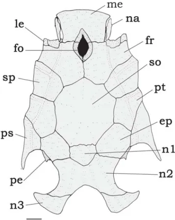
Fig. 8. Neurocranium of Tatia aulopygia, INPA 11080, 80.0 mm SL. Dorsal view. Abbreviations: ep, epioccipital; fo, single
cranial fontanel, fr, frontal; le, lateral ethmoid; me, mesethmoid; na, nasal; n1, first nuchal plate, n2; second nuchal plate; n3,
third nuchal plate; pe, posterior epioccipital process; ps,
Distribution.Tatia aulopygia occurs in the Madeira river drainage of the Amazon basin. Most records are from upper reaches, in the Guaporé and Mamoré rivers (Fig. 6).
Remarks.A short cranial fontanel, limited to the frontal in T. aulopygia, is unique among known Tatia species. In specimens less than 60 mm SL, however, the cranial fontanel is larger, its opening extends between the mesethmoid and frontals (exemplified by MNHN 1988-994). An ontogenetic shift may occur, as adult specimens have a much reduced opening. A reduced cranial fontanel is found in adult Centromochlus perugiae and C. romani, but in these spe-cies the opening is contained by the mesethmoid and frontal (see Soares-Porto, 1998, fig. 4 for comparison).
Tatia aulopygia is somewhat similar in color pattern to three congeners: T. neivai,T. dunni, and T. intermedia. All three species also have the caudal fin with spots or vertical bars, with T. aulopygia most closely resembling T. neivai. Diagnostic features aside, T. aulopygia is further distin-guished from T. neivai by having a large first nuchal plate (Fig. 8, vs. reduced in T. neivai).Tatia aulopygia is further distinguished from T. dunni by having a shallower snout, depth 41.5-45.9% HL (vs. 47.0-51.8% in T. dunni).Tatia aulopygia is further distinguished from T. intermedia by a narrower interorbital distance, 53.2-58.9% HL (vs. 60.1-63.6% HL in T. intermedia).
Material examined. 17 specimens (21.8-159.0 mm SL). Syntypes.
Brazil: NMW 47331, 1 (50.0 mm SL) and NMW 47333, 1 (55.0 mm SL), Guaporé river (syntypes of Centromochlus aulopygius).
Non-type specimens. Bolivia: Beni: AMNH 39818, 2 (25.8-27.8
mm SL), Itenez river; INHS 37034, 1 (75.2 mm SL), Matos river, Apere river drainage, Amazon basin; MNHN 1988-994, 1 (56.3 mm SL) (R), Mamoré river drainage; UMMZ 204834, 1 (24.6 mm SL), Baures river, about 500 m upstream from mouth of Itenez
river. Brazil: Amazonas: ZMA 114.280, 2 (39.6-46.8 mm SL) (R), Madeira river drainage at Humaitá. Rondônia: FMNH 58015, 3 (21.8-43.8 mm SL), Guaporé river in Maciel; INPA 11078, 1 (76.2 mm SL), INPA 11079, 1 (159.0 mm SL) and INPA 11080, 3, 1 CS (80.0-104.6 mm SL) Guaporé river.
Tatia boemia Koch & Reis, 1996
Fig. 1, 10-12
Tatia boemia Koch & Reis, 1996: 86, fig. 2 [type locality: Brazil, Rio Grande do Sul, Esmeralda, Pelotas river, road Anita Garibaldi to Pinhal da Serra]. Burgess & Finley, 1996:166 [reference]. Soares-Porto, 1998: 333 [citation]. Ferraris, 2003:476 [checklist]. Ministério do Meio Ambiente, 2004: 140 [endangered species]. Ferraris, 2007: 77 [checklist].
Diagnosis.Tatia boemia is distinguished from other species ofTatia by its unique color pattern of dorsum and dorsolat-eral sides greyish-brown with small dark chromatophores. Chromatophores become sparse and faint towards the ven-tral portion of the body (cf. Koch & Reis, 1996). The species is also distinguished by the following combination of charac-ters: nasal ossified with wide medial flanges partially sutured to lateral border of mesethmoid; pectoral fin with four branched rays; ribs attached to consecutive post-Weberian vertebrae; with two vertebrae alternatingly unribbed and ribbed (Fig. 1). Additional features useful for distinguishing species include: first nuchal plate somewhat elliptical to rounded; eye 17.4-22.8% HL; caudal-fin lobes of equal length in mature females, upper lobe slightly elongated in mature males; number of vertebrae 34.
Description. Measured adult specimens 52.4-64.5 mm SL;
morphometric data presented in Table 3. Body slim, head slightly depressed dorso-ventrally. Head large, robust, out-line of head in dorsal view elliptic, broader than long. Dorsal outline of trunk from dorsal-fin base to caudal peduncle gradu-ally compressed posteriorly. Lateral profile of head from snout tip to above opercular margin slightly convex to pectoral-fin insertion. Ventral profile of head and abdomen flat. Ventral profile of body concave posterior to anal fin.
Head integument thin, cranial roof visible; well-developed adipose eye lid; eye latero-dorsally located in anterior por-tion of head; mouth terminal, upper lip extended postero-laterally as well-developed fleshy rictal fold; snout margin rounded; anterior nostril tubular, located on anterior border of snout, above lip; posterior nostril large, rounded, limited by small skin flap; transverse distance between anterior nos-trils slightly shorter than distance between posterior ones. Maxillary barbel moderate in size, extending beyond poste-rior tip of postcleithral process, reaching vertical through middle of dorsal fin; four mental barbels, tips not reaching pectoral-fin base, arranged in arc along ventral surface of jaw; inner mental barbel about 50.0-61.0% length of outer mentals. Postcleithral process well developed, almost
reach-Fig. 9. Male modified anal fin of Tatia aulopygia, INPA 11080, 80.0 mm SL. Left side lateral view. Abbreviations: ac, antrorsely
curved denticulation; b4, branched fourth ray; b8, branched
eighth ray; dd, deferent duct; rc, retrorsely curved
denticula-tion;sf, skin flap; ui, unbranched first ray; uii, unbranched
ing vertical through middle of dorsal fin. Caudal peduncle deep, its depth about 13.6-14.2% SL.
Rostral border of cranium broad with large mesethmoid; premaxilla underneath with synchondral articulation; cranial fontanel narrow, elliptical, bounded by mesethmoid and fron-tal (Fig. 11); nasal ossified, with medial flanges partially su-tured to lateral margin of mesethmoid; autopalatine tubular, oriented obliquely to longitudinal axis of body; maxilla small, shorter than autopalatine; prevomer expanded with well de-veloped arrow-shaped lateral processes; jaws of equal size; premaxilla and dentary with three to four rows of conical teeth; first nuchal plate somewhat elliptical; second nuchal plate laterally concave, partially in contact or not with supraoccipi-tal; third nuchal plate relatively straight, projected laterally. Epioccipital process small.
Suspensorium, hyoid arch, branchial skeleton and oper-cular bones as in generic description.Suprapreopercle present as short canal bone. Six branchiostegal rays articulated with hyoid arch: four with anterior ceratohyal and two with poste-rior ceratohyal. Branchial skeleton as for genus. Basibranchial
2 forming osseous rod with broad cartilaginous anterior tip, separated from shorter basibranchial 3.
Six infraorbital bones in complete series. Infraorbital 1 broad, with moderately developed ventro-lateral process, around anterior border of eye; remaining infraorbitals thin, reduced to canalicular portions. Infraorbital 2 smallest, close to infraorbital 1, followed by three elongate canal bones, form-ing bottom orbital rim; infraorbital 5 small, formform-ing posterior orbit. Lateral line on body with ossified canal bones until vertical through pelvic fin.
Dorsal fin I,5 (n=8); dorsal-fin spine with 13-16 antrorse serrations along entire anterior margin; posterior margin smooth. Pectoral fin I,4 (n=8); pectoral-fin spine with 19-22 antrorse serrations along anterior margin, small serrations close to spine base; 14-15 retrorse serrations along posterior margin; serrations along both margins progressively larger towards spine tip. Pelvic-fin i,5 (n=8), margin rounded. Adi-pose fin large, origin on vertical through middle anal-fin base. Anal fin iii,7 (n=8); anal-fin pterygiophores in eight rod-like proximal radials and seven cartilaginous distal radials (Fig. 1).
Fig. 10.Tatia boemia,(a) MZUSP 47921, paratype, male, 64.5 mm SL. (b)MZUSP 47921, paratype, female, 52.4 mm SL, rio
Caudal fin forked, lobes with rounded tips, 8+9 principal rays, 19-21 upper procurrent, 19-20 lower procurrent rays (n=8). First nine post-Weberian vertebrae ribbed. Tenth vertebrae correspond to a gap, with no ribs attached, plus one rib at-tached to 11th vertebrae (Fig. 1). Post-Weberian vertebrae 34
(n=3).
Color in alcohol.Color pattern diagnostic within Tatia, as stated in the original description by Koch & Reis (1996): Dor-sal surface of head, back and upper sides, greyish-brown with many chromatophores surrounding small lighter (depigmented) areas; sides becoming paler ventrally as chro-matophores become progressively more widely spaced; lower sides and ventral surfaces yellowish. Dorsal-fin spine dark. Lips, anterior nostril and chin whitish. Barbels, posterior part of nuchal shield, pectoral, pelvic, anal, and adipose fins un-pigmented. Caudal fin largely pale with small dark spots.
Sexual dimorphism. Based on examination of gonads, T. boemia attains sexual maturity above 52.4 mm SL. In mature females a genital papilla is not evident. In mature males a genital papilla is visible, thick, with a short deferent duct. The
anal fin of the mature males (Fig. 12) is strongly modified with the three unbranched and first branched rays enlarged and thickened. The first unbranched ray is non-segmented and shortest, about half-the length of second unbranched ray (Fig. 12). First unbranched anal-fin ray is immediately pre-ceded by a tegumentary keel (Fig. 12, tk). The second un-branched ray has an intermediate size between the neighbor-ing first and third rays. Third unbranched and first branched are the longest rays forming a fin tip (Fig. 12, uiii). Third un-branched with the three distal segments smaller, antrorsely curved (Fig. 12, ac). First branched ray with four distal seg-ments retrorsely curved (Fig. 12, rc). The posterior branched rays are progressively shorter.
The hemal spines 15-17 are associated with anal-fin pterygiophores in males and become thick during maturity. Female hemal spines 15-18 are associated with pterygiophores and undifferentiated (Fig. 1).
There is a discrete sexual dimorphism regarding the cau-dal-fin margin in mature males of T. boemia. The upper cau-dal-fin lobe is slightly elongate, about 10.0% longer than the lower lobe, whereas mature females have equal lobes.
Distribution.Tatia boemia is endemic to upper reaches of the Uruguay river drainage (Fig. 6). It is the most southernly
Range Mean SD N
Standard length 52.4-64.5 56.6 5 Percents of standard length
Body depth 21.7-26.3 23.1 1.45 5 Body width 18.6-20.0 19.2 0.47 5 Caudal peduncle depth 13.6-14.2 13.9 0.46 5 Caudal peduncle length 23.5-26.0 24.8 1.82 5 Predorsal length 29.7-32.6 31.1 0.91 5 Preanal length 71.2-74.8 72.8 1.82 5 Prepelvic length 52.2-57.2 54.0 1.71 5 Dorsal origin to pectoral origin 21.3-24.5 22.4 1.07 5 Dorsal origin to pelvic origin 31.0-36.7 34.0 1.86 5 Pectoral origin to pelvic origin 35.3-39.6 37.2 1.98 5 Prepectoral length 18.6-23.3 21.0 1.59 5 Dorsal-fin base length 8.8-10.7 9.6 0.56 5 Adipose-fin base length 11.0-13.0 12.0 0.70 5 Anal-fin base length 3.7-10.7 7.5 2.85 5 Dorsal-fin spine length 15.0-18.7 16.1 1.17 5 Pectoral-fin spine length 19.8-21.8 21.5 0.75 5 Postcleithral process length 16.6-19.4 18.5 0.85 5 First branched pelvic-fin ray 9.9-14.5 12.9 1.50 5 Longest anal fin ray 6.5-10.4 8.7 1.50 5 Maxillary barbell length 30.5-36.1 33.4 1.78 5 Outer mental barbel length 7.8-10.3 9.0 0.96 5 Mental barbel length 4.3-6.1 5.2 0.56 5 Head length 22.0-25.2 23.2 1.21 5
Percents of head length
Head width 74.1-78.9 76.5 1.98 5 Snout depth 51.4-55.6 53.0 1.81 5 Interorbital distance 52.9-58.2 55.5 1.95 5 Left internarial width 23.1-24.6 24.1 0.69 5 Anterior internarial distance 35.9-37.3 36.4 0.66 5 Posterior internarial distance 39.4-44.7 41.5 1.99 5 Snout length 37.7-43.6 39.9 1.98 5 Orbital diameter 17.4-22.8 20.7 1.79 5 Mouth width 39.0-43.2 41.2 1.89 5
Table 3. Morphometric data for Tatia boemia. SD = standard deviation.
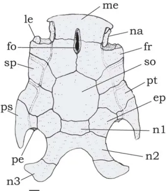
Fig. 11. Neurocranium of Tatia boemia, MCP 12949, paratype, 59.9 mm SL. Dorsal view. Abbreviations: ep, epioccipital; fo,
single cranial fontanel; fr, frontal; le, lateral ethmoid; me,
mesethmoid;na, nasal; n2; second nuchal plate; n3, third
tributed species of Tatia. Together with T. neivai both are the only species inhabiting the La Plata basin (Koch & Reis, 1996).
Remarks. Tatia boemia is the only auchenipterid catfish listed in Brazil as an endangered species (Ministério do Meio Ambiente, 2004). As it is a typically nocturnal catfish, the local people call T. boemia under the common name “boa noite”, meaning good night (Walter R. Koch, pers. comm.).
InT. boemia the first (anterior) nuchal plate is variable in size. First nuchal plate is sometimes assymmetrical, permiting contact between supraoccipital and second nuchal plate (as in Fig. 11), or symmetrical, bordered by supraoccipital and second (middle) nuchal plate. We observed variation in first nuchal plate size in other auchenipterid species, such as Centromochlus perugiae and Glanidium leopardus. Ante-rior (first) nuchal plate either fully developed or reduced is reported in a few doradid species as Oxydoras niger and Doras fimbriatus (Birindelli et al., 2007: 680), and the species are considered polymorphic regarding this character. Poly-morphism seems to be also the case in the above mentioned auchenipterids including T. boemia.
Tatia boemia is presumed to be sister to T. neivai, both sharing the presence of a single vertebrae without ribs pre-ceding the last ribbed vertebrae (character 17, fig. 14 of Soares-Porto, 1998). This ribless vertebra (number 9 in T. neivai, 10 in T. boemia) has each transverse process with a reduced costal facet. The last rib pair is small and attached to the hemal arch of vertebra 10 (T. neivai) or 11 (T. boemia). In all other Tatia species the ribs are attached to consecutive post-Weberian vertebrae.
Material examined. 8 specimens (33.1-64.1 mm SL). Paratypes.
Brazil: Rio Grande do Sul: MCP 12949, 6, 1 CS, (33.1-61.1 mm SL) and MZUSP 47921, 2 (52.4-64.5 mm SL) (R), Esmeralda, Pelotas river, road Anita Garibaldi to Pinhal da Serra (paratypes of Tatia boemia).
Tatia brunnea Mees, 1974
Figs. 13-15
Tatia brunnea Mees, 1974: 84, fig. 21 [type locality: Suriname, Compagnie stream]. Mees, 1983: 46 [French Guiana: streams Balaté & Awahakiki]. Mees, 1985: 242 [Suriname, Loë stream]. Mees, 1988: 411 [French Guiana: Sinnamary, Petit-Saut]. Soares-Porto, 1998: 331-350 [citation]. Kobayagawa, 1991:104 [reference]. Wallace, 2002: 297 [Negro river]. Ferraris, 2003:475 [checklist]. Ferraris, 2007: 77 [checklist].
Tatia cf. intermedia. Mees, 1983: 46, fig. 2 [Blanche stream, Acarouany, French Guiana]. Le Bail et al., 2000:68 [refer-ence].
Tatia aulopygia. Goulding et al., 1988: 180 [Negro river]. Tatia sp. cf. brunnea. Burgess, 1989: 595, pl. 113 [tropical
South America].
Tatia intermedia. Burgess, 1989: 242 [Guianas]. Soares-Porto, 1998: 333 [citation].
Diagnosis.Tatia brunnea is distinguished from its conge-ners by the male anal fin with sharp pointed tip; the first unbranched anal-fin ray divided into 3-4 segments; and the last branched ray reduced (Fig. 15). Tatia brunnea differs from most of its congeners, except T. aulopygia, T. intermedia andT. gyrina by having a wide mouth, width 54.0-59.7% HL (vs. 39.0-53.3% HL). Additional characteristics for recogni-tion of T. brunnea are diagnostic in combination: Nasal ossi-fied with wide medial flanges partially sutured to lateral mar-gin of mesethmoid; ribs 9-10; post-Weberian vertebrae 34-36. Additional features useful for distinguishing T. brunnea in-clude details in coloration, such as: border of mouth whitish, contrasting with dark head; posterior border of nuchal shield usually whitish or pale; pectoral-fin spine usually with trans-verse bands; and caudal fin usually whitish with scattered dark brown blotches.
Description. Measured adult specimens 54.6-97.4 mm SL;
morphometric data presented in Table 4. Body deep, head depressed dorso-ventrally. Head robust, outline of head in dorsal view somewhat elliptic, broader than long. Trunk from dorsal-fin base to caudal peduncle gradually compressed lat-erally. Outline of head in dorsal view from snout tip to oper-cular margin slightly convex until pectoral-fin insertion. Ven-tral profile of head and abdomen slightly convex. VenVen-tral pro-file of body gently curved, concave behind anal-fin origin.
Head integument thin, cranial roof visible; well-developed adipose eye lid; eye latero-dorsally located in anterior por-tion of head; mouth terminal, upper lip extended postero-laterally as well-developed fleshy rictal fold; anterior nostril tubular, located on anterior border of snout, above lip; poste-rior nostril large, rounded, limited by small skin flap; trans-verse distance between anterior nostrils proportionally the same distance between posterior ones in HL. Maxillary bar-bel short, extending close to posterior tip of postcleithral pro-cess, sometimes larger; mental barbels short, tips not
reach-Fig. 12. Male modified anal fin of Tatia boemia, MCP 12949, paratype, 59.9 mm SL. Left side lateral view. Abbreviations:
ac, antrorsely curved denticulation; b1, branched first ray; b7, branched seventh ray; dd, deferent duct; rc, retrorsely
curved denticulation; tk, tegumentary keel; ui, unbranched
first ray; uii, unbranched second ray; uiii, unbranched third
ing pectoral-fin base, arranged in arc along ventral surface of jaw; inner mental barbel about 55.0-65.0% length of outer mental barbel. Postcleithral process almost reaching vertical through middle or end of dorsal fin. Caudal peduncle deep, depth about 14.3-17.4% SL.
Rostral border of cranium broad with mesethmoid as long as broad; premaxilla underneath with synchondral articula-tion; cranial fontanel elliptical, bounded by mesethmoid and frontal (Fig. 14); nasal ossified with narrow medial flanges, not sutured to mesethmoid in immature specimens, but su-tured to mesethmoid in adults; autopalatine tubular, oriented obliquely to longitudinal axis of body; maxilla about same size of autopalatine; prevomer expanded anteriorly with well developed arrow-shaped lateral processes; jaws of equal size; premaxilla and dentary with three to four rows of conical teeth. First nuchal plate trapezoid; second nuchal plate slightly con-cave along lateral margins; third nuchal plate curved, pro-jected laterally, with broad tip. Epioccipital process very small. Suspensorium, hyoid arch, branchial skeleton and oper-cular bones as in generic description. Suprapreopercle present as short canal bone. Five to six branchiostegal rays
articu-lated with hyoid arch: three or four with anterior ceratohyal and two with posterior ceratohyal; last two flattened and ex-panded; basibranchial 2 forming osseous rod with broad carti-laginous anterior tip, separated from shorter basibranchial 3.
Five infraorbital bones in incomplete series. Infraorbital 1 thin with short ventro-lateral process; remaining infraorbitals thin, reduced to canalicular portions; infraorbital 2 smallest, followed by non-ossified portion of canal below eye and by two posterior canal bones much close to sphenotic, forming posterior orbital rim. Lateral line on body with ossified canal bones posteriorly to vertical of pelvic fin origin.
Dorsal fin I,5 (n=24); dorsal-fin spine with 13-14 antrorse serrations along anterior margin; posterior margin smooth. Pectoral fin I,4-5 (n=24); pectoral-fin spine with 19-21 antrorse serrations along anterior margin; 12-14 retrorse serrations along posterior margin; serrations along both margins pro-gressively larger towards spine tip. Pelvic-fin i,5 (n=24); mar-gin rounded. Adipose fin large, orimar-gin on vertical through middle anal-fin base. Anal fin iii, 7, rarely iii, 8 (n=24); anal-fin pterygiophores in seven to eight rod-like proximal radials and six to seven cartilaginous distal radials. Caudal fin forked,
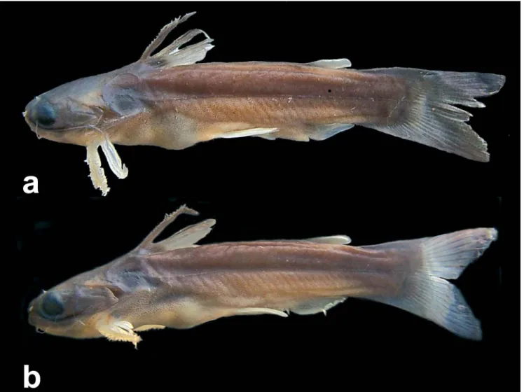
Fig. 13. Tatia brunnea.(a)AMNH 58390, paratype, male, 54.6 mm SL, Kamaloe Stream, Suriname. (b) MZUSP 81250, female,
lobes with rounded tips, 8+9 principal rays, 18-22 upper procurrent, 14-20 lower procurrent rays (n=24). Pleural ribs 9-10, attached to consecutive vertebrae. Post-Weberian verte-brae 34-36 (n=13).
Color in alcohol. Color pattern considered as diagnostic in
original description by Mees (1974: 84): color dark brown in fresh specimens, with vague pale areas; older specimens with wavy longitudinal bands and dots of dark brown, alternating with pale brown areas; dorsal shield usually distinctly paler than body; pectoral, dorsal and adipose fins spotted with brown, dorsal fin largely dark brown; ventrals and anal fins hyaline, caudal fin hyaline with large irregular blackish brown dots. Contour of lips and anterior nostrils usually whitish. Pectoral fin usually with transversal dark bands. Live coloration in T. dunni aquarium specimen illustrated by Burgess (1989: pl. 113). Large preserved specimens (over 90 mm SL) with dimin-ished color pattern. These individuals bear spots, blotches or even stripes generally much less defined.
Color variation. Regional variation in pigmentation was
ob-served in T. brunnea (as exemplified in Fig. 13). Suriname specimens attain smaller adult size (50-72 mm SL) and have
irregular stripes or blotches along body (Fig. 13a). Specimens from Negro river attain larger size (60-97.4 mm SL), and are mottled with dark and light brown areas (Fig. 13b). Some are completely dark brown along sides of body, with caudal-fin lobes irregularly striped (MZUSP 81139).
In the headwaters of upper Negro river, draining the Cerro de Neblina Mountains, the catfishes attain the largest size observed for the species, 116 mm SL, and have large irregular stripes on sides of body. A single population of T. brunnea was found in central Amazon, in the Trombetas river. These fishes have dark brown bands at the center of each caudal-fin lobe, a coloration pattern also observed in some specimens from the Negro river (INPA 15989).
Sexual dimorphism. Based on examination of gonads, T. brunneaattains sexual maturity above 54.6 mm SL. The up-per Amazon population, however, is found to consist of larger individuals and maturation was attained above 70 mm SL. In mature females a genital papilla is not evident. The genital papilla of mature male is visible, with an emergent deferent duct. The anal fin of mature males (Fig. 15) is strongly modi-fied, with three thickened unbranched rays. The first un-branched ray is the shortest ray, about three-quarter length of second unbranched ray. First unbranched ray is segmented,
Fig. 14. Neurocranium of Tatia brunnea, RMNH 26198, paratype, 39.1 mm SL. Dorsal view. Abbreviations: ep,
epioccipital;fo, single cranial fontanel, fr, frontal; le, lateral
ethmoid;me, mesethmoid; na, nasal; n1, first nuchal plate, n2; second nuchal plate; n3, third nuchal plate; pe, posterior
epioccipital process; ps, posttemporal-supracleithrum; pt,
pterotic;so, supraoccipital; sp, sphenotic. Scale bar =1.0 mm.
Table 4. Morphometric data for Tatia brunnea. SD = standard deviation.
Range Mean SD N
Standard length 54.6-97.4 62.1 24 Percents of standard length
Body depth 15.7-23.8 19.6 1.75 24 Body width 17.5-23.4 20.0 1.46 24 Caudal peduncle depth 14.3-17.4 15.5 1.31 24 Caudal peduncle length 24.2-29.4 27.0 1.55 24 Predorsal length 25.2-34.8 31.2 1.64 24 Preanal length 63.1-71.4 66.4 2.22 24 Prepelvic length 46.2-53.4 50.2 1.65 24 Dorsal origin to pectoral origin 20.4-24.7 22.3 1.23 24 Dorsal origin to pelvic origin 26.6-33.2 29.9 1.72 24 Pectoral origin to pelvic origin 30.1-37.3 33.4 1.59 24 Prepectoral length 18.1-22.8 20.0 1.15 24 Dorsal-fin base length 7.8-12.5 9.9 1.30 24 Adipose-fin base length 8.4-15.8 11.1 1.92 24 Anal-fin base length 4.4-9.4 7.4 2.04 24 Dorsal-fin spine length 13.4-22.1 17.4 1.82 24 Pectoral-fin spine length 16.8-28.0 23.3 2.22 24 Postcleithral process length 15.7-21.8 18.5 1.58 24 First branched pelvic-fin ray 10.1-14.9 12.5 1.55 24 Longest anal fin ray 5.3-12.5 8.9 1.63 24 Maxillary barbel length 28.0-34.0 31.0 1.82 24 Outer mental barbel length 7.2-10.5 8.7 0.74 24 Mental barbel length 4.3-6.6 5.4 0.53 24 Head length 21.0-26.1 23.2 1.24 24
Percents of head length
usually with 3-5 separated dermal segments (lepidotrichia). A segmented first unbranched ray was observed in most Suriname specimens, but not in all, and may be associated to regional differentiation. The first unbranched anal-fin ray is immediately preceded by a tegumentary keel (Fig. 15, tk). The second unbranched ray has an intermediate size between the neighboring first and third rays. Third unbranched is the long-est ray forming a long sharp pointed fin tip together with the first branched (Fig. 15, uiii). Third unbranched distal seg-ments are antrorsely curved (Fig. 15, ac). First branched ray bearing retrorsely curved distal segments (Fig. 15, rc). Poste-rior branched rays are normally developed and progressively shorter; with last ray reduced (Fig. 15, b7).
Hemal spines 16-19 interdigitate with the anal-fin pterygiophores; hemal spines 15-17 or 16-18 are thickened in mature males, but undifferentiated in females. The caudal-fin lobes have the same length in mature females, whereas upper lobe is elongated in mature males.
Distribution.Tatia brunnea was described from the Suriname and Marowijne-Maroni river basins in Suriname. It was re-corded in French Guiana from the Maroni and Sinnamary river basins. In Brazil it occurs in the Negro river drainage and at a single locality in Central Amazon basin, in the Trombetas river drainage (Fig. 6).
Remarks. Until recently, T. brunnea was thought to be re-stricted to Suriname (Ferraris, 2003), although it was previ-ously recorded in the Negro river as well (Wallace, 2002). The overall coloration of T. brunnea ressembles that of T. dunni, from upper Amazon. Details in coloration of caudal fin helps to distinguish between these two species, as in T. brunnea the caudal fin is whitish with dark spots or bands (vs. darker with whitish blotches in T. dunni). Additional distinctions include a wide head in T. brunnea, 86.6-93.4% HL (vs. narrow inT. dunni, 76.0-80.1% HL); a wide mouth, 54.0-59.7% HL (vs. narrow, 48.1-52.3% HL); and a male modified anal fin with sharply pointed distal tip (vs. short rounded distal tip). Tatia brunnea occurs in sympatry with T. intermedia in some riv-ers in Suriname and French Guiana and also in the Trombetas river.
Material examined. 128 specimens (16.2-97.4 mm SL).
Holo-type. Suriname: RMNH 26196, 56.5 mm SL, Compagnie stream
(holotype of Tatia brunnea).Paratypes. Suriname: AMNH 58390,
1 (54.6 mm SL) (R), Kamaloe stream, right margin of Marowijne river. RMNH 26197, 3 (47.6-60.0 mm SL), Compagnie stream; RMNH 26198, 3, 1 CS (35.1-40.3 mm SL), Kwambaolo stream, near dam; ZMA 105.526, 4 (26.5-36.6 mm SL), Gran river, 63 Km south of Affobakka; ZMA 105.860, 1 (41.5 mm SL) (R), Sara stream, about 27 Km south of dam; ZMA 105.849, 7 (27.0-52.2 mm SL) (R), Maka stream, tributary of Lawa river, Marowijne district (paratypes of Tatia brunnea). Non-type specimens: Brazil:
Amazonas: ANSP 165747, 2, igarapé Castanho, Negro river; CAS 76790, 2 (70.3-80.2 mm SL), Cuieras river; INPA 14228, 2 (96.9-97.4 mm SL), Urubu river, igarapé of Gavião, Farm Esteio, Negro river basin; INPA 15989, 1 (66.6 mm SL), Presidente Figueiredo, Urubu river, Negro river basin; INPA 16577, 1 (89.0 mm SL),
Jauaperi river, igarapé Cambina, Negro river basin; MCZ 52670, 2 (28.2-37.0 mm SL), Cuieras river in isolated pool; MZUSP 9352, 1 (18.6 mm SL), Central lake, left margin of Negro river between Camanaú and Apeú rivers; MZUSP 31075, 1 (34.0 mm SL), Negro river, Barcelos, island lake; MZUSP 44126, 1 (38.3 mm SL); MZUSP 44258, 2 (23.4-36.7 mm SL), Negro river, Anavilhanas archipaelago; ZMA 119.949, 1 (58.0 mm SL), Negro river and tributaries; MZUSP 81139, 1 (78.0 mm SL), Tiquié river, between communities of Caruru and Boca de Sal, Negro river drainage; MZUSP 81177, 2 (36.9-59.4 mm SL), Tiquié river, mouth of igarapé Açaí, near São Pedro community, Negro river drainage; MZUSP 81250, 10, 1 CS (44.6-81.7 mm SL), Tiquié river, between communities of São Pedro and Caruru upstream from waterfalls, Negro river drainage; ZMA 119.949, 1 (58.0 mm SL), Negro river and tributaries. Pará: MNRJ 15332, 1 (32.7 mm SL), MNRJ 15333, 1 (44.4 mm SL) and MNRJ 15334, 3, 1 CS (31.1-43.4 mm SL), igarapé Saracazinho, tributary of Batata lake, Porto Trombetas. French Guiana: RMNH 28570, 3 (29.6-50.0 mm SL), Awahakiki river; RMNH 28569, 2 (34.3-41.4 mm SL) and RMNH 28571, 4, 1 CS (25-37 mm SL), Balaté stream; RMNH 30494, 1 (70.0 mm SL), Petit-Saut, Sinnamary. Suriname: AMNH 58391, 2 (65.3-75.4 mm SL), Suriname river near Botopasi; RMNH 27530, 3 (26.7-28.0 mm SL), upper Loë river, tributary of Litani river; RMNH 28568, 2 (42.5-45.0 mm SL), stream below Acarouany; RMNH 28654, 1 (51.5 mm SL), stream below Bivouac downstream from Lombok waterfalls; RMNH 28655, 1 (54.0 mm SL), tributary at right margin of Nickerie river below Blanche Marie Falls; RMNH 28656, 2 (50.0-81.0 mm SL), tributary at right margin of Kaboeri stream, Corantjn river basin; RMNH 28658, 2 (45.1-74.0 mm SL), tributary at right margin of Kabalebo river, about 8 Km below Avanavero waterfalls; USNM 226124, 1, stream south of Matapi, Nickerie district; USNM 226125, 5 (16.2-23.3 mm SL), tributary of Corantijn river, north of Tiger Falls, Nickerie district; ZMA 105.831, 3 (34.4-60.2 mm SL), tributary of Nickerie river south of Stondansie Vallen.
Tatia caxiuanensis, new species
Figs. 16-18
Holotype.MPEG 9859, male (32.6 mm SL), Brazil, Pará: município
de Melgaço, Estação Científica Ferreira Pena, Curuá river, Caxiuanã, 01o44’53”S 51o27’13”W, 1 Nov 1999, R. Barthem and team.
Fig. 15. Male modified anal fin of Tatia brunnea, RMNH 26196, holotype, 56.5 mm SL. Left side lateral view. Abbrevia-tions:ac, antrorsely curved denticulation; b1, branched first
ray;b7, branched seventh ray; dd, deferent duct; rc, retrorsely
curved denticulation; tk, tegumentary keel; ui, unbranched
first ray; uii, unbranched second ray; uiii, unbranched third