Financial Support from Fundação para a Ciência e Tecnologia (FCT) – Ph.D. grant SFRH/BD/43207/2008.
Work performed at:
Control of Gene Expression Laboratory Instituto de Tecnologia Química e Biológica
Av. da República (EAN)
2781-901 Oeiras – Portugal Tel: +351-21-4469548
Doutora Cecília Maria Pais de Faria de Andrade Arraiano – Investigadora Coordenadora, Instituto de Tecnologia Química e Biológica, Universidade Nova de Lisboa.
Presidente (President of the Jury):
Doutora Claudina Amélia Marques Rodrigues Pousada -
Professora
Catedrática Convidada do Instituto de Tecnologia Química e Biológica da
Universidade Nova de Lisboa, por delegação.
Vogais (Members of the Jury)
:
Doutor Phil J. Mitchell – Professor, University of Sheffield, Reino Unido.
Doutora Luísa Maria Ferreira Romão Loison – Investigadora Principal com Habilitação, Instituto Nacional de Saúde Dr. Ricardo Jorge.
Doutor Francisco Javier Enguita Lombardo – Professor Auxiliar da Faculdade de Medicina da Universidade de Lisboa.
“The noblest pleasure is the joy of understanding.”
I would like to thank all the people that directly and indirectly contributed and supported this work. This Ph.D. dissertation would not have been possible without the support of:
Fundação para a Ciência e Tecnologia (FCT) for the financial support crucial of
my Ph.D. fellowship (SFRH / BD / 43207 / 2008).
Instituto de Tecnologia Química e Biológica (ITQB) for providing me the
facilities and resources for the execution of this work.
Professor Cecília Arraiano for giving me the opportunity to start this big
adventure, for all the support and endless enthusiasm. I am also thankful for all the opportunities to expose my work in several national and international meetings and for giving me the opportunity to spend some time working in other laboratories, which was very important to expand my scientific knowledge.
Ana Barbas for all the support during my Ph.D. and for giving me the confidence
to perform the practical work without being afraid to fail.
Michal Malecki for his interest in my research, the fruitful work discussions and
for reading this Dissertation.
To all the past and present colleagues and friends in the lab for making it such an enjoyable place to be and work at: Ana, Andreia, Cátia, Clémentine, Inês Guinote, Inês Silva, Joana, Margarida Matos, Margarida Saramago, Michal,
Mauro, Patrick, Raquel Santos, Ricardo Moreira, Ricardo Santos, Rute, Sandra,
Professor Ambro van Hoof and his group at Department of Microbiology and
Molecular Genetics, University of Texas Health Science Center – Houston, Texas, for kindly receiving me in his laboratory, and for all the guidance and comments since the beginning of this work. A special thanks to Ale Klauer and Daneen Schaeffer for their help and support during my stay there.
Thank you to our collaborators in Centro de Biologia Molecular “Severo Ochoa” (CSIC-UAM), Campus Universidad Autonoma de Madrid, Spain, Dr. Paulino Gómez-Puertas and Dr. Eduardo López-Viñas.
To all my colleagues in the other labs in the Institute. Thank you for having participated in this work by discussing science during coffee breaks and lunch. A special thanks to Dr. Collin McVey, for helping and advising me in all the questions I could have about proteins.
Thank you to my Mom for being unconditionally supportive.
Thank you to my Dad, an absence that is always present. We have had some Victories together!
Thank you to my brother and my sister in law, Sara, for giving me the right words and incentives to follow my path.
Thank you to all my friends and family, Grandparents, Uncles, Aunts and Cousins. It is impossible to acknowledge you all, one by one. A special thanks to my Aunt Teresa, Uncle Nuno and Aunt Gaby, who have always shown a special interest in my studies.
Table of Contents
ABSTRACT ... XIII
RESUMO... XIX
LIST OF PUBLICATIONS... XXV
DISSERTATION OUTLINE ... XXIX
ABBREVIATIONS ... XXXIII
CHAPTER 1 ... 1
CHAPTER 2 ... 33
CHAPTER 3 ... 67
CHAPTER 4 ... 101
CHAPTER 5 ... 139
CHAPTER 6 ... 159
The cellular concentration of a given RNA is the result of the balance between its synthesis and degradation. RNA degradation plays a fundamental role in RNA metabolism, since the fast turnover of mRNA is important for the control of gene
expression. One of the key components of RNA metabolism are enzymes from the RNase II family. Members of this family are highly processive hydrolytic enzymes that play important roles in RNA decay, processing and quality control
pathways. RNase II is the prototype of this family of exoribonucleases and RNase II homologues can be found in all domains of life. Members of this family share sequence identity, a similar domain organization and a highly conserved domain
responsible for exonucleolytic activity, the RNB domain. While in Escherichia coli, members of this family function as individual proteins, in yeast some of them function in the context of a multiprotein complex called the exosome.
The first objective of this PhD work was to characterize a domain at the
N-terminus of Rrp44, whose function was unknown in the beginning of this study. In order to address the specific role of this domain in the activity of the enzyme, Rrp44 truncated mutants were constructed and their ribonucleolytic
activity analyzed. A mutant containing only the PIN domain allowed us to demonstrate the presence of an additional endonuclease active site in Rrp44, whose activity is abolished when we mutate a conserved acidic residue important
for catalysis. Moreover, we were able to further characterize this activity since we
demonstrated that it is sequence independent, but has a preference for 5’ monophosphorylated substrates. Decades after the discovery of the major 3’-5’
exonuclease activity in eukaryotes, this work has demonstrated the presence of an endonuclease active site in Rrp44, and consequently, in the exosome. These
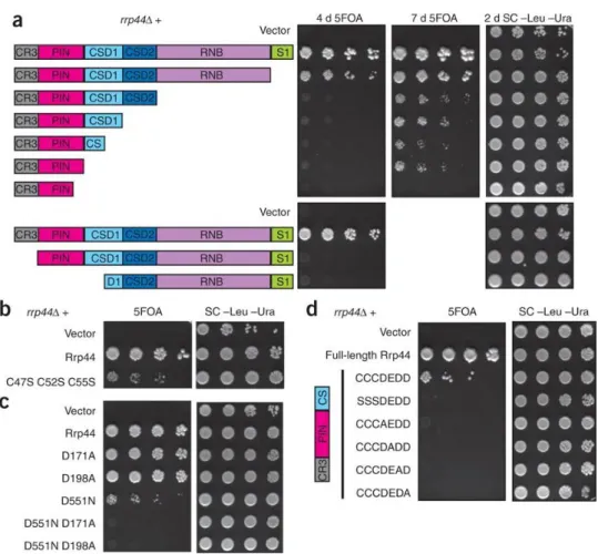
In the second part of this Doctoral project, the aim was to study the specific role of the CR3 motif at the N-terminus of Rrp44. This region had been previously identified in Drosophila melanogaster, but its function was unknown.
Previous data suggest that this region is needed for a large number of exosome functions. Also, previous structural data suggest that it could coordinate a metal ion. To evaluate the role of the N-terminus, a set of mutations in this region were
constructed. These Rrp44 mutant proteins were compared to the wild-type protein regarding their endoribonucleolytic activity. These results, coupled with genetic analysis, demonstrated that this region coordinates a metal ion and
regulates Rrp44 endonucleolytic activity and its interaction with the exosome. Additionally, these results helped to explain why mutating the CR3 region results in numerous exosome defects, both in the nucleus and the cytoplasm.
In the third part of this work, we focused on the functional characterization of three residues located in the highly conserved RNB catalytic domain: Tyr595, Gln892 and Gly895. To address experimentally their function we
carried out point mutations and tested their role in Rrp44 activity. When we mutated residues Gln892 and Tyr595 and tested their activity in vitro, the enzymes became 2 fold more active. We also showed that when we mutate
Tyr595, the final degradation product of Rrp44 changed from 4 to 6 nucleotides. This result confirms that this residue is responsible for the stacking of the RNA substrate in the catalytic cavity, as was predicted by the structure of Rrp44.
Furthermore, we also showed that a strain with a mutation in this residue has a growth defect and affects RNA processing and degradation. This result led us to
hypothesize that Tyr595 has an important biological role. This study sheds light on the model previously proposed for Rrp44 activity and mode of action, and gives new insights in RNA processing and degradation by the exosome. Since
understanding the activity of Rrp44 is essential to understanding the activity of the exosome.
To finalize this study we also studied the prototype of this family of exoribonucleases, E. coli RNase II. A thermosensitive allele that was generated in vitro, rnb500, has been essential for the determination of the cellular function of
RNase II. However, little was known about the mutation responsible for such phenotype. During this work we have sequenced the DNA from the rnb500 allele and identified two substitutions in the sequence of this allele. To determine which
mutation is responsible for the thermolability of this allele, we introduced each point mutation individually or both substitutions in the rnb gene. RNase II mutant proteins were compared to the wild-type enzyme regarding their
exoribonucleolytic activity at the non-permissive temperature. Furthermore, we also performed in vivo complementation assays using a temperature-sensitive E.
coli strain. These assays helped us to identify the mutation responsible for this phenotype - Cys284Tyr. This work also unravelled an unpredicted disulphide bridge in the RNase II structure, which is responsible for a relevant covalent
interaction between residues Cys284 and Cys399. The localization of these residues is conserved in other organisms, which led us to propose that the knowledge of these mutations can be an useful tool for studying RNase II
enzymes function in other organisms.
The results of this Dissertation allowed for the functional characterization of the N-terminal region of Saccharomyces cerevisiae Rrp44 enzyme, namely the
PIN domain and the CR3 region. These studies also contribute to a better understanding of Rrp44 activity and mode of action and, consequently, the
involvement of the eukaryotic exosome in several processes of RNA metabolism.
A concentração celular de RNA é o resultado do balanço entre a sua síntese e
degradação. A degradação de RNA desempenha um papel fundamental no
metabolismo de RNA, uma vez que a reciclagem rápida de mRNA é importante
para o controlo da expressão génica. Um dos componentes chave do metabolismo
de RNA são enzimas da família da RNase II. Os membros desta família são
enzimas processivas e hidrolíticas que desempenham papéis importantes na
degradação, processamento e controlo de qualidade do RNA. A enzima RNase II
é o protótipo desta família de exoribonucleases e enzimas homólogas podem ser
encontradas em todos os domínios da vida. Membros desta família têm uma
sequência e estrutura semelhantes, e partilham um domínio catalítico
característico, o domínio RNB. Enquanto que em Escherichia coli, os membros
desta família de proteínas funcionam individualmente, em levedura algumas
delas funcionam no contexto de um complexo multiproteico para a degradação
do RNA – o exossoma.
O primeiro objetivo deste trabalho de Doutoramento foi caracterizar um
domínio de função desconhecida na proteína Rrp44. A sua atividade
ribonucleolítica foi analisada, a fim de caracterizar o papel deste domínio na
atividade da enzima. Mutantes truncados da proteína Rrp44 foram construídos.
Um mutante contendo apenas o domínio PIN permitiu-nos demonstrar a
presença de um sítio ativo com atividade endonucleolítica na proteína Rrp44.
Quando um resíduo conservado importante para a catálise é mutado a atividade
é abolida. Adicionalmente, demonstrámos que a atividade deste domínio não
depende da sequência do substrato mas tem uma preferência para substratos 5'
monofosforilados. Este trabalho demonstrou pela primeira vez a presença de um
sítio ativo com atividade endonucleolítica na proteína Rrp44 e,
no modo de ação do exossoma, nomeadamente no mecanismo de recrutamento e
degradação de RNA, forçando o modelo do exossoma a ser reavaliado.
Na segunda parte do presente projeto de Doutoramento, o objetivo foi
estudar o papel específico do motivo CR3 no terminal N da proteína Rrp44. Esta
região tinha sido previamente identificada em Drosophila melanogaster mas a sua
função era desconhecida. Dados anteriores sugeriram que esta região é necessária
para um grande número de funções do exossoma. Além disso, dados estruturais
da proteína sugerem que esta poderia coordenar um ião metálico. Foram
construídas várias mutações nesta região para avaliar o papel do N-terminal no
funcionamento da enzima. A atividade endoribonucleolítica das proteínas
mutantes foi comparada com a proteína do tipo selvagem. Estes resultados,
juntamente com a análise genética, demonstraram que esta região coordena um
ião de metal, regula a atividade endonucleolítica da proteína Rrp44 e a sua
interação com o exossoma. Além disso, estes resultados ajudam a explicar porque
é que uma mutação nesta região ocasiona problemas na degradação pelo
exossoma, tanto no núcleo como no citoplasma.
Na terceira parte deste trabalho, debruçámo-nos na caracterização
funcional de três resíduos localizados no domínio catalítico altamente
conservado, RNB: Tyr595, Gln892 e Gly895. Para abordar experimentalmente a
sua função realizámos mutações pontuais e testámos o seu papel na atividade da
enzima Rrp44. Quando mutámos os resíduos Gln892 e Tyr595 e testámos a sua
atividade in vitro, as enzimas tornaram-se 2 vezes mais ativas. Mostrámos
também que quando se altera o resíduo Tyr595, o produto da degradação final
muda de 4 para 6 nucleótidos. Este resultado confirma que este resíduo é
responsável pelo posicionamento do RNA na cavidade catalítica, tal como foi
uma estirpe com uma mutação neste resíduo tem defeitos no crescimento e afeta
o processamento e degradação do RNA. Este resultado levou-nos a sugerir que o
resíduo Tyr595 tem um papel biológico importante. Estes resultados esclarecem o
modelo anteriormente proposto para a atividade e modo de ação da enzima
Rrp44, e dá novas perspetivas no processamento e degradação de RNA pelo
exossoma. Uma vez que a proteína Rrp44 é a única subunidade catalítica, a
compreensão da atividade da enzima Rrp44 é essencial para a compreensão da
atividade do exossoma.
Para finalizar este estudo, também estudámos o protótipo desta família
de exoribonucleases, a enzima RNase II de E. coli. Um alelo termosensível
produzido por mutagénese (rnb500) tem sido essencial para a determinação da
função celular da proteína RNase II. No entanto, pouco se sabe sobre a mutação
responsável por tal fenótipo. No âmbito deste trabalho, sequenciámos o DNA
deste alelo e identificámos duas substituições na sequência do alelo rnb500. Para
determinar qual a mutação responsável pela termolabilidade deste alelo,
introduzimos cada mutação pontual individualmente ou ambas as substituições
no gene rnb. A atividade exoribonucleolítica de proteínas mutantes da RNase II
foi comparada com a da enzima do tipo selvagem, à temperatura não permissiva.
Além disso, também realizámos ensaios de complementação in vivo utilizando a
estirpe termosensível de E. coli. Estes ensaios ajudaram-nos a identificar a
mutação responsável por este fenótipo - Cys284Tyr. Este trabalho também
desvendou uma ponte dissulfídrica não prevista na estrutura da proteína RNase
II, a qual é responsável por uma interação covalente entre os resíduos Cys284 e
Cys399. A localização desses resíduos é conservada em outros organismos, o que
nos levou a propôr que o conhecimento dessas mutações pode ser uma
ferramenta útil para estudar enzimas homólogas da RNase II em outros
Os resultados desta Dissertação permitiram a caracterização funcional da
região N-terminal enzima Rrp44 de Saccharomyces cerevisiae, isto é, o domínio PIN
e a região CR3. Estes estudos também contribuiram para uma melhor
compreensão da atividade da enzima Rrp44 e seu modo de ação.
Consequentemente, avançámos na compreensão do papel do exossoma
eucariótico em diversos processos de metabolismo do RNA. Além disso, os
resultados obtidos a partir desta Dissertação podem ser aplicados para uma
melhor compreensão dos mecanismos de degradação em outros membros desta
Scientific articles in international peer reviewed journals
Reis FP, López-ViñasE, Gómez-PuertasP and Arraiano CM. Identification of the temperature-sensitive mutation that impairs the function in vivo and in vitro of RNase II. Manuscript in preparation.
Reis FP, Barbas A, Klauer AA, Schaeffer D, López-ViñasE, Gómez-PuertasP, van Hoof A and Arraiano CM. Modulating the RNA processing and decay by the exosome: altering Rrp44/Dis3 activity and end-product. Submitted.
Reis FP, Pobre V, Silva IJ, Malecki M, and Arraiano CM. The RNB Family of Exoribonucleases – Putting the ‘Dis’ in Disease. Wiley Interdisciplinary Reviews (WIREs) RNA.Accepted.
Schaeffer D, Reis FP, Johnson SJ, Arraiano CM, van Hoof A. 2012. The CR3 motif of Rrp44p is important for interaction with the core exosome and exosome function. Nucleic Acids Res 40:9298-9307.
Arraiano CM, Andrade JM, Domingues S, Guinote IB, Malecki M, Matos RG, Moreira RN, Pobre V, Reis FP, Saramago, M, Silva, IJ and Viegas, S.C. 2010. The critical role of RNA processing and degradation in the control of gene expression.
FEMS Microbiol Rev34:883-923.
Schaeffer D, Tsanova B, Barbas A, Reis FP, Dastidar EG, Sanchez-Rotunno M, Arraiano CM, van Hoof A. 2009. The exosome contains domains with specific endoribonuclease, exoribonuclease and cytoplasmic mRNA decay activities.
Nature Structural &Molecular Biology16:56-62.
Domingues S, Matos RG, Reis FP, Fialho AM, Barbas A and Arraiano CM. 2009. Biochemical characterization of the hydrolytic exoribonucleases from the human pathogens Salmonella typhimurium and Streptococcus pneumonia. Biochemistry 48:11848-57.
Book chapters
Matos RG, Pobre V, Reis FP, Malecki M, Andrade JM and Arraiano CM. 2011.
Structure and Mechanisms of 3’ to 5’ Exoribonucleases. In: Nicholson AW, ed.
This Dissertation is divided into six chapters.
Chapter one consists of a general introduction to contextualize the reader in the subject of this Dissertation. It includes a brief overview of the degradation of
RNA, with a particular emphasis in Escherichia coli RNase II and Saccharomyces
cerevisiae Rrp44, the main focus of this Dissertation.
The results of this Doctoral work are presented in the following chapters. Each
chapter has its own Introduction, Results, Discussion, Materials and Methods,
References and Acknowledgments sections.
Chapter two consists of an article published in the Nature Structural & Molecular Biology journal in which the author of this Dissertation played an important
contribution. In this chapter several mutant proteins of Rrp44, including a
truncated protein containing the PIN domain but lacking the RNB domain, were
constructed, purified and their ribonucleolytic activity analyzed. This work led to
important conclusions regarding the role of the PIN domain.
In Chapter three the role of the CR3 region at the N-terminus was studied. Biochemical data and genetic analysis showed that this region of Rrp44 forms a
CR3 motif of unpredicted functions and characterized its mechanistic role. This
chapter is presented as a manuscript that was published in the Nucleic Acid
Research journal. The author of this Dissertation played an important contribution.
Chapter four characterizes the role of specific residues in Rrp44 active site. Based on previous studies in the corresponding residues in E. coli RNase II, several
residues were mutated. The mutations examined were Y595A, G895A, G895E,
vitro and in vivo. The analysis of these Rrp44 mutants brought new insights and
improved the RNA degradation model previously proposed. The author of this
Dissertation played a major contribution.
Chapter five focuses on the thermosensitive E. coli rnb500 allele. Sequence analysis, biochemical in vitro assays and in vivo complementation assays at the
nonpermissive temperature, led to the determination of the mutation responsible
for this phenotype. The author of this Dissertation played a major contribution.
5-FOA 5-fluoroorotic acid Aadenine
Å angstrom Amp ampicillin
ATP adenosine triphosphate bp base pair
°C degree Celsius C cytosine
CDS cold shock domain Cm chloramphenicol cpm counts per minute
CR3 cysteine-rich with three cysteines CUTs cryptic unstable transcripts deletion
Da dalton
DTT dithiothreitol
DNA deoxyribonucleic acid
E. coli Escherichia coli
EDTA ethylenediaminetetraacetic acid eEF eukaryotic translation elongation factor EF-Tu elongation factor thermo unstable
eRF eukaryotic release factor ETS external transcribed spacer G guanine
g relative centrifugal force Gal galactose
GST glutathione S-transferase h hour
HEPES hydroxyethyl piperazineethanesulfonic
acid
His histidine
IPTG IsoPropyl--D-thiogalactopyranoside
Kb kilobase kDa kilodalton L liter
LA luria agar
LB luria- bertani broth M molar/molarity (mol/L) mg milligram
g microgram l microliter M micromolar
Mg magnesium
ml milliliter min minute mM milliMolar
mmol millimole Mn manganese Mol mole
Mpp6 M-phase phosphoprotein 6
mRNA messenger RNA MW molecular weight nM nanoMolar
nmol nanomole nt nucleotide
OB oligonucleotide binding
OD optical density oligo oligonucleotide
o.n. over night
32P phosphorus 32 radionucleotide
PAA polyacrylamide
PAGE polyacrylamide gel electrophoresis PAP I Poly(A) Polymerase I
PCR polymerase chain reaction PIN PilT N-terminus
pmol picomol
PMSF phenylmethanesulfonylfluoride
PNPase polynucleotide phosphorylase Poly(A) polyadenylate
RNA ribonucleic acid
RppH RNA pyrophosphohydrolase rRNA ribosomal RNA
snRNA small nuclear RNA
snoRNA small nucleolar RNA
ssRNA single stranded RNA dsRNA double stranded RNA RNase ribonuclease
rpm rotations per minute rRNA ribosomal RNA
Rrp ribosomal RNA-processing protein
s second
SC synthetic complete
S. cerevisiae Saccaromyces cerevisiae SDS sodium dodecyl sulphate
SRP signal recognition particle T thymine
Tris trishydroxymethylaminomethane tRNA transfer RNA
ts thermosensitive U uracil
UV ultraviolet radiation vol volume
v/v volume/volume wt wild-type w/v weight/volume
Chapter 1
Introduction
Part of this chapter was based on:
Reis FP, Pobre V, Silva IJ, Malecki M, and Arraiano CM. The RNB Family of Exoribonucleases – Putting the ‘Dis’ in Disease. Wiley Interdisciplinary Reviews (WIREs) RNA.Accepted.
and
Introduction ... 5
RNA Degradation ... 5
Ribonucleases ... 5 Model of mRNA Degradation in Prokaryotes ... 6 Model of mRNA Degradation in Eukaryotes ... 8
RNase II Family of Enzymes ... 9
Functional Domains ... 10 Exonuclease Activity ... 11 Endonuclease Activity ... 13 Substrate Specificity ... 15
Exosome ... 17
Structure ... 17 Cofactors and Functions ... 19 Nuclear Cofactors and Functions ... 19
Cytoplasmic Cofactors and Functions ... 21
Aim of this Dissertation ... 22
Introduction
The continuous synthesis (transcription) and degradation of RNA allow the
metabolic changes that are required for cells to grow, divide and adapt to new
environmental conditions. Transcription is important to determine RNA
steady-state levels, but the control of RNA stability has been shown to be critical for the
regulation of gene expression. RNA degradation is essential for the turnover of
mRNA, but is also important for RNA surveillance and processing (Houseley &
Tollervey, 2009). Incorrectly synthesized RNAs that would be detrimental for the
cell have to be removed. In this way RNA surveillance plays a key role in the
fidelity of gene expression (Fig. 1).
Figure 1. Schematic representation of the involvement of RNA degradation in the control of gene expression. Steady-state levels of RNA in the cells are the result of DNA transcription and RNA turnover. However, the control of gene expression is also dependent on the existence of functional RNAs (RNA processing) and on the fidelity of gene expression (RNA surveillance).
RNA Degradation
Ribonucleases
Ribonucleases (RNases) are the major players of RNA decay processes. However,
they are usually assisted by conserved ancillary mRNA-modifying enzymes,
RNases can act alone or they can cooperate in RNA degradation
complexes. Many of them are essential but others exhibit a functional overlap and
are interchangeable, making redundancy a general feature. Many of them are also
multifunctional, since they can precisely process some RNA species and
completely degrade others.
In terms of mode of action they can be divided into endonucleases, which
cleave the RNA internally, or exonucleases, which cleave the RNA from one of
the extremities. They can be distributive, releasing the substrate at each catalytic
cycle, or processive, remaining bound to the substrate that they degrade.
Ribonucleases can act hydrolytically if they use a water molecule to attack the
RNA phosphodiester bond releasing nucleotide monophosphates or
phosphorolytically, if they use inorganic phosphate to break the RNA backbone
releasing mononucleotide 5' diphosphate products.
Model of mRNA Degradation in Prokaryotes
The conventional pathway for RNA degradation in Escherichia coli is initiated
with an endonucleolytic cleavage at one or more internal sites on the RNA
molecule by the action of endoribonuclease (Apirion, 1973), RNase Eor RNase III
(Fig. 2). While RNase E cleaves single-stranded RNA (ssRNA) (Mackie, 1992),
RNase III cleaves double-stranded RNA (dsRNA) (Robertson et al., 1968). RNase
E has a significant preference for 5’-monophosphate over the triphosphorylated
extremities of primary transcripts (Mackie, 1998). RNA pyrophosphohydrolase
(RppH) enzyme facilitates RNase E cleavage by releasing a pyrophosphate from
the 5’ end of triphosphorylated transcript, liberating a 5’ monophosphate
molecule (Deana et al., 2008). After endoribonucleolytic cleavages, decay
intermediates are rapidly degraded by 3’–5’ exoribonucleases, RNase II, RNase R
and PNPase (Coburn & Mackie, 1998; Kushner, 2002). The small
finally degraded to mononucleotides by oligoribonuclease (Ghosh & Deutscher,
1999).
Figure 2.Model of mRNA degradation in Escherichia coli. Decay is initiated by one or more endonucleolytic cleavages, followed by the complete degradation of the substrate by exonucleases to oligo- and mononucleotides. The addition of poly(A) tails helps the digestion of structured RNAs. Adapted from Arraiano et al. (2010).
In cases where secondary structures exist, poly(A) polymerase (PAP I)
has an important role. The addition of poly(A) tails to the short 3’ overhang
provides a platform to which exoribonucleases can bind, increasing the
susceptibility of RNA to degradation (Coburn & Mackie, 1998). Cycles of
polyadenylation and exoribonucleolytic digestion can overcome RNA secondary
structures. Also, RNase R, unlike RNase II and PNPase (Spickler & Mackie, 2000),
is efficient against highly structured RNAs (Cheng & Deutscher, 2005), provided
a 3’ toehold is available. Alternatively, the association of PNPase with a
degradation of RNA duplexes (Coburn & Mackie, 1998; Khemici & Carpousis,
2004).
In bacteria, the exoribonucleolytic activity was believed to be just in the 3’
to 5´direction (Zuo & Deutscher, 2001). However, this dogma was broken, since it
was observed that there is an enzyme in Bacillus subtilis (RNase J1) with both 5’ to
3’ exonucleolytic and endonucleolytic activity (Mathy et al., 2007).
Model of mRNA Degradation in Eukaryotes
Eukaryotes and prokaryotes have evolved distinct RNA degradation pathways.
RNA degradation in eukaryotes is much more complex and involves more factors
than those in prokaryotes. The eukaryotic cell is divided into two main parts, the
nucleus and the cytoplasm, and RNA degradation takes place in both
compartments. Compartmentalization causes considerable change in the fate of
mRNA: eukaryotic mRNAs have to survive in the cell much longer than
prokaryotic messengers, and the molecule synthesized in the nucleus has to be
transported to the cytoplasm for protein production.
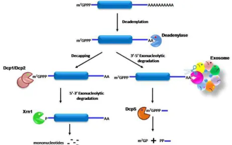
The predominant cytoplasmic mRNA decay pathway is the
deadenylation-dependent RNA degradation (Fig. 3). mRNA decay is initiated
with the shortening of the poly(A) tail at the 3’ end by deadenylation enzymes
(Muhlrad & Parker, 1992; Decker & Parker, 1993). In yeast, two deadenylase
complexes were described, the major Ccr4/NOT complex and Pan2/Pan3 (Brown
& Sachs, 1998; Tucker et al., 2001). After deadenylation, there are two possible
degradation pathways for the transcripts. One is the removal of the 5’ cap
structure of the transcripts by the Dcp1/Dcp2 decapping complex, exposing the
RNA molecule to the cytoplasmic Xrn1 5’-3’ exoribonuclease (Hsu & Stevens,
1993; Muhlrad et al., 1994). The other pathway is the 3’-5’ exoribonucleolytic
degradation by a large protein complex, termed the exosome (Anderson &
oligonucleotide is degraded by the scavenger decapping enzyme DcpS (Liu et al.,
2002).
Figure 3. Model of mRNA degradation in eukaryotes. In general, decay is initiated by deadenylation and is followed by one of two pathways: the complete degradation of the substrate by decapping and 5’-3’ exonucleases or 3’-5’ degradation by the exosome. Adapted from Arraiano et al. (2010).
Thus far, we have described the general mRNA decay pathway in
eukaryotes. Nevertheless, there are specialized mRNA decay pathways, namely,
deadenylation independent decapping (Muhlrad & Parker, 1994), accelerated 3’
to 5’ degradation (van Hoof et al., 2002) and endonuclease cleavage (Doma &
Parker, 2006).
RNase II Family of Enzymes
Enzymes from the RNase II family are important in RNA processing, quality
control and degradation. Enzymes of this family are sequence-independent
hydrolytic exoribonucleases that processively digest RNA in the 3’ to 5’ direction,
yielding 5’-nucleoside monophosphates (Zuo & Deutscher, 2001).
This family of enzymes is present in all domains of life and has important
with phosphorolytic nuclease PNPase, are responsible for the majority of
exonucleolytic RNA degradation (Andrade et al., 2009; Matos et al., 2011). The
best studied member in eukaryotes is Rrp44 (also called Dis3), the catalytic
component of the exosome (Liu et al., 2006; Dziembowski et al., 2007). Members
of this family have been described to be essential for growth (Mitchell et al., 1997),
involved in mitotic control (Lim et al., 1997) and cancer (Chapman et al., 2011;
Rose et al., 2011; Astuti et al., 2012; Ding et al., 2012; Reis et al., 2012), and
required for virulence in several organisms (Cheng et al., 1998; Erova et al., 2008).
Functional Domains
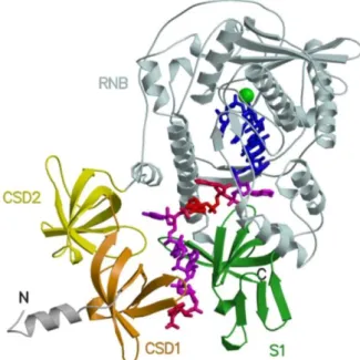
The crystal structures of E. coli RNase II and of a catalytically inactive mutant in
complex with RNA, revealed the presence of four domains (Frazão et al., 2006;
Zuo et al., 2006). Three of these domains have a common oligonucleotide
binding-fold (OB binding-fold) characteristic of nucleic-acid-binding proteins (Murzin, 1993), two
cold-shock domains (CSD1 and CSD2) and an S1 RNA-binding domain, that are
grouped on one side of the 3D structure of the molecule. The well-conserved
catalytic domain (RNB domain) is on the other side of the molecule (Frazão et al.,
2006). The function of each domain had been previously determined and it was
shown that the RNB domain is responsible for the catalytic activity, whereas CSD
and S1 domains are responsible for the binding to RNA (Amblar et al., 2006).
In addition to the domains described for E. coli RNase II and RNase R
(RNase II-like region), Rrp44/Dis3 contains an additional domain at the
N-terminus (Fig. 4). Our group and two others have independently shown that the
PIN domain of yeast Rrp44 confers endoribonuclease activity to this enzyme
(Lebreton & Seraphin, 2008; Schaeffer et al., 2009; Schneider et al., 2009). This
domain also plays an important structural role by establishing a link of yeast
Figure 4. Domain organization of RNase II, RNase R and yeast Rrp44. Members of this family have a similar modular domain organization. The N-terminal region is variable but shares CSD1 (orange) and CSD2 (yellow), followed by a RNB domain (light cyan) and an S1 domain (green). At the N-terminus, yeast Rrp44 also contains a catalytic PIN domain (light blue). Ribonucleolytic active sites are indicated (red circle). Adapted from Chlebowski et al. (2010).
Two crystallographic structures of Saccharomyces cerevisiae Rrp44 have
been solved: the structure of a catalytically inactive mutant of Rrp44 lacking the
PIN domain in complex with RNA (Lorentzen et al., 2008) and the full length
Rrp44 in complex with two other subunits of the exosome (Bonneau et al., 2009).
The individual domains of Rrp44 are similar in structure to the corresponding
domains of RNase II, with 85% of the residues of the RNB domain and more than
70% of the three OB-fold domains superposing in their -carbon positions. The
most striking difference between Rrp44 and RNase II lies in the relative position
of the binding domains to the catalytic domain, with implications on the route of
RNA access to the catalytic site (Lorentzen et al., 2008).
Exonuclease Activity
The exonucleolytic active site of the RNase II family of enzymes resides in the
RNB domain (Amblar & Arraiano, 2005). This domain is only present in the
members of this family and contains four highly conserved motifs (I to IV; Mian,
1997). Crystal structures of E. coli RNase II (Fig. 5) and S. cerevisiae Rrp44 in
complex with RNA shed light on the mechanisms of RNA cleavage, translocation
and enzyme processivity, as discussed below (Frazão et al., 2006; Lorentzen et al.,
2008). The structure of RNase R was not solved yet. Thus, most of the knowledge
RNase II/Rrp44 structures and from models proposed (Barbas et al., 2008;
Domingues et al., 2009).
Figure 5. Structure of RNase II in complex with RNA. Representation of RNase II–RNA complex, showing domains CSD1 (orange), CSD2 (yellow), RNB (light cyan) and S1 (green), the Mg2+ ion as a sphere (green) and RNA in sticks. Adapted from Frazão et al.
(2006).
E. coli RNase II and S. cerevisiae Rrp44 active sites lie buried deep within
the RNB domain, where four conserved acidic residues coordinate two
magnesium ions that facilitate a two-metal ion catalysis mechanism (Steitz &
Steitz, 1993; Frazão et al., 2006; Lorentzen et al., 2008). Mutation of one of those
conserved residues abolishes the exonucleolytic activity of the enzyme: D209N in
RNase II (Amblar & Arraiano, 2005), D280N in RNase R (Matos et al., 2009) and
D551N in yeast Rrp44 (Dziembowski et al., 2007).
Single-stranded RNA is threaded to the catalytic cavity through the
anchor region, constituted by the RNA-binding domains: CSD2 and S1 domains
in RNase II and CSD1 and RNB domains in Rrp44. The first five nucleotides
residues of the enzyme that stabilize the RNA in the active site, promoting its
translocation between successive cleavage events (Frazão et al., 2006; Lorentzen et
al., 2008). When the size of the RNA is not sufficient for the packing of the bases
in the catalytic cavity, the translocation of the RNA stops and the final end
product is released. In RNase II, residue Glu542 is in close proximity with the
leaving nucleotide, and it was proposed that it helps to release the last nucleotide
after cleavage (Frazão et al., 2006). However, its substitution by an alanine
induced a subtle conformational change in the RNB domain, as predicted by
computational modeling, leading to a 110-fold increase in RNase II activity
(Barbas et al., 2009). This result shows that Glu542 is involved in the tuning of
RNase II activity.
Processivity of RNB family enzymes is achieved by the simultaneous
contacts of RNA at two different regions of the enzyme: the catalytic region and
the anchor region (Cannistraro & Kennell, 1994; Frazão et al., 2006; Lorentzen et
al., 2008). In RNase II, 10 nucleotides is the shortest RNA fragment able to retain
contacts with both regions (Frazão et al., 2006). Shorter RNA fragments establish
fewer contacts with the protein and the enzyme becomes distributive
(Cannistraro & Kennell, 1994). Mutational analysis and biochemical data
supported the structural results, since the substitution of Tyr253 in the catalytic
site was shown to alter the size of the end-product from 4 to 10 nucleotides
(Barbas et al., 2008). This mutation causes the loosening of the RNA substrate at
the catalytic region and the RNA fragment is released (Barbas et al., 2008). In
RNase R, Tyr324 was also found to be responsible for setting the size of the
end-product (Matos et al., 2009).
Endonuclease Activity
Rrp44/Dis3 also has endoribonuclease activity conferred by the PIN domain
domains were named for their homology with the N-terminus domain of a
bacterial protein involved in the biosynthesis of type IV pili (PilT N terminus;
Wall & Kaiser, 1999). The PIN domain family has representatives in all domains
of life and are found in proteins with different functions, some of which with
nuclease activity (Wall & Kaiser, 1999; Arcus et al., 2004). Members of this family
with nuclease activity are found in bacterial toxin-antitoxin systems (Arcus et al.,
2005), are involved in eukaryotic ribosomal RNA processing (Fatica et al., 2004;
Bleichert et al., 2006), nuclear quality control of mRNPs (Skruzny et al., 2009) and
nonsense-mediated decay (Glavan et al., 2006).
The PIN domain consists of 100 amino acids and shows a similar fold to
the catalytic core of T4 RNase H, with four conserved acidic residues involved in
the coordination of a metal ion (Bhagwat et al., 1997; Makarova et al., 1999;
Clissold & Ponting, 2000; Arcus et al., 2004). In Rrp44, the activity of this domain
is highest in the presence of manganese ions and digests linear and circular
substrates (Lebreton & Seraphin, 2008; Schaeffer et al., 2009; Schneider et al.,
2009). Mutation of acidic residues in the active site impairs its activity, in vivo and
in vitro (Fatica et al., 2004; Bleichert et al., 2006; Glavan et al., 2006; Lebreton &
Seraphin, 2008; Schaeffer et al., 2009; Schneider et al., 2009). Interestingly, there
are three different versions of Dis3 in some eukaryotes that differ in the
conservation of the acidic residues and the presence or absence of the PIN
domain. In humans, there are three homologues: hDIS3, hDIS3L1 and hDIS3L2
(Staals & Pruijn, 2010; Tomecki et al., 2010). Two of them, hDIS3 and hDIS3L1,
were proved to interact with the exosome ring complex. hDIS3 is mainly
nucleoplasmic and has endonucleolytic activity, while hDIS3L1 is strictly
cytoplasmic and has no endonucleolytic activity. In humans and
Schizosaccharomyces pombe there is a third Dis3 homologue - Dis3L2 (Malecki et al.,
Submitted; Astuti et al., 2012). Although this protein does not have the PIN
Submitted; Astuti et al., 2012). The lack of the PIN domain suggests that hDis3L2
does not interact with the exosome ring, and makes it structurally more similar to
bacterial RNase R/II enzymes (Staals et al., 2010).
Substrate Specificity
The activity of the enzymes of RNase II family is sequence independent and they
release nucleoside 5′ monophosphates and short oligonucleotides with 2-5 nt in
length (Cannistraro & Kennell, 1994; Cheng & Deutscher, 2002; Dziembowski et
al., 2007). However, each enzyme has different characteristics.
While yeast Rrp44 is able to digest through dsRNA, provided there is a
single-stranded 3’ overhang with at least four nucleotides (Lorentzen et al., 2008),
bacterial RNase II stalls around seven nucleotides before it reaches a
double-stranded region (Spickler & Mackie, 2000). These differences can be rationalized
with the different routes of access to the catalytic active site, proved by the
crystallization data (Lorentzen et al., 2008).
A molecular mechanism model was proposed for the unwinding of
secondary structures by yeast Rrp44. Accordingly, the unwinding of the RNA is
achieved by pulling the 3’ tail into a channel to the catalytic active site that is
specific for ssRNA, leading to the separation of duplex regions at the entrance
pore (Lorentzen et al., 2008). The driving force would rely on the chemical energy
released upon hydrolysis of the RNA chain instead of ATP hydrolysis, as in
classical helicases (Lorentzen et al., 2008). Recently it was shown, by
single-molecule fluorescence analysis, that this energy is accumulated during multiple
single nucleotide step hydrolysis reactions until about four base pairs are
unwound in a burst (Lee et al., 2012). Surprisingly, kinetic analysis data shows
that RNA unwinding determines the overall RNA degradation rate (Lee et al.,
Similarly to Rrp44, wild-type RNase R is able to digest through dsRNA,
provided there is a single-stranded 3’ overhang with at least 5 nucleotides (Cheng
& Deutscher, 2005). However, a truncated RNase R protein without CSDs and/or
the S1 domain is able to degrade double-stranded substrates (Matos et al., 2009).
This result shows that the binding domains (CSD1, CSD2 and S1) block the
entrance of the substrates into the catalytic channel. Nevertheless, it seems
reasonable that the underlying mechanism is similar to Rrp44 (Lorentzen et al.,
2008; Lee et al., 2012). Only the solving of the RNase R structure will elucidate its
mode of action.
RNase II can bind RNA and DNA, although it is not able to cleave DNA.
Furthermore, it is inhibited by DNA oligonucleotides since they bind to its
specific binding site (Cannistraro & Kennell, 1994). In the RNase II structure the
O2’-ribose oxygens of nucleotides 8, 10 and 12 are engaged in hydrogen bonds
with the side chains of conserved residues Glu390, Asp201 and Tyr313 (Frazão et
al., 2006). These interactions are important for the proper orientation of RNA and
catalysis by RNase II. Instead of three, Rrp44 displays five interactions with 2’ OH
groups consistent with an higher affinity for ssRNA than for a corresponding
ssDNA oligonucleotide (Lorentzen et al., 2008). Similarly, RNase R binds poorly
to a DNA oligonucleotide (Cheng & Deutscher, 2002; Vincent & Deutscher, 2006).
RNase II-family members activity does not depend on the RNA sequence,
however they show different degrading efficiencies depending on the substrate.
For instance, while RNase II and RNase R are more active on the homopolymer
poly(A) substrate (Cheng & Deutscher, 2002), Rrp44 seems to be more active on
AU-rich sequences and generic substrates (Liu et al., 2006). These preferences
Exosome
Rrp44/Dis3 is part of a multiprotein complex known as the eukaryotic exosome.
This complex was first identified in S. cerevisiae as a complex of 3’ to 5’
exoribonucleases required for 3’ processing of the 5.8S rRNA (Mitchell et al.,
1997). Later, it was shown to be evolutionary conserved, to participate in several
aspects of cellular RNA metabolism (Allmang et al., 1999a; Allmang et al., 1999b)
and to have an endonuclease cleavage site (Lebreton & Seraphin, 2008; Schaeffer
et al., 2009; Schneider et al., 2009). Nuclear and cytoplasmic forms of the exosome
share 10 essential subunits (Mitchell et al., 1997; Allmang et al., 1999b).
Structure
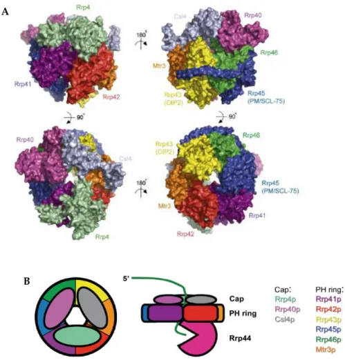
The eukaryotic core exosome is composed of a six subunits ring structure (Rrp41,
Rrp42, Rrp43, Rrp45, Rrp46 and Mtr3) that is capped on one side by three other
subunits (Csl4, Rrp4 and Rrp40; Fig. 6; Liu et al., 2006).
The subunits that form the ring belong to the PDX family of
exoribonucleases, which also includes E. coli RNase PH, PNPase and the archaeal
exosome (Mian, 1997; Zuo & Deutscher, 2001). Members of this family activate an
inorganic phosphate to cleave the substrate in the 3’ to 5’ direction, and release 5'
nucleoside diphosphate products (Zuo & Deutscher, 2001). Surprisingly, the
eukaryotic exosome lacks the phosphorolytic activity that is characteristic of this
family, and in most eukaryotes can induce RNA degradation only when
cooperating with the active component Rrp44/Dis3 (Liu et al., 2006; Dziembowski
et al., 2007). This suggests that instead of having a catalytic role, the ring acts as a
protein scaffold that may have a role in the recruitment of exosome cofactors and
substrates.
The proteins that form the cap contain RNA binding domains, namely, S1
domain (K-homology; Grishin, 2001). The cap is thought to stabilize the exosome
and to bind RNA. The core exosome is architecturally similar to the archaeal
exosome complex (Buttner et al., 2005; Lorentzen et al., 2005; Lorentzen et al.,
2007) and to the bacterial PNPase (Symmons et al., 2000).
Figure 6. Overall structure of the eukaryotic core exosome. A. Surface representation of the human core exosome, depicting opposing views from sides (on the top), top (bottom left) and bottom (bottom right). Adapted from Liu et al. (2006). B. Schematic representation of the eukaryotic exosome, top view (left) and side view of the exosome (right). Adapted from Schaeffer et al. (2009).
In yeast, Rrp44 interacts with the nine-protein ring-shaped complex to
generate a ribonucleolytically active exosome (Liu et al., 2006; Dziembowski et al.,
A
2007). Biochemical and structural studies showed that even if the Rrp44 protein
by itself is able to degrade RNA, it seems that the substrates that are delivered to
this nuclease first have to pass the channel in the exosome ring structure
(Bonneau et al., 2009; Malet et al., 2010; Wasmuth & Lima, 2012). We have
recently shown that three cysteines at the N-terminus of Rrp44, together with a
conserved histidine from the PIN domain, affect the binding to the exosome
(Schaeffer et al., 2012). However, in fission yeast, Dis3L2 is able to degrade RNA
without binding the exosome (Malecki et al., Submitted).
Cofactors and Functions
The eukaryotic exosome is involved in RNA processing, degradation and
surveillance reactions. It is present in two different compartments of the cell,
where it has different specialized functions and associates with auxiliary
components. Exosome cofactors are thought to direct the exosome to specific
RNA substrates and to activate the exosome (Houseley et al., 2006). However, it is
not yet known how this activation occurs.
Nuclear Cofactors and Functions
The nuclear exosome has a dual role, the processing and degradation of RNA.It
is involved in the processing of different species of RNA precursors, including
snRNA, snoRNA and rRNA (Mitchell et al., 1997; Allmang et al., 1999a; Allmang
et al., 1999b; van Hoof et al., 2000). In addition, the exosome also participates in
the degradation of defective pre-RNAs, which are not correctly processed, spliced
or assembled into ribonucleoprotein complexes (Bousquet-Antonelli et al., 2000;
Libri et al., 2002; Torchet et al., 2002; Kadaba et al., 2004; LaCava et al., 2005). The
nuclear exosome is also implicated in the control of expression levels of mRNA
(Kuai et al., 2005), and in the removal of processing byproducts and a myriad of
The main interactors of the nuclear exosome are Rrp6 (Briggs et al., 1998)
and the TRAMP complex (for Trf4/5-Air1/2-Mtr4; LaCava et al., 2005; Vanacova et
al., 2005).
Rrp6 is a distributive hydrolytic 3’ to 5’ exoribonuclease that shares
sequence similarity to E. coli RNase D (Zuo & Deutscher, 2001). Although not
essential for cell viability (Briggs et al., 1998), Rrp6 has a critical role in RNA
processing and surveillance pathways (Briggs et al., 1998; Allmang et al., 1999a;
Allmang et al., 1999b; van Hoof et al., 2000; Thomsen et al., 2003; Kuai et al., 2004).
In yeast, Rrp6 is only associated with nuclear exosome complexes (Allmang et al.,
1999b), while in humans small fractions were also found in the cytoplasm
(Lejeune et al., 2003; van Dijk et al., 2007; Tomecki et al., 2010). It is not clear how
the interaction with the core exosome is mediated. Different models place Rrp6
near the cap proteins or the ring proteins (Lehner & Sanderson, 2004; Cristodero
et al., 2008), suggesting a distinct recruitment pathway. These data are in
agreement with biochemical data showing that the activity of Rrp6 is not affected
upon association with the exosome core (Liu et al., 2006). Interestingly, Rrp6 is
able to function in vivo without interacting with the core exosome (Callahan &
Butler, 2008). Additionally, Rrp6 binds to Rrp47 (C1D in humans) and the
M-phase phosphoprotein 6, Mpp6 (Mitchell et al., 2003; Stead et al., 2007; Milligan et
al., 2008; Butler & Mitchell, 2010; Costello et al., 2011). These nuclear proteins are
involved in RNA binding and preferentially interact with structured and
pyrimidine-rich sequences, respectively (Stead et al., 2007; Milligan et al., 2008).
Both of these proteins are required for 3’ processing of 5.8S rRNA in yeast and
depletion of Rrp47 causes similar processing defects on some substrates, to those
caused by lack of Rrp6 (Mitchell et al., 2003; Milligan et al., 2008).
The TRAMP complex is responsible for nuclear surveillance of many
different RNAs. It is composed of a non-canonical poly(A) polymerase (Trf4 or
(Mtr4; LaCava et al., 2005; Vanacova et al., 2005). The TRAMP complex is thought
to activate the exosome by recruiting the exosome to the aberrant RNA substrates
through the addition of short poly(A) tails and by stimulating the exonucleolytic
activity of Rrp6 and Rrp44(LaCava et al., 2005; Vanacova et al., 2005; Schneider et
al., 2007; Callahan & Butler, 2010). Instead of stabilizing the RNA, poly(A) tails
recruit and activate the exosome leading to rapid degradation. The role of
polyadenylation in stimulating RNA degradation by 3’ to 5’ exoribonucleases,
resembles bacterial mRNA decay (Dreyfus & Regnier, 2002). Mtr4 is thought to
unwind secondary structures and to recruit the exosome to TRAMP substrates,
since its depletion leads to RNA hyperadenylation in vivo (Houseley & Tollervey,
2006). Mtr4 also functions as a cofactor of the exosome independently of the
TRAMP complex andis required for both RNA degradation and maturation (de
la Cruz et al., 1998).
Cytoplasmic Cofactors and Functions
In the cytoplasm, the exosome is strictly involved in mRNA degradation, where it
degrades normal and aberrant transcripts. In this compartment, the transcripts
are translated to proteins. Therefore, it is very important to check the translational
abilities of RNAs and remove incorrect molecules that can cause the production
of aberrant proteins (Doma & Parker, 2007). The types of aberrant mRNAs
targeted for degradation by the exosome include transcripts that lack termination
codons (nonstop decay surveillance pathway; Frischmeyer et al., 2002; van Hoof
et al., 2002), transcripts that stall translation elongation (no-go decay surveillance
pathway; Doma & Parker, 2006) or contain premature termination codons
(nonsense mediated decay surveillance pathway; Mitchell & Tollervey, 2003).
The cytoplasmic exosome binds the Ski complex (Ski2, Ski3 and Ski8) and
Ski7. Ski2 is closely related to Mtr4 and is also a member of the DExH box RNA
(eukaryotic translation elongation factor 1A), eRF3 (eukaryotic release factor 3)
and the prokaryoticEF-Tu (elongation factor thermo unstable; Benard et al., 1999)
proteins, and is thought to recognize specific RNA substrates and to recruit the
exosome to mRNA (Frischmeyer et al., 2002; van Hoof et al., 2002; Mitchell &
Tollervey, 2003). Ski7 interacts with the Ski complex and the exosome (Araki et
al., 2001), bridging the interactions between both complexes.
Aim of this Dissertation
Enzymes from the RNase II family are involved in RNA metabolism and are
conserved both in prokaryotes and eukaryotes. While in E. coli, members of this
familyfunction as individual proteins, S. cerevisiae Rrp44 functions in the context
of the exosome. When this study started, not much was known about Rrp44 and
its involvement in RNA metabolism.The objectives proposed for this Dissertation
were focused on the biochemical characterization of S. cerevisiae Rrp44 and E. coli
RNase II, with the aim of learning more about these enzymes and their
implications in RNA metabolism.
Although in the beginning of this work the existence of 5 domains in
Rrp44 was already known, only the domains present in RNase II were
functionally characterized. Therefore, in the first part of this work we focused on
the study of a well conserved PIN domain in yeast Rrp44, which function was
unkown. In order to address the specific role of this domain in the activity of the
enzyme, we constructed and purified several Rrp44 mutant proteins, and
analyzed their ribonucleolytic activity.
In the second part, we wanted to study the role of the CR3 motif at the
N-terminus of Rrp44. This region had been previously identified in Drosophila
this region we have constructed and purified several truncated proteins, and have
studied how this region affects the endonucleolytic activity of the enzyme.
Continuing our study of yeast Rrp44, we have investigated the specific
role of certain residues in the evolutionary conserved RNB domain. The residues
selected had been previously in vitro biochemically characterized in E. coli RNase
II. We have constructed proteins with point mutations to test the biochemical
function of different residues and analyze their physiological implications in
yeast.
We were also interested in determining the mutation(s) responsible for
the thermosensitive phenotype of E. coli RNase II (Donovan & Kushner, 1986),
both in vivo and in vitro. The localization of this(these) mutation(s) can be an
useful tool for studying RNase II enzymes and function in other organisms.
This knowledge will contribute to better understand the involvement of
these enzymes in RNA metabolism.
References
Allmang C, Kufel J, Chanfreau G, Mitchell P, Petfalski E, Tollervey D. 1999a.
Functions of the exosome in rRNA, snoRNA and snRNA synthesis. Embo
J18:5399-5410.
Allmang C, Petfalski E, Podtelejnikov A, Mann M, Tollervey D, Mitchell P. 1999b. The yeast exosome and human PM-Scl are related complexes of 3' --> 5' exonucleases. Genes & development13:2148-2158.
Amblar M, Arraiano CM. 2005. A single mutation in Escherichia coli ribonuclease
II inactivates the enzyme without affecting RNA binding. FEBS J272
:363-374.
Amblar M, Barbas A, Fialho AM, Arraiano CM. 2006. Characterization of the functional domains of Escherichia coli RNase II. J Mol Biol360:921-933. Anderson JS, Parker RP. 1998. The 3' to 5' degradation of yeast mRNAs is a
general mechanism for mRNA turnover that requires the SKI2 DEVH box
protein and 3' to 5' exonucleases of the exosome complex. EMBO J
17:1497-1506.
Andrade JM, Pobre V, Silva IJ, Domingues S, Arraiano CM. 2009. The role of 3'-5'
Apirion D. 1973. Degradation of RNA in Escherichia coli. A hypothesis. Mol Gen Genet122:313-322.
Araki Y, Takahashi S, Kobayashi T, Kajiho H, Hoshino S, Katada T. 2001. Ski7p G protein interacts with the exosome and the Ski complex for 3'-to-5' mRNA decay in yeast. EMBO J20:4684-4693.
Arcus VL, Backbro K, Roos A, Daniel EL, Baker EN. 2004. Distant structural homology leads to the functional characterization of an archaeal PIN
domain as an exonuclease. J Biol Chem279:16471-16478.
Arcus VL, Rainey PB, Turner SJ. 2005. The PIN-domain toxin-antitoxin array in mycobacteria. Trends Microbiol13:360-365.
Arraiano CM, Andrade JM, Domingues S, Guinote IB, Malecki M, Matos RG, Moreira RN, Pobre V, Reis FP, Saramago M, Silva IJ, Viegas SC. 2010. The critical role of RNA processing and degradation in the control of gene
expression. FEMS Microbiol Rev34:883-923.
Astuti D, Morris MR, Cooper WN, Staals RH, Wake NC, Fews GA, Gill H, Gentle D, Shuib S, Ricketts CJ, Cole T, van Essen AJ, van Lingen RA, Neri G, Opitz JM, Rump P, Stolte-Dijkstra I, Muller F, Pruijn GJ, Latif F, Maher ER. 2012. Germline mutations in DIS3L2 cause the Perlman syndrome of
overgrowth and Wilms tumor susceptibility. Nat Genet44:277-284.
Barbas A, Matos RG, Amblar M, Lopez-Vinas E, Gomez-Puertas P, Arraiano CM. 2008. New insights into the mechanism of RNA degradation by ribonuclease II: identification of the residue responsible for setting the
RNase II end-product. J Biol Chem 283:13070-13076.
Barbas A, Matos RG, Amblar M, Lopez-Vinas E, Gomez-Puertas P, Arraiano CM. 2009. Determination of key residues for catalysis and RNA cleavage specificity: one mutation turns RNase II into a "SUPER-ENZYME". J Biol
Chem284:20486-20498.
Benard L, Carroll K, Valle RC, Masison DC, Wickner RB. 1999. The ski7 antiviral protein is an EF1-alpha homolog that blocks expression of non-Poly(A)
mRNA in Saccharomyces cerevisiae. J Virol73:2893-2900.
Bhagwat M, Meara D, Nossal NG. 1997. Identification of residues of T4 RNase H
required for catalysis and DNA binding. J Biol Chem272:28531-28538.
Bleichert F, Granneman S, Osheim YN, Beyer AL, Baserga SJ. 2006. The PINc domain protein Utp24, a putative nuclease, is required for the early
cleavage steps in 18S rRNA maturation. Proc Natl Acad Sci U S A
103:9464-9469.
Bonneau F, Basquin J, Ebert J, Lorentzen E, Conti E. 2009. The yeast exosome functions as a macromolecular cage to channel RNA substrates for degradation. Cell139:547-559.
Bousquet-Antonelli C, Presutti C, Tollervey D. 2000. Identification of a regulated
Briggs MW, Burkard KT, Butler JS. 1998. Rrp6p, the yeast homologue of the human PM-Scl 100-kDa autoantigen, is essential for efficient 5.8 S rRNA 3' end formation. J Biol Chem273:13255-13263.
Brown CE, Sachs AB. 1998. Poly(A) tail length control in Saccharomyces
cerevisiae occurs by message-specific deadenylation. Mol Cell Biol
18:6548-6559.
Butler JS, Mitchell P. 2010. Rrp6, Rrp47 and cofactors of the nuclear exosome. Adv
Exp Med Biol702:91-104.
Buttner K, Wenig K, Hopfner KP. 2005. Structural framework for the mechanism
of archaeal exosomes in RNA processing. Mol Cell20:461-471.
Cairrão F, Arraiano C, Newbury S. 2005. Drosophila gene tazman, an orthologue
of the yeast exosome component Rrp44p/Dis3, is differentially expressed
during development. Dev Dyn232:733-737.
Callahan KP, Butler JS. 2008. Evidence for core exosome independent function of
the nuclear exoribonuclease Rrp6p. Nucleic Acids Res36:6645-6655.
Callahan KP, Butler JS. 2010. TRAMP complex enhances RNA degradation by the
nuclear exosome component Rrp6. J Biol Chem285:3540-3547.
Cannistraro VJ, Kennell D. 1994. The processive reaction mechanism of ribonuclease II. J Mol Biol243:930-943.
Chapman MA, Lawrence MS, Keats JJ, Cibulskis K, Sougnez C, Schinzel AC, Harview CL, Brunet JP, Ahmann GJ, Adli M, Anderson KC, Ardlie KG, Auclair D, Baker A, Bergsagel PL, Bernstein BE, Drier Y, Fonseca R, Gabriel SB, Hofmeister CC, Jagannath S, Jakubowiak AJ, Krishnan A, Levy J, Liefeld T, Lonial S, Mahan S, Mfuko B, Monti S, Perkins LM, Onofrio R, Pugh TJ, Rajkumar SV, Ramos AH, Siegel DS, Sivachenko A, Stewart AK, Trudel S, Vij R, Voet D, Winckler W, Zimmerman T, Carpten J, Trent J, Hahn WC, Garraway LA, Meyerson M, Lander ES, Getz G, Golub TR. 2011. Initial genome sequencing and analysis of multiple
myeloma. Nature471:467-472.
Cheng ZF, Deutscher MP. 2002. Purification and characterization of the Escherichia coli exoribonuclease RNase R. Comparison with RNase II. J Biol Chem277:21624-21629.
Cheng ZF, Deutscher MP. 2005. An important role for RNase R in mRNA decay.
Mol Cell17:313-318.
Cheng ZF, Zuo Y, Li Z, Rudd KE, Deutscher MP. 1998. The vacB gene required for virulence in Shigella flexneri and Escherichia coli encodes the
exoribonuclease RNase R. J Biol Chem273:14077-14080.
Chlebowski A, Tomecki R, Lopez ME, Seraphin B, Dziembowski A. 2010.
Catalytic properties of the eukaryotic exosome. Adv Exp Med Biol 702