www.cbpv.org.br/rbpv
Review Article
ISSN 0103-846X (Print) / ISSN 1984-2961 (Electronic)
Braz. J. Vet. Parasitol., Jaboticabal, v. 26, n. 3, p. 253-279, july-sept. 2017
Doi: http://dx.doi.org/10.1590/S1984-29612017045
All about neosporosis in Brazil
Tudo sobre neosporose no Brasil
Camila Koutsodontis Cerqueira-Cézar
1; Rafael Calero-Bernal
1; Jitender Prakash Dubey
1; Solange Maria Gennari
2*
1 Animal Parasitic Diseases Laboratory, Beltsville Agricultural Research Center, United States Department of Agriculture, AgriculturalResearch Service, Beltsville, MD, United States of America
2 Departamento de Medicina Veterinária Preventiva e Saúde Animal, Faculdade de Medicina Veterinária e Zootecnia, Universidade de São Paulo – USP, São Paulo, SP, Brasil
Received January 22, 2017 Accepted June 28, 2017
Abstract
Neospora caninum
is protozoan parasite with domestic and wild dogs, coyotes and grey wolves as the definitive hosts
and many warm-blooded animals as intermediate hosts. It was cultivated and named in 1988. Neosporosis is a major
disease of cattle and has no public health significance. Since 1990’s
N. caninum
has emerged as a major cause of abortion
in cattle worldwide, including in Brazil.
N. caninum
also causes clinical infections in several other animal species.
Considerable progress has been made in understanding the biology of
N. caninum
and there are more than 200 papers
on this subject from Brazil. However, most of the reports on neosporosis from Brazil are serological surveys. Overall,
little is known of clinical neosporosis in Brazil, particularly cattle. The few reports pertain to sporadic cases of abortion
with no information on epidemics or storms of abortion. The objective of the present review is to summarize all reports
from Brazil and suggest topic for further research, including prevalence of
N. caninum
oocysts in soil or in canine feces,
and determining if there are additional definitive hosts, other than the domestic dog. There is need for a national survey
in cattle using defined parameters. Future researches should focus on molecular characterization of
N. caninum
strains,
possibility of vaccine production and relationship between wildlife and livestock epidemiology.
Keywords:
Neospora caninum
, neosporosis, domestic animals, wild animals, Brazil.
Resumo
Neospora caninum
é um protozoário parasita que possui os canídeos domésticos e selvagens, coiotes e lobos cinzentos
como hospedeiros definitivos e vários animais de sangue quente como hospedeiros intermediários. Foi cultivado e nomeado
em 1988. A neosporose é uma das principais doenças em bovinos e não tem significância em saúde pública. Desde 1990,
N. caninum
tem emergido como uma das principais causas de aborto em bovinos em todo o mundo, inclusive no Brasil.
N. caninum
também causa infecções clínicas em várias outras espécies animais. Consideráveis avanços foram feitos na
compreensão da biologia desse parasita e há mais de 200 trabalhos sobre o assunto no Brasil. No entanto, a maioria
dos relatos de neosporose do Brasil são relacionados a sorologia. Em geral, pouco se sabe sobre a neosporose clínica no
Brasil, particularmente em bovinos. Os poucos relatos referem-se a casos esporádicos de aborto sem informações sobre
epidemias ou surtos de aborto. O objetivo da presente revisão é resumir todos os relatos sobre
N. caninum
no Brasil e
sugerir tópicos para pesquisas futuras, incluindo a prevalência de oocistos de
N. caninum
no solo ou em fezes caninas e
determinar se há hospedeiros definitivos adicionais, exceto o cão doméstico no país. Uma pesquisa nacional em bovinos
usando parâmetros definidos seria de grande importância. Pesquisas futuras deveriam ser focadas na caracterização de
cepas de
N. caninum
, possibilidade de produção de vacinas e a relação epidemiológica entre a vida selvagem e o gado.
Palavras-chave:
Neospora caninum
, neosporose, animais domésticos, animais selvagens, Brasil.
Cerqueira-Cézar, C.K. et al. Braz. J. Vet. Parasitol.
254
Introduction
Neosporosis is relatively a newly recognized disease. In 1988,
the etiologic agent of neosporosis was cultivated and named,
Neospora caninum
. It is ancestrally and morphologically related
to
Toxoplasma gondii
. Since 1990’s
N. caninum
has emerged as a
major cause of abortion in cattle worldwide, including in Brazil.
N. caninum
also causes clinical infections in several other animal
species. Considerable progress has been made in understanding
the biology of
N. caninum
. A recent book on neosporosis
(DUBEY et al., 2017) listed more than 2100 citations and most
reports (>200) from Brazil. Many of the reports from Brazil are
scattered in local journals. The objective of the present review is
to summarize reports from Brazil and suggest topic for further
research. To minimize citations, only references from Brazil are
listed in bibliography.
Basic Biology
As stated earlier,
N. caninum
and
T. gondii
are morphologically
similar but biologically different coccidians. Both parasites have a
wide host range but unlike
T. gondii
,
N. caninum
is not considered
zoonotic. Canids (domestic and wild canids dogs, coyote, and wolf)
are the definitive hosts of
N. caninum
whereas felids (domestic
and wild Felidae) are definitive hosts for
T. gondii
. Neosporosis
is a major disease of cattle whereas cattle are considered resistant
to
T. gondii.
Transplacental transmission is a major route of
propagation of
N. caninum
in cattle while, although medically
important, is not the major route of transmission for
T. gondii
.
There is only one species of
Toxoplasma
,
T. gondii
, but the genus
Neospora
has two species,
N. caninum
and
N. hughesi
; only horses
are reported as an intermediate host for
N. hughesi.
History of Neosporosis in Brazil
The
in vitro
cultivation of
N. caninum
in 1988 in USA made it
possible to develop diagnostic tests for neosporosis (DUBEY et al.,
2017). However, unlike
T. gondii
, it is difficult to culture
N. caninum
(see later discussion) and only one isolate (NC-1) was available
initially at the USDA laboratory in Beltsville, Maryland, USA.
Thus, commercial tests were not developed for the diagnosis of
neosporosis for several years. Therefore, no research was performed
on this subject in Brazil until mid 1990’s.
In BA, Gondim et al. (1999b, 2001) first recognized clinical
neosporosis in an aborted bovine fetus and first isolated viable
N. caninum
(NC-Bahia) from the brain of a naturally-infected
adult dog presenting incoordination and hind-limb paresis.
Corbellini et al. (2002) first recognized neosporosis as an important
cause of bovine abortion. They documented lesions consistent
with protozoal infection in 22 (47.8%) of 46 fetuses submitted
from 12 farms in RS. From those, 18 specimens of fetuses with
encephalitis reacted to
N. caninum
antisera.
Brazilan Contributions to the General
Biology of
N. caninum
Viable N. caninum isolates and genetic diversity
Unlike
T. gondii
, little is known of genetic diversity of
N. caninum
mainly because there are only few viable isolates available
worldwide. One reason for this is the difficulty to isolate viable
N. caninum
from animal tissues, especially from latently infected
animals. Table 1 summarizes all viable isolates of
N. caninum
from
animals in Brazil. The greatest success of isolating
Neospora
from
asymptomatic animals has been achieved by feeding brain tissue
from naturally-infected animals to dogs and then examining dog
feces for oocyst excretion. By doing so,
N. caninum
was isolated
from buffaloes, sheep and cattle.
In addition, to provide these
Neospora
isolates for biological and
genetic studies to researchers in other countries (DUBEY et al.,
2017), García-Melo in association with researchers from Spain
did the first microsatellite typing data for Nc-Goiás isolated from
clinically healthy cattle. Although the isolate had most of the
alleles already identified, unique alleles were described for this
strain at the MS5 and MS10 loci, using 12 microsatellite markers
(GARCÍA-MELO et al., 2009). The recent isolate from brain
tissue from a naturally-infected cattle (OLIVEIRA et al., 2017)
was also found to have unique microsatellite alleles as MS5, MS6a,
MS8 and MS10. A dog fed brain tissue from a naturally-infected
cattle excreted
N. caninum
oocysts for 14 days starting seven days
post infection (dpi), with an average number of 102 oocysts/g
of feces. DNA isolated from cell culture derived tachyzoites was
characterized using microsatellites. No new alleles were found and
comparison showed closest relation to multilocus genotyping with
strains from Spain and Argentina. Comparison with Brazilian
strains (NC-Bahia; NC-Goiás) revealed variation in three and
four of the nine markers used.
Additionally, the Brazilian NC-Bahia strain isolated from the
dog was included among the
N. caninum
strains characterized
by researchers outside Brazil (AL-QASSAB et al., 2009, 2010;
REGIDOR-CERRILLO et al., 2013).
Transmission
Neosporosis in Brazil
v. 26, n. 3, july-sept. 2017 255
(n=4), liver (n=3), and a group remaining as control (n=3), no
infected. None of the control dogs excreted oocysts and three dogs
that received brain, two that received masseter, two that received
heart and one that received liver excreted
N. caninum
-like oocysts,
from day seven to day 17 after ingestion of tissues. All dogs
that received brain and liver excreted only
N. caninum
oocysts.
The results were confirmed by polymerase chain reaction (PCR)
using Nc5 and ITS-1 amplification and indicated that a variety
of visceral, neural, and muscular tissues are infected naturally
with
N. caninum
.
How dogs become infected with
N. caninum
in nature is not fully
understood. Fecal transmission of
N. caninum
in dogs appears to be
less important than carnivorism. Until the study by Bandini et al.
(2011) it was unknown if the dogs could be infected via ingestion of
oocysts. They fed four dogs with 1,000, 5,000 or 10,000
N. caninum
oocysts; none of the four dogs excreted
N. caninum
–like oocysts in
their feces during the observation period of 30 days. However, the
two dogs fed with 10,000 oocysts seroconverted and the two dogs
fed with 1,000 or 5,000 oocysts did not. Neither parasite DNA
nor parasite stages were demonstrable in tissues of the seropositive
dogs euthanized six months after feeding oocysts. These findings
suggest that fecal transmission may not be an important mode
of transmission of the parasite for the definitive host but results
need confirmation.
Tachyzoites are important in transplacental transmission
of
N. caninum
infection. Cavalcante et al. (2012) confirmed
transplacental transmission of
N. caninum
in dogs. Six bitches in
two groups were inoculated with a very high dose (10
8tachyzoites).
In group I, three bitches were inoculated during the third week of
gestation, and in group II, three bitches were inoculated at the sixth
week of gestation. The bitches were allowed to whelp naturally.
Dams and their pups were tested by immunohistochemistry
(IHC), serology, and PCR. In group I, six of the ten pups died
within 48 hour of birth. In group II, seven of the 13 pups died
between five and ten days of birth.
N. caninum
DNA was detected
by nested PCR in two pups (hearts of both and liver of one) from
group I, and one pup in central nervous system (CNS) and lymph
node from group II. The dams and the pups that survived were
clinically normal.
N. caninum
was not demonstrable in tissues of
any of pups and their dams.
Studies in Brazil showed that
N. caninum
can be transmitted
transplacentally in water buffaloes (CHRYSSAFIDIS et al., 2014,
2015). The authors conducted an important experiment in six
buffaloes and seven cows inoculated intravenously with
N. caninum
tachyzoites at 70 day of gestation. Three buffaloes and five cows
were inoculated with a Brazilian
N. caninum
strain (NC-Bahia);
only one cow aborted but all fetuses became infected. The other
two cows and three buffaloes were inoculated with the NC-1
strain; all fetuses died by 35 dpi as determined by ultrasound and
N. caninum
DNA was detected in fetal tissues.
Chryssafidis et al. (2011) also showed, for the first time, that
N. caninum
can be transmitted transplacentally in naturally infected
buffaloes; they found
N. caninum
DNA in one brain of the nine
fetuses from buffaloes slaughtered in an abattoir, aiming Nc5 and
ITS-1 DNA regions. Although, viable or intact parasite has not
been demonstrated in naturally infected fetuses from buffaloes, this
is the first indication of transplacental transmission in this host.
Table 1. Isolation of N. caninum from naturally infected animals from Brazil.Host Source Bioassay Isolate
designation
Genetic
data Remarks Reference
Animals Cell culture
Cattle
Clinical, blind, neurological calf,
3-months old
SW mice, Vero BCN/PR3 No
Tissue cysts were found in the brains of SW mice that were
immunocompetent
Locatelli- Dittrich et al.
(2003) Aborted fetus,
7 months of gestation
Mice, gerbils Vero BCN/PR1 Yes
Initial isolation in immunosuppressed mice. Tachyzoites infective to immunocompetent mice and gerbils
Locatelli- Dittrich et al.
(2004) Asymptomatic,
4 months old KO mice MARC-145 Nc-Goiás 1b Yes Not pathogenic to BALB/c mice
García-Melo et al. (2009) Adult slaughtered
in abattoir
Dog, SW
mice, gerbils Vero NC-SP1 Yes Isolated from dog bioassay
Oliveira et al. (2017)
Dog Collie, 7 years old, clinical Gerbil Vero (COS-1) NC-Bahia Yes Isolate pathogenic to gerbils Gondim et al. (2001, 2004)
Sheep
Two 4 months old, clinically
normal
Gerbil, SW and vesper mice, dogs
Vero Not stated No
Mice remained healthy. Necropsied 60 dpi. Tissue cysts in brains of both
gerbils.
Pena et al. (2007)
Water buffalo
6 seropositive from abattoir
Dogs, gerbil,
KO mice CV1
NCBrBuf-1
No Both gerbils and KO remained asymptomatic
Rodrigues et al. (2004) NCBrBuf-2
Cerqueira-Cézar, C.K. et al. Braz. J. Vet. Parasitol.
256
In addition to bradyzoites and oocysts, tachyzoites can also
be infectious orally. Four to five day old gerbils were successfully
infected by oral inoculation of tachyzoites (FERREIRA et al., 2016).
All 17 gerbils died of neosporosis between eight and 17 dpi (one
died on each of days 8, 9, 15, 16, and 17, four died on day 12, and
five died on day 15 (personal communication to JPD on October
24, 2016) with 4 x 10
5NC-1 tachyzoites.
N. caninum
DNA was
found in heart, lung, spleen, kidney, liver and CNS of gerbils.
Environmental resistance of oocysts
Oocyst is the environmentally resistant stage. Researchers in
Brazil found that
N. caninum
oocysts were rendered noninfectious
by heating to 100 °C for one minute, and 10% sodium hypochlorite
for one hour but not at 60 °C for one minute, and by other
commonly used disinfectants (ALVES et al., 2011). Aqueous 2%
sulfuric acid has been commonly used to store
N. caninum
oocysts at 4 °C; how long oocysts remain viable under these
conditions is not known.
N. caninum
oocysts remained viable
for 108 days but not for 46 months when stored in 2% sulfuric
acid at 4 °C (UZÊDA et al., 2007a).
Diagnostic tests
Recently, Fehlberg et al. (2017) reported on successful
development of a high resolution melting PCR method to
distinguish
Neospora, Sarcocystis
and
Toxoplasma
using a single
pair of primers targeting 18S rDNA.
Epidemiology
More serological studies for
N. caninum
infection in animals
have been conducted in Brazil than rest of the world.
The results, however, are not comparable because of different
serological assays used different cut-offs employed, different
antigens used. For example, in the indirect fluorescent antibody
test (IFAT) and the
Neospora
agglutination test (NAT) whole
tachyzoites are used detecting antibodies to surface proteins whereas
in the enzyme-linked immunoabsorbent assay (ELISA) different
antigens are used, some of them using crude tachyzoite extract
while others used soluble antigens (Table 2a). The standardization
of serological tests was based on studies in other countries and a
full discussion of these is beyond the scope of this review—this
subject was recently reviewed (DUBEY et al., 2017). One of the
problems with serological testing is the availability of standardized
sera from experimentally infected animals. Immunoblotting (IB)
was employed in some studies where no such sera were available
(Table 2b).
Little has been done in Brazil to characterize
Neospora
recombinant antigens for the serological diagnosis or vaccine
development, except the report of Bezerra et al. (2017) who
characterized one surface protein, by cloning the sequence and
named it as NcSRS67, which has no orthologue with
Toxoplasma
gondii
, only with
Hammondia hammondi
.
In Tables 3-12 serological reports in different hosts are
summarized. We listed all reports that we found.
Salient features are commented for some of these surveys.
Dogs:
Numerous surveys from different regions of Brazil are
summarized in Table 3. As stated earlier results of these types of
surveys are not strictly comparable. However, 45 of 49 surveys
used IFAT as a diagnostic technique, and most of them employed
the cut-off value of 1:50, facilitating comparisons of occurrence
values. In three surveys, IFAT and ELISA were compared for
serological diagnosis of
N. caninum
in dog sera and IFAT was
superior to indirect ELISA used (SILVA et al., 2007; HIGA et al.,
2000; RAIMUNDO et al., 2015).
In some studies, risk factors were evaluated (Table 3). The age
of dogs was a statistically significant factor in nine reports; older
dogs were more likely to be seropositive (OLIVEIRA et al.,
2004; FERNANDES et al., 2004; AZEVEDO et al., 2005;
ANDREOTTI et al., 2006; CUNHA et al., 2008; MINERVINO et al.,
2012; NOGUEIRA et al., 2013; BALTHAZAR et al., 2013;
RAIMUNDO et al., 2015). Gender and breed were not associated
with presence of antibodies. In two surveys mixed breed dogs
constituted a risk factor for the infection (BRUHN et al., 2012;
RAIMUNDO et al., 2015). The diet in some studies was related
with access to street and prevalence was higher in dogs that had
outdoor access than in pets with little or no outdoor scavenging
(GENNARI et al., 2002; CAÑÓN-FRANCO et al., 2003;
BENETTI et al., 2008; SICUPIRA et al., 2012). However, in some
studies such an association was not found (MINEO et al., 2004;
JESUS et al., 2006; PLUGGE et al., 2011). Vertical transmission
was also studied (TAQUES et al., 2016), with 41 stillborn puppies
from 23 bitches. By PCR and IFAT, five (21.7%) bitches were
positive and 22 (53.6%) stillborn were positive by PCR, utilizing
ITS-1 DNA region/locus, being 17 from positive bitches and five
from negative ones. Although the prevalence of positive stillborn
was higher from positive bitches, no conclusions were made.
Epidemiologically, contact between cattle and dogs has been
identified as a possible risk factor that deserves attention (Table 3).
Dogs from peri-urban or rural areas had more chance to be infected
by
N. caninum
(IFAT 1:50) than urban dogs in the surveys from
MG (FERNANDES et al., 2004), PR (PLUGGE et al., 2008),
RS (CUNHA et al., 2008), and BA (SICUPIRA et al., 2012),
and this risk factor is normally associated with proximity to cattle;
access and ingestion of fetal membranes, carcasses, and prey.
Leishmania infantum chagasi
is an important parasite of dogs
in Brazil; and immunosuppression caused by
Leishmania
spp.
may enhance the susceptibility of dogs to
N. caninum
infection
(CRINGOLI et al., 2002). Serological surveys correlating
N. caninum
and
Leishmania
spp., had been conducted (GENNARI et al.,
2006; ANDREOTTI et al., 2006; GUIMARÃES et al., 2009;
GRECA et al., 2010; VALADAS et al., 2010a; LOPES et al., 2011;
PAULAN et al., 2013; SEABRA et al., 2015; CONSTANTINO et al.,
2016). In dogs from endemic cities of visceral leishmaniosis such
as Araçatuba, SP (GENNARI et al., 2006), Campo Grande, MS
(ANDREOTTI et al., 2006), and Teresina, PI (LOPES et al.,
2011), positive association was found (Table 3).
Neosporosis in Brazil
v. 26, n. 3, july-sept. 2017 257
Table 2a. ELISA techniques used to confirm presence of N. caninum antibodies.
Sera source Antigen ELISA type* Results in table Reference
Dogs
Antigen incorporated into iscoms A, C, I
3
Mineo et al. (2004) NC-1 strain maintained in bovine
monocytes cells; soluble antigen B, C Silva et al. (2007)
NC-1 tachyzoites strain in Vero cells A, C Raimundo et al. (2015)
Cattle
NC-1 tachyzoites strain in Vero cells,
lysed in SDS buffer J
4
Chahan et al. (2003)
Commercial E Andreotti et al. (2004)
Commercial F Paz et al. (2007)
Commercial E Melo et al. (2001)
Commercial E Melo et al. (2004)
Antigen incorporated into iscoms I Mineo et al. (2006)
Commercial E Locatelli-Dittrich et al. (2001)
Commercial E Locatelli-Dittrich et al. (2008)
Commercial E Marques et al. (2011)
Commercial E Nascimento et al. (2014)
NC-1 strain in Vero cells; soluble
antigen B, C Ramos et al. (2016)
Commercial E Munhoz et al. (2006, 2009)
Commercial D Vogel et al. (2006)
Commercial E Sartor et al. (2003)
Commercial M Sartor et al. (2005)
Buffalo
Commercial E
5
Viana et al. (2009)
Commercial D Vogel et al. (2006)
Commercial kit G, H Silva et al. (2014)
Sheep
NC-1 strain in Vero cells A, L
6
Andreotti et al. (2009) NC-1 strain in Vero cells, soluble
antigen A, C Rossi et al. (2011)
Commercial D Vogel et al. (2006)
Captive deer Commercial E 11 Zimpel et al. (2015)
Horse
NC-1 strain in HeLa cells A, C
12
Pivoto et al. (2014) NC-1 strain in bovine turbinate cell
monolayers C, K Hoane et al. (2006)
*A = WT = Whole tachyzoite extract; B = SA = soluble antigen; C = IH = In house; D = CHEKIT = CHEKIT Neospora, indirect ELISA, detergent lysate of tachyzoites, IDEXX Laboratories, The Netherlands; E = IDEXX = IDEXX HerdChek Neospora caninum antibody, indirect ELISA, sonicate lysate of tachyzoites, IDEXX Laboratories, USA; F = VMRD = Neospora caninum cELISA competitive ELISA GP65 surface antigen of tachyzoites VMRD, USA; G = Horse IgG = Horse IgG(T) ELISA Quantitation Set; Bethyl Laboratories; H = Horse IgM = Horse IgM ELISA, Kamiya Biomedical Company, Seattle; I = ISCOM = Detergent extracted tachyzoite antigen incorporated in immune stimulating complex particles; J = NcSAG1 = Recombinant surface antigen; K = NhSAG1 = Recombinant NhSAG1; L = rNcSRS2 = Recombinant antigen protein; M = NS/ND = not stated/ not done.
Table 2b. Immunoblotting technique used to confirm presence of N. caninum antibodies.
Sera
source Antigen* Sera tested previously
Results in
Table Reference
Dog NC-1 tachyzoites lysed in SDDS-PAGE buffer IFAT, 1:25; indirect ELISA 3 Silva et al. (2007) Cattle NC-1 tachyzoites lysed in SDDS-PAGE buffer NcSAG1 ELISA, 60/66 sera positive for ELISA 4 Chahan et al. (2003) Sheep NC-1 tachyzoites lysed in 4% sodium dodecyl sulfate buffer IFAT, 1:50; discordant results 6 Rossi et al. (2011)
Cat Detergent extracted whole NC-1 tachyzoites NAT, 1:80 9 Dubey et al. (2002)
Donkey Whole NC-1 tachyzoites Sera positive by IFAT, 1:100 12 Galvão et al. (2015)
Cer
queira-Cé
zar
, C.K. et al.
B
raz. J.
V
et. P
arasitol.
258
Table 3. Serological studies of N. caninum in dogs from Brazil.
State Type No. tested No. positive % Positive Test Cut-off Remarks Reference
Alagoas Urban 128 5 3.8 IFAT 1:50 Age, sex, breed, area, habitat Sousa et al. (2012)
Rural 99 5 4.8 IFAT 1:50
Amazon region Indian
commu-nities 325 32 9.8 IFAT 1:50 Age, sex, T. gondii, others Minervino et al. (2012)
(MT and TO)
Bahia Stray 250 28 11.2 IFAT 1:50 Age, sex, others Jesus et al. (2006)
Owned 165 22 13.3 IFAT 1:50
Bahia
Urban 156 4 2.6 IFAT 1:50 Area, contact with other dogs, feeding habits, others
Sicupira et al. (2012) Rural 41 6 14.6 IFAT 1:50 Area, contact with other dogs, feeding habits, others
Peri-urban 214 28 13.1 IFAT 1:50 Area, contact with other dogs, feeding habits, others
Espírito Santo Rural 187 22 11.7 IFAT 1:50 Sex, T. gondii Acosta et al. (2016)
Goiás Zoonosis Center 72 26 36.1 IFAT 1:50 Sex, origin Boaventura et al. (2008)
Hospital (owned) 125 39 31.2 IFAT 1:50 Sex, origin
Maranhão Stray 100 45 45.0 IFAT 1:50 Sex Teixeira et al. (2006)
Mato Grosso Clinics 60 27 45.0 IFAT 1:50 Age, sex, diet, access to streets Benetti et al. (2008)
Mato Grosso Rural 37 25 67.6 IFAT 1:50 Benetti et al. (2009)
Mato Grosso do Sul Pet 245 65 26.5 IFAT 1:50 Age, sex Oliveira et al. (2004)
Mato Grosso do Sul Rural 40 12 30.0 IFAT 1:100 Andreotti et. al. (2004)
Mato Grosso do Sul Urban 345 93 27.2 IFAT 1:50 Age, sex, Leishmania Andreotti et al. (2006)
Minas Gerais Clinical 163 11 6.7 IFAT 1:25 T. gondii Mineo et al. (2001)
Minas Gerais
Urban 300 32 10.7 IFAT 1:50 Age, breed, sex, area
Fernandes et al. (2004)
Peri urban 58 11 18.9 IFAT 1:50
Rural 92 20 21.7 IFAT 1:50
Minas Gerais Clinic 275 22 7.9 ELISA A,C Age, breed, sex, habitat, T. gondii, others Mineo et al. (2004)
Stray 94 12 12.8 ELISA I
Minas Gerais Clinic, stray 300 32 10.7 IFAT 1:50 Age, breed, sex, T. gondii Silva et al. (2007)
Clinic, stray 300 105 35.0 ELISA B,C
Minas Gerais Clinics 228 7 3.1 IFAT 1:50 Age, breed, sex T. gondii, Leishmania, Babesia canis Guimarães et al. (2009)
Minas Gerais Rural 240 36 15.0 IFAT 1:50 Age, breed, origin, sex, others Bruhn et al. (2012)
Minas Gerais RuralUrban 182421 1558 13.78.2 IFATIFAT 1:501:50 Age, diet, hunting, area Nogueira et al. (2013) Bovine abortion, area
Pará Rural 72 8 11.1 IFAT 1:50 Sex, area T. gondii, Leishmania Valadas et al. (2010b)
Urban-stray 57 8 14.0 IFAT 1:50
Paraíba Urban Domestic 286 24 8.4 IFAT 1:50 Age, sex, breed, others, habitat,T. gondii Azevedo et al. (2005)
Paraná Dairy farms 134 29 21.6 IFAT 1:50 Age, sex, breed Souza et al. (2002)
Paraná Neurological 98 0 0.0 IFAT 1:50 T. gondii Giraldi et al. (2002)
Paraná Sheep farms 24 7 29.1 IFAT 1:50 Age, sex, breed, T. gondii Romanelli et al. (2007)
Paraná
Urban 181 23 12.7 IFAT 1:50 Area, habitat
Plugge et al. (2008)
Peri urban 178 28 15.7 IFAT 1:50
Rural 197 50 25.3 IFAT 1:50
Paraná Rural 129 32 25.0 IFAT 1:50 Dogs and cattle Locatelli-Dittrich et al. (2008)
N
eospor
osis in B
razil
v. 26, n. 3, july-sept. 2017
259
State Type No. tested No. positive % Positive Test Cut-off Remarks Reference
Paraná Owned 127 14 11.0 IFAT 1:50 T. gondii, neurologic signs Plugge et al. (2011)
Stray 20 3 15.0 IFAT 1:50
Paraná Stray 26 3 11.5 IFAT 1:25 Leishmania spp, T. gondii,Trypanosoma cruzi Constantino et al. (2016)
Pernambuco
Figueredo et al. (2008)
Paulista Domiciled 289 75 26.0 IFAT 1:50 T. gondii, origin
Amaraji Domiciled 168 44 26.2 IFAT 1:50
Garanhuns Domiciled 168 58 34.5 IFAT 1:50
Pernambuco Rural villages 56 0 0 IFAT 1:50 T.gondii antibodies surveyed Arraes-Santos et al. (2016)
Piauí 530 17 3.2 IFAT 1:50 Age, sex, breed, T. gondii,Leishmania Lopes et al. (2011)
Piauí Rural villages 71 5 7.0 IFAT 1:50 T.gondii antibodies surveyed Arraes-Santos et al. (2016)
Rio de Janeiro Urban - Clinic 402 34 8.4 IFAT 1:50 Age, neurologycal signs, others Balthazar et al. (2013)
Rio Grande do Sul Rural 230 47 20.4 IFAT 1:50
Age, sex, area, type of farm, carcasses and
abor-tions not removed, others Cunha et al. (2008)
Urban 109 6 5.5 IFAT 1:50
Rondônia Domiciled (Street access) 157 13 8.3 IFAT 1:50 Age, sex, diet, street access Cañón-Franco et al. (2003)
Rondônia Farms 174 22 12.6 IFAT 1:50 Age, diet, abortion, stillbirth, others Aguiar et al. (2006)
São Paulo Hospital 203 44 21.6 IFAT 1:50 T. gondii, neurological signs Higa et al. (2000)
São Paulo Rural and urban 295 25 8.4 IFAT 1:50 Age, sex, T. gondii, neurologic signs Varandas et al. (2001)
São Paulo Domiciled 500 49 10.0 NAT 1:25 Age, sex, breed, habitat Gennari et al. (2002)
Street 611 151 25.0 NAT 1:25
São Paulo Urban 204 36 17.6 IFAT 1:50 Age, sex, T. gondii,Leishmania spp., Gennari et al. (2006)
São Paulo Urban 108 17 15.7 IFAT 1:50 Age, sex, diet, others, T. gondii Bresciani et al. (2007b)
São Paulo Urban 865 223 25.8 IFAT 1:50 Age, sex, area Moraes et al. (2008)
Rural 65 11 16.9 IFAT 1:50
Peri urban 33 11 33.3 IFAT 1:50
São Paulo Rural (stray) 100 14 14.0 IFAT 1:25 Leishmania spp. Greca et al. (2010)
São Paulo Sheep farms 42 2 4.8 IFAT 1:25 Type of raising (chained or free), access to raw meat or offal Machado et al. (2011)
São Paulo Neurologic 50 7 14% IFAT 1:25 T. gondii Langoni et al. (2012)
São Paulo Domiciled 342 17 4.9 IFAT 1:25 Age, sex, breed, T. gondii Langoni et al. (2014)
São Paulo Rural 93 6 6.5 IFAT 1:50 Leishmania, T. gondii, Ehrlichia spp., Babesia canis Paulan et al., (2013)
São Paulo KennelClinic 167133 3736 22.127.0 IFATIFAT 1:251:25 T. gondii, Leishmania Seabra et al. (2015)
T. gondii, Leishmania
Tocantins
Rural 99 43 43.4 ELISA A,C Age, breed, area, T. gondii
Raimundo et al. (2015)
31 31.3 IFAT 1:25
Urban 105 45 42.9 ELISA A,C
31 29.5 IFAT 1:25
Bold=statistically significant risk factor, Area = urban, peri urban, rural; Habitat=stray, domiciled.
Cer
queira-Cé
zar
, C.K. et al.
B
raz. J.
V
et. P
arasitol.
260
Table 4. Serologic studies of N. caninum antibodies in cattle from Brazil.
State No. tested Type No. herds No.
positive % positive Test Cut-off Remarksa Reference
Bahia 447 Dairy 14 63 14.0 IFAT 1:200 - Gondim et al. (1999a)
Goiás 444 Dairy 11 135 30.4 IFAT 1:250 - Melo et al. (2006)
Goiás 30 Mixed 1 13 43.3 IFAT 1:250 - Melo et al. (2006)
Goiás 456 Beef 9 135 29.6 IFAT 1:250 - Melo et al.(2006)
Maranhão 812 Dairy 27 412 50.7 IFAT 1:200 - Teixeira et al. (2010)
Mato Grosso 932 Dairy 24 farms 499 53.5 IFAT 1:200 - Benetti et al. (2009)
Mato Grosso do Sul 197 NS 6 66 33.5 ELISA J - Chahan et al. (2003)
Mato Grosso do Sul 87 Beef NS 26 29.9 IFAT 1:25 - Ragozo et al. (2003)
Mato Grosso do Sul 23 Dairy NS 5 21.7 IFAT 1:25 - Ragozo et al. (2003)
Mato Grosso do Sul 90 Beef (history of abortions) NS 38 43.0 ELISA E - Andreotti et al.(2004)
Mato Grosso do Sul 60 Heifers 18 30.0 ELISA E - Andreotti et al. (2004)
Mato Grosso do Sul 91 Healthy 7 7.7 ELISA E - Andreotti et al. (2004)
Mato Grosso do Sul 2448 NS 205 449 14.9 IFAT 1:50 Production system (Dairy/beef) Oshiro et al. (2007)
Mato Grosso do Sul 275 Beef 2 farms 81 29.5 cELISA F - Paz et al. (2007)
Mato Grosso do Sul 392 Dairy/beef 4 farms 43 9.1 IFAT 1:50 - Mello et al. (2008)
Mato Grosso do Sul 1098 Beef 1 farm 687 62.5 IFAT 1:50 Reproductive failure, 15% higher in seropositive cows. Andreotti et al. (2010)
Minas Gerais 584 Dairy 18 109 18.7 ELISA E - Melo et al. (2001)
Minas Gerais 126 Dairy 2 43 34.4 IFAT 1:25 - Ragozo et al. (2003)
Minas Gerais 36 Beef NS 4 11.1 IFAT 1:25 - Ragozo et al. (2003)
Minas Gerais 576 Dairy 18 106 18.4 ELISA E - Melo et al. (2004)
Minas Gerais 243 Dairy 2 41 16.8 ELISA I - Mineo et al. (2006)
Minas Gerais 559 Dairy 18 510 91.2 IFAT 1:200 Farm size, number of cows lactating, milk produc-tion per day Guedes et al. (2008)
Minas Gerais 575 Dairy Abattoir 559 97.2 IFAT 1:200 Farm size, number of cows lactating, milk produc-tion per day Guedes et al. (2008)
Minas Gerais 503 Dairy Abattoir (fetuses) 64 12.7 IFAT 1:25 Farm size, number of cows lactating, milk produc-tion per day Guedes et al. (2008)
Minas Gerais 1,204 Dairy 40 farms 260 21.6 IFAT 1:200 Reproductive failure Bruhn et al. (2013)
Pará 500 beef 500 260 52 IFAT 1:128 Region, T. gondii Silva et al. (2017)
Pará 40 Dairy 4 farms 7 17.5 IFAT 1:100 - Minervino et al. (2008)
Pará 120 Beef 12 23 19.2 IFAT 1:100 - Minervino et al. (2008)
Paraná 172 Dairy 1 60 34.8 ELISA E Abortion Locatelli-Dittrich et al. (2001)
Paraná 15 Beef NS 4 26.7 IFAT 1:25 - Ragozo et al. (2003)
Paraná 75 Dairy NS 16 21.3 IFAT 1:25 - Ragozo et al. (2003)
Paraná 623 Dairy 23 farms 89 14.3 IFAT 1:25 Breed, presence of dogs, age, feed Guimarães et al. (2004)
Paraná 385 Dairy 90 farms 45 12.0 IFAT 1:200 - Ogawa et al. (2005)
N
eospor
osis in B
razil
v. 26, n. 3, july-sept. 2017
261
State No. tested Type No. herds No.
positive % positive Test Cut-off Remarks
a Reference
Paraná 1263 NS 77 farms 423 33.0 ELISA E - Locatelli-Dittrich et al. (2008)
Paraná 159 Beef Abattoir 24 15.1 ELISA E - Marques et al. (2011)
Paraná 309 Dairy 15 farms 63 20.4 IFAT 1:100 Feed, wild animal access, artificial insemination Martins et al. (2012)
Paraná 76 Beef 4 23 30.3 ELISA E - Nascimento et al.(2014)
Pernambuco 469 Dairy 20 farms 163 31.7 IFAT 1:200
Veterinary assistance, nutritional condition, presence of wetlands, manipulation of newborn calves, destination of cows that had aborted, abor-tion history, aborabor-tions period.
Silva et al. (2008)
Pernambuco 316 Dairy 25 munici-palities
31 19.6 IFAT 1:200
Transplacental transmission Ramos et al. (2016)
57 36.0 ELISA B, C
Rio Grande do Sul 223 (abortion history) Dairy 5 25 11.2 IFAT 1:200 Reproductive failure Corbellini et al. (2002)
Rio de Janeiro 75 Dairy NS 17 22.7 IFAT 1:25 - Ragozo et al. (2003)
Rio de Janeiro 75 Beef NS 5 6.7 IFAT 1:25 - Ragozo et al. (2003)
Rio de Janeiro 563 Dairy 57 farms 131 23.2 ELISA E Breed Munhoz et al. (2006, 2009)
Rio Grande do Sul 70 Dairy NS 13 18.6 IFAT 1:25 - Ragozo et al. (2003)
Rio Grande do Sul 70 Beef NS 15 21.4 IFAT 1:25 - Ragozo et al. (2003)
Rio Grande do Sul 1,549 Dairy 60 farms 276 17.8 IFAT 1:200 several Corbellini et al. (2006)
Rio Grande do Sul 781 Dairy/beef NS 89 11.4 ELISA D - Vogel et al. (2006)
Rondônia 1011 Dairy 50 114 11.2 IFAT 1:25 Farm size, number of cows Aguiar et al.(2006)
Rondônia 584 Beef 11 farms 56 9.5 IFAT 1:25 - Aguiar et al. (2006)
Rondônia 514 Mixed 25 farms 50 9.7 IFAT 1:25 - Aguiar et al. (2006)
Rondônia 621 Dairy 63 farms 66 10.6 IFAT 1:100 Abortion, birth of weak calves Boas et al. (2015)
Santa Catarina 1518 Dairy 72 farms 466 30.6 IFAT 1:100 Presence of dogs Fávero et al. (2017)
Santa Catarina 130 Dairy 29 farms 57 43.8 IFAT 1:200 Age, no. of pregnancies Klauck et al. (2016)
São Paulo and
Minas Gerais 600 NS NS 101 16.8 IFAT 1:200 Area Costa et al. (2001)
São Paulo 150 NS NS 41 27.3 IFAT 1:25 - Ragozo et al. (2003)
São Paulo 521 Dairy NS 82 15.9 IFAT 1:200 - Sartor et al. (2003)
São Paulo 521 Dairy NS 159 30.5 ELISA E - Sartor et al. (2003)
São Paulo 777 Beef 8 Farms 121 15.5 IFAT 1:200 - Hasegawa et al. (2004)
São Paulo 505 Beef 11 Herds 101 20.0 ELISA M Production system Sartor et al. (2005)
São Paulo 408 Dairy 6 herds 145 35.5 ELISA M N. caninum significatively higher in dairy cattle Sartor et al. (2005)
São Paulo 1027 Dairy 3 farms 107 10.4 IFAT 1:100 High degree of association between serological status of dams and daughterN. caninum Cardoso et al. (2012a)
Tocantins 192 Dairy 10 farms 48 25.0 IFAT 1:200 Martins et al. (2011)
aStatistically significant risk factors.
Cerqueira-Cézar, C.K. et al. Braz. J. Vet. Parasitol.
262
Table 5. Serological studies of N. caninum in buffaloes from Brazil.
State No. of farms No. tested No. positive % positive Test Cut-off References
Bahia 4 117 42 35.9 IFAT 1:200 Gondim et al. (2007)
Pará 3 196 139 70.9 IFAT 1:25 Gennari et al. (2005)
Pará 26 500 195 39 IFAT 1:128 Silva et al. (2017)
Pará 4 212 187 88.2 ELISA E Viana et al. (2009)
Paraíba 14 136 26 19.1 IFAT 1:200 Brasil et al. (2015)
Rio Grande do Sul 164 24 14.6 ELISA D Vogel et al. (2006)
São Paulo 11 222 117 53.0 NAT 1:40 Fujii et al. (2001a, b)
142 64.0 IFAT 1:25
São Paulo 12 411 230 56.0 IFAT 1:200 Souza et al. (2001)
São Paulo 5 192 169 88.0 IFAT 1:50 Chryssafidis et al. (2015)
Northern Brazil-13 provinces 4,796 2,665 55.5 ELISA G, H Silva et al. (2014)
2,345 48.8 IFAT 1:40
Table 6. Serological studies of N. caninum in sheep from Brazil.
State Type No. tested No. positive % positive Test Cut-off Remarks Reference
Alagoas 26 farms 343 33 9.6 IFAT 1:50 Small farms, water supply Faria et al. (2010)
Federal
District 32 farms 1028 90 8.8 IFAT 1:50 Titers up to 51,200
Ueno et al. (2009)
Maranhão 5 farms 64 3 4.7 IFAT 1:25 Food supplement, reproductive
failure
Moraes et al. (2011)
Mato Grosso
do Sul 1 farm 441
136 30.8 IFAT 1:50
Comparison of techniques Andreot-ti et al. (2009)
141 32.0 ELISA A, L
Minas Gerais 12 farms 334 27 8.1 IFAT 1:50 Abortion history Salaberry et al. (2010)
Minas Gerais 2 farms 155 73 47.1 IFAT 1:50 Age Rossi et al.
(2011)
41 26.4 ELISA A, C
Minas Gerais 63 farms 488 64 13.1 IFAT 1:50 Age, area Andrade et al. (2012)
Paraná 9 farms 305 29 9.5 IFAT 1:50 Sex, age, breed, reproductive fails, presence of dogs li et al. (2007)
Romanel-Pernambuco 23 farms 81 52 64.2 IFAT 1:50 Age Tembue et al. (2011)
Pernambuco villagesRural 179 39 21.8 IFAT 1:50 Region, age, sex, breed
Arraes-Santos et al.
(2016)
Piauí villagesRural 153 8 5.2 IFAT 1:50 Region, age, sex, breed
Arraes-Santos et al.
(2016)
Rio Grande
do Norte 35 farms 409 7 1.8 IFAT 1:50
Sex, presence of dogs, reproduc-tive fails
Soares et al. (2009a)
Rio Grande
do Sul 4 counties 62 2 3.2 ELISA D
Vogel et al. (2006)
Rondônia 15 farms 141 41 29.0 IFAT 1:50
Titers up to 1:25,600; Reproduc-tive problems, presence of dogs, source of water
Aguiar et al. (2004)
São Paulo breedsMeat 597 55 9.2 IFAT 1:50 Age and presence of dogs Figliuolo et al. (2004a)
São Paulo 16 farms 1497 120 8.0 IFAT 1:25 Water supply, presence of dogs,
reproductive problems
Machado et al. (2011)
São Paulo 8 farms 382 49 12.8 IFAT 1:25 Tested for T. gondii Langoni et al. (2011)
São Paulo/ Rio Grande do Sul
Abattoir 596 353 59.2 IFAT 1:25 Sex, breeding system, breed,
area, age
Neosporosis in Brazil
v. 26, n. 3, july-sept. 2017 263
Table 7. Serological studies of antibodies to N. caninum in goats from Brazil.
State Type No. tested No. positive % positive Test Cut-off Remarks Reference
Bahia 9 herds 384 58 15.1 IFAT 1:100 Breed Uzêda et al. (2007b)
Maranhão 5 farms 46 8 17.4 IFAT 1:25 Reproductive failure Moraes et al. (2011)
Minas Gerais 90 herds 667 71 10.7 IFAT 1:50 Maximum titer 1:3,200 Andrade et al. (2013)
Paraíba Abattoir 306 10 3.3 IFAT 1:50 Gender Faria et al. (2007)
Pernambuco 23 farms 319 85 26.6 IFAT 1:50 Age Tembue et al. (2011)
Pernambuco Rural Villages 174 5 2.9 IFAT 1:50 Region, breed, age, sex
Arraes-Santos et al.
(2016)
Piauí Rural Villages 202 4 2.0 IFAT 1:50 Region, breed, age, sex
Arraes-Santos et al.
(2016)
Rio Grande do
Norte 14 farms 381 4 1.0 IFAT 1:50
Gender, reproductive fails, presence of dogs
Lima et al. (2008)
Santa Catarina 57 cities 654 30 4.6 IFAT 1:50 Age, abortion, diet Topazio et al. (2014)
São Paulo 19 farms 394 25 6.4 IFAT 1:50 Maximum titer 1:12,800; Age, presence of dogs Figliuolo et al. (2004b)
São Paulo 17 farms 923 161 17.7 NAT 1:25
Presence of dogs, Age, gender, reproductive problems
Modolo et al. (2008) Bold=statistically significant risk factors.
Table 8. Serological studies of N. caninum antibodies in swine from Brazil.
State Type No.
tested
No. positive
%
positive Test Cut-off Remarks Reference
Paraíba Abattoir 130 4 3.1 IFAT 1:50 Sex, Also tested for T. gondii Azevedo et al. (2010)
Paraíba Abattoir 190 6 3.2 IFAT 1:50 Also tested for T. gondii Feitosa et al. (2014b)
Mato Grosso do Sul Free living, wild 83 9 10.8 IFAT 1:50 Sex, age Soares et al. (2016)
Bold=statistically significant risk factor.
Table 9. Serological studies of N. caninum in cats from Brazil.
State Type No.
tested
No. positive
%
positive Test Cut-off Remarks Reference
Bahia Indoor, outdoor 272 8 2.9 IFAT 1:50 Also tested Sarcocystis neurona Meneses et al. (2014)
Maranhão Outdoor access 200 54 27.0 IFAT 1:25 Also tested T. gondii Braga et al. (2012)
Mato Grosso do Sul
Free roaming and
domiciled 151 10 6.6 IFAT 1:50
Also tested T. gondii and L.
infantum Sousa et al. (2014)
Paraíba Free roaming and domiciled 201 0 0.0 IFAT 1:50 - Feitosa et al. (2014a)
Pernambuco Rural villages 32 2 6.2 IFAT 1:50 Region, breed, age, sex Arraes-Santos et al. (2016)
Piauí Rural villages 3 0 0.0 IFAT 1:50 Region, breed, age, sex Arraes-Santos et al. (2016)
São Paulo Free roaming and domiciled 502 60 11.9
NAT 1:40 NAT + (1+ at 1:800)
Dubey et al. (2002) 10 IB +
IB B, C Also tested S. neurona
São Paulo Domiciled 400 98 24.5 IFAT 1:16 4 IFAT +
1:256 Bresciani et al. (2007a)
Cerqueira-Cézar, C.K. et al. Braz. J. Vet. Parasitol.
264
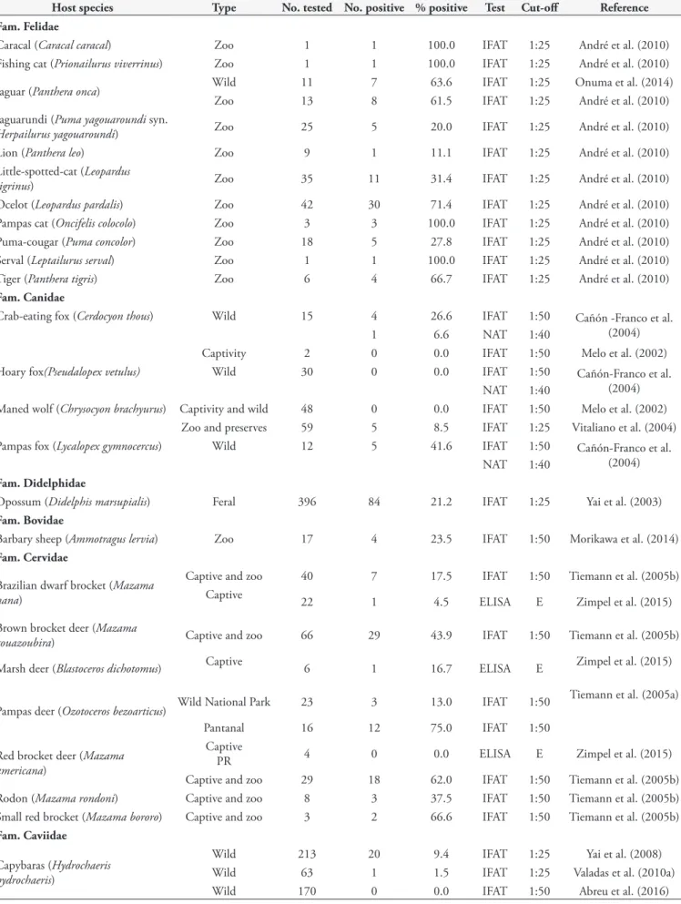
Table 10. Serological studies of N. caninum in wild animals from Brazil.
Host species Type No. tested No. positive % positive Test Cut-off Reference
Fam. Felidae
Caracal (Caracal caracal) Zoo 1 1 100.0 IFAT 1:25 André et al. (2010)
Fishing cat (Prionailurus viverrinus) Zoo 1 1 100.0 IFAT 1:25 André et al. (2010)
Jaguar (Panthera onca) Wild 11 7 63.6 IFAT 1:25 Onuma et al. (2014)
Zoo 13 8 61.5 IFAT 1:25 André et al. (2010)
Jaguarundi (Puma yagouaroundi syn.
Herpailurus yagouaroundi) Zoo 25 5 20.0 IFAT 1:25 André et al. (2010)
Lion (Panthera leo) Zoo 9 1 11.1 IFAT 1:25 André et al. (2010)
Little-spotted-cat (Leopardus
tigrinus) Zoo 35 11 31.4 IFAT 1:25 André et al. (2010)
Ocelot (Leopardus pardalis) Zoo 42 30 71.4 IFAT 1:25 André et al. (2010)
Pampas cat (Oncifelis colocolo) Zoo 3 3 100.0 IFAT 1:25 André et al. (2010)
Puma-cougar (Puma concolor) Zoo 18 5 27.8 IFAT 1:25 André et al. (2010)
Serval (Leptailurus serval) Zoo 1 1 100.0 IFAT 1:25 André et al. (2010)
Tiger (Panthera tigris) Zoo 6 4 66.7 IFAT 1:25 André et al. (2010)
Fam. Canidae
Crab-eating fox (Cerdocyon thous) Wild 15 4 26.6 IFAT 1:50 Cañón -Franco et al.
(2004)
1 6.6 NAT 1:40
Captivity 2 0 0.0 IFAT 1:50 Melo et al. (2002)
Hoary fox(Pseudalopex vetulus) Wild 30 0 0.0 IFAT 1:50 Cañón-Franco et al.
(2004)
NAT 1:40
Maned wolf (Chrysocyon brachyurus) Captivity and wild 48 0 0.0 IFAT 1:50 Melo et al. (2002)
Zoo and preserves 59 5 8.5 IFAT 1:25 Vitaliano et al. (2004)
Pampas fox (Lycalopex gymnocercus) Wild 12 5 41.6 IFAT 1:50 Cañón-Franco et al.
(2004)
NAT 1:40
Fam. Didelphidae
Opossum (Didelphis marsupialis) Feral 396 84 21.2 IFAT 1:25 Yai et al. (2003)
Fam. Bovidae
Barbary sheep (Ammotragus lervia) Zoo 17 4 23.5 IFAT 1:50 Morikawa et al. (2014)
Fam. Cervidae
Brazilian dwarf brocket (Mazama nana)
Captive and zoo 40 7 17.5 IFAT 1:50 Tiemann et al. (2005b)
Captive 22 1 4.5 ELISA E Zimpel et al. (2015)
Brown brocket deer (Mazama
gouazoubira) Captive and zoo 66 29 43.9 IFAT 1:50 Tiemann et al. (2005b)
Marsh deer (Blastoceros dichotomus) Captive 6 1 16.7 ELISA E Zimpel et al. (2015)
Pampas deer (Ozotoceros bezoarticus) Wild National Park 23 3 13.0 IFAT 1:50
Tiemann et al. (2005a)
Pantanal 16 12 75.0 IFAT 1:50
Red brocket deer (Mazama
americana)
Captive
PR 4 0 0.0 ELISA E Zimpel et al. (2015)
Captive and zoo 29 18 62.0 IFAT 1:50 Tiemann et al. (2005b)
Rodon (Mazama rondoni) Captive and zoo 8 3 37.5 IFAT 1:50 Tiemann et al. (2005b)
Small red brocket (Mazama bororo) Captive and zoo 3 2 66.6 IFAT 1:50 Tiemann et al. (2005b)
Fam. Caviidae
Capybaras (Hydrochaeris hydrochaeris)
Wild 213 20 9.4 IFAT 1:25 Yai et al. (2008)
Wild 63 1 1.5 IFAT 1:25 Valadas et al. (2010a)
Neosporosis in Brazil
v. 26, n. 3, july-sept. 2017 265
Table 11. Serological studies of N. caninum antibodies in horses from Brazil.
State No.
tested Type
No.
positive % positive Assay
Cut-off titer
or test Remarks Reference
Mato Grosso 200 Healthy 30 15.0 IFAT 1:50 Highest titer 1:400 in 1 horse. Laskoski et al. (2015)
Pará 411 Healthy horses 28 6.8 IFAT 1:50 No risk factors detected. Norlander (2014)
Paraná 72 Mares 28 38.8 IFAT 1:50 2 foals had pre-colostral antibodies
Locatelli-Dittrich et al.
(2006)
Paraná 14 Pregnant mares 12 85.7 IFAT 1:50 Highest titer 1:400
Hoffmann Kormann et al.
(2008)
Paraná 97 Healthy horses 14 14.4 IFAT 1:50 Highest titer 1:200 in 2 horses Villalobos et al. (2012)
Paraná and Santa
Catarina 112
Mares from 5
breeding farms 14 12.5 IFAT 1:50
25.7% (9/35) prevalence in mares with reproductive problem versus 6.4% (5/77) without problems. Highest titer only 1:50.
Abreu et al. (2014)
Rio Grande do Sul 241 Cart horses and Crioula breed 34 15.9 IFAT 1:50 - Toscan et al. (2011)
Rio Grande do Sul 181 Pregnant mares 39 21.5 ELISA A, C
9.3% of their paired foals had pre-colostral anti-Neospora
antibodies.
Pivoto et al. (2014)
Rio Grande do Sul 197 Abattoir 77 39.1 IFAT 1:50 Tested for spp. and Sarcocystis
T. gondii
Portella et al. (2017)
Rio Grande do Sul, São Paulo, Rio de Janeiro
101 Race horses 0 0.0 NAT 1:25 - Dubey et al. (1999)
Santa Catarina 615 Healthy 25 4.1 IFAT 1:50
72 with history of neurological and reproductive problems.
Moura et al. (2013)
São Paulo 325 Healthy 19 5.8 IFAT 1:50 Highest titer 1:400 Villalobos et al. (2006)
483 Diseased 73 15.1
São Paulo 26 History of ataxia. 15 57.6 IFAT 1:2 26 cerebrospinal fluids
negative.
Stelmann et al. (2011)
South 203 Mares 129 63.3 IFAT 1:50
Of 129, 34.8% gave birth to seropositive foals.
Antonello et al. (2012)
10 states 961 Old horses from abattoirs 24 2.5 ELISA C, K - Hoane et al. (2006)
Table 12. Detection of N. caninum antibodies in avian species from Brazil.
Host Tested %positive DNA or Antibodies Test Cut-off / Method Reference
Chicken (Gallus gallus domesticus)
200 (outdoor) 47 (23.5) Antibodies IFAT 1:50
Costa et al. (2008)
200 (indoor) 3 (1.5) Antibodies IFAT 1:50
10 positive 6 (60.0) DNA PCR 3/4 of brain Direct PCR for Nc5
100 farm chickens 17 (17.0) Antibodies IFAT 1:50 Gonçalves et al. (2012)
6 (6.0) DNA PCR Np21/Np6
Sparrow (Passer domesticus) 40 3 (7.5) DNA PCR Nested PCR of Nc5 and Heart and brain. Gondim et al. (2010) sequencing of ITS
Cerqueira-Cézar, C.K. et al. Braz. J. Vet. Parasitol.
266
In one survey, 802 serum samples of female cattle from 55 dairy
and beef farms from six Brazilian states (SP, RJ, MG, PR, RS, MS)
were assayed by IFAT (cut-off 1:25) and 23.6% were seropositive;
association between positivity to
N. caninum
and state of origin, age
and production purpose was analyzed using uniform methodology
(RAGOZO et al., 2003). Although seroprevalence was higher in
animals older than 24 months, this difference was not statistically
significant. Conclusion is not definitive because of the selection
of low cut-off of 1:25. Among the six states, RJ had the lowest
prevalence and MG the highest and dairy cattle had higher
prevalence than beef cattle. In another large study from MS,
N. caninum
antibodies were found in cattle from 143 of 205 herds
(IFAT 1:50); the cows were older than two years (OSHIRO et al.,
2007). Overall seroprevalence was 14.9% (Table 4).
Even after using a higher cut-off titer (IFAT 1:200) than used most
of the surveys listed in Table 4, high prevalence were recorded from
MG, where
N. caninum
antibodies were detected in 23 of 24 herds
with individual seroprevalence of 21.6% (BRUHN et al., 2013); and
MT, where antibodies to
N. caninum
were found in 499 (53.5%)
of 932 cattle samples, with at least one positive in each farm
(BENETTI et al., 2009). In another report from MG, very high
(91.2%; 510/559) prevalence of antibodies to
N. caninum
was
found among 18 farms (GUEDES et al., 2008), revealing that
N. caninum
is well spread in the southern region of the state.
They also recorded 97.2% (559/575) prevalence in cows from
an abattoir.
There is an unconfirmed report that the quality of milk might
be affected by neosporosis (MELO et al., 2001); an association was
reported between the type of milk, classified as A, B and C according
to its quality, produced in the farms and positivity to
N. caninum
antibodies, with higher occurrence in farms of MG that produce
milk grade A/B than grade C; and the authors discussed about
the production technology used indifferent farms, animals stress
and commercialization. One possible cause is that the production
of types A/B milk is higher and requests more from the animals,
generating stress, which could be responsible for the higher
prevalence.
An association between seroprevalence and age of cattle (older
animals presenting higher prevalence) was detected, while feed
produced on the farm was negatively associated with
N. caninum
infection in PR (GUIMARÃES et al., 2004). Also in PR, production
of food in the farm, absence of artificial insemination and access
of domestic and wild animals to feed facilities were associated
with infection (MARTINS et al., 2012).
A strong association of
N. caninum
infection was found in
farms of RS, which had presence of dogs close to the livestock and
which also fed calves with colostrum pooled from several cows
(CORBELLINI et al., 2006). In this study, the size of the farm
was inversely associated with the presence of positive animals.
An association between beef herds and infection by
N. caninum
was reported in the Amazon region, RO, while comparing
beef, dairy and mixed herds (AGUIAR et al., 2006). In that
region, farms with more than 25 animals were also a risk factor.
However, reproductive problems, contact with forest areas and
presence of dogs were not associated with the coccidian infection.
Significant association related to animal management was
found in PE (SILVA et al., 2008). Veterinary assistance, nutritional
status, presence of wetlands, manipulation of the newborn calves
(use of gloves when handling aborted fetuses), and destination
of cows that aborted (higher in the ones that were treated with
antibiotics than the ones that were discarded) were risk factors
for the infection.
Pure bred Holstein cows had a higher exposure rate than
mixed breeds (MUNHOZ et al., 2009) and an association
between reproductive abnormalities (Table 4) (repeated estrus,
repeated miscarriages and temporary anestrus) and seropositivity
to
N. caninum
has been reported (BRUHN et al., 2013).
N. caninum
is considered a primary pathogen and not
influenced by concurrent BHV1, BVDV infections in dairy herds
(MELO et al., 2004; MINEO et al., 2006).
Antibodies can fluctuate during pregnancy as reported,
and increase in titer is not always associated with abortion
(CARDOSO et al., 2009). A prospective longitudinal study was
carried out in three farms in SP, during two consecutive years
and the reproductive parameters were analyzed in those herds
(CARDOSO et al., 2012a). In only one of the three herds the
relative risk of abortion between
N. caninum
positive and negative
cows was higher in the positive animals. No difference was
observed regarding gestational age at abortion, repeated abortion,
number of inseminations, and calving interval. A high association
between
N. caninum
serological status of dams and daughters
were observed in a longitudinal study carried out in three farms
from SP, confirming the importance of vertical transmission, but
there was no difference in the culling rate between positive and
negative cows (CARDOSO et al., 2012b). A significant relationship
between seropositivity of cattle and their offspring was also found
in PE (RAMOS et al., 2016), which had a rate of transplacental
transmission of 72.2% (13/18) for adults and 69.2% (9/13) for
heifers by IFAT and 43.5% (17/39) for adults and 50.0% (9/18)
for heifers by ELISA, concluding that vertical transmission is the
major form of infection in this region.
Buffaloes:
Studies with serum samples from Brazilian buffaloes
(Table 5) showed occurrence of
N. caninum
antibodies varying from
14.6% to 88%. However, despite the high occurrence values, no
reproductive disorders were reported in those groups (FUJII et al.,
2001a, b; GENNARI et al., 2005; GONDIM et al., 2007)
Sheep:
Seroprevalences ranged from 1.8% to 64.2%
(Table 6). Some surveys stated as risk factors: abortion in the
flock, presence of dogs, extensive husbandry systems and breed
of the sheep. Great part of the studies also evaluated the presence
of
T. gondii
infection, which usually showed a higher prevalence.
In 2017, Filho et al. (2017) studied the vertical transmission rate
of 50 naturally infected sheep, analysed by in house ELISA for six
months. The initial prevalence of infection was 26.0% (13/50)
and by the end of the study it had increased to 72% (36/50),
being the vertical transmission rate 11%, which one sheep out of
nine from a group gave birth to two infected ewes (IFAT 1:25).
Neosporosis in Brazil
v. 26, n. 3, july-sept. 2017 267