Antigenotoxic effects of
Mandevilla velutina
(Gentianales, Apocynaceae)
crude extract on cyclophosphamide-induced micronuclei in Swiss mice
and urethane-induced somatic mutation and recombination
in
Drosophila melanogaster
Regildo Márcio Gonçalves da Silva
1, Neila Coelho de Sousa
1, Ulrich Graf
2and Mário Antônio Spanó
11
Laboratório de Mutagênese, Instituto de Genética e Bioquímica, Universidade Federal de Uberlândia,
Uberlândia, MG, Brazil.
2Institute of Animal Sciences, Swiss Federal Institute of Technology Zurich, Schwerzenbach, Switzerland.
Abstract
AMandevilla velutina crude extract was investigated using the mouse micronucleus test (MNT) and the Drosophila melanogaster somatic mutation and recombination test (SMART) using standard (ST) and high bioactivation (HB) crosses. The MNT used 10 mg, 20 mg or 40 mg per 100 g of body weight (bw) of extract with and without 0.2 mg per 100 g bw peritoneal cyclophosphamide. There was no genotoxicity in the negative control or extract only groups and, compared to the cyclophosphamide control, there was a significant reduction in micronucleated polychromatic eryth-rocytes in all the groups given extract plus cyclophosphamide. For SMART larvae were fed 5 or 10 mg mL-1
of extract for seven days with and without 0.89 mg mL-1
of urethane given on day seven. The ST and HB flies showed no signifi-cant differences in spots between the negative control and the extract only groups. The number of urethane-induced spots was reduced by the highest concentration of extract for the ST flies and by both concentrations of extract for the HB flies. The results suggest thatM. velutina extract is not genotoxic but is antigenotoxic.
Key words:antigenotoxicity, genotoxicity tests, micronucleus assay, SMART, wing spot test. Received: September 20, 2007; Accepted: November 8, 2007.
Introduction
The herbaceous plantMandevilla velutina(Mart. Ex Stadelm.) Woodson (Gentianales, Apocynaceae), a mem-ber of the dogbane family (Apocynaceae) which also in-cludes the periwinkle generaCatharanthusandVinca, is found in the southern Atlantic Forrest (Mata Atlântica in Portuguese) and southeastern Brazil. An infusion of the rhi-zomes of this plant is employed in folk medicine as a popu-lar remedy to treat snakebites, possibly by inhibition of phospholipases A2 (PLA2), and as a general anti-inflammatory agent (Hirschmann and Dearias, 1990; Mors, 1991; Bentoet al., 2003; Biondoet al., 2003).
The selective antagonism ofM. velutinacrude extract against bradykinin and its derivatives has been studied by various authors (Calixtoet al., 1985; Calixto and Yunes, 1986; Calixtoet al., 1987; Calixto and Yunes, 1990) and some active compounds in the extracts have been chemi-cally characterized. The pregnane glycoside compounds
isolated fromM. velutinahave been shown to be effective in antagonizing bradykinin (BK) responses in a variety of preparations and also exhibit potent and long-lasting anal-gesic, anti-inflammatory and anti-oedematogenic activities against a variety of inflammatory and fever-inducing phlo-gistic agents but was more effective in inhibiting processes involving kinins (Calixto and Yunes, 1991; Henriqueset al., 1991; Maraschinet al., 2000).
Yuneset al.(1993) investigatedM. velutinaand iso-lated and purified the pregnanic steroid velutinol A, a po-tent anti-inflammatory agent and bradykinin antagonist which has also detected inM. velutinacell cultures (Ma-raschinet al., 2001). Velutinol A has also been shown to se-lectively block edema responses (Mattoset al., 2006a) and produces peripheral antinociceptive action in some models of acute and persistent inflammatory pain by interacting with kinin B-1-receptor mediated effects (Mattos et al., 2006b). A review summarizing recent advances in the iden-tification and the potential therapeutic properties of non-peptide antagonists for kinin B-1 receptors is presented by Camposet al. (2006). Bento et al.(1996) used quantita-tive-1H-1H nuclear Overhauser enhancement (NOE) data
www.sbg.org.br
Bioactive triterpenoids which have been isolated from M. velutina include the bradykinin antagonist MV 8613, the anti-inflammatory agent and bradykinin antago-nist MV 8612 and the lipoxygenase inhibitor MV 8608 (Brito and Brito, 1993), with the latter two compounds also having exhibited anti-inflammatory and anti-oedemato-genic effects in mice (Calixto et al., 1991; Zanini et al., 1992) and inhibited PLA2-induced paw edema in rats (Ne-veset al., 1993). Santoset al.(2003) reported that MV8612 caused graded and complete inhibition of bradykinin-induced thermal hyperalgesia (i.e.an increased sensitivity to pain) in mice and inhibited both the neurogenic and in-flammatory pain responses to formalin. In addition, Silva and Hamaguchi (1997) demonstrated that different concen-trations ofM. velutinacrude extract are capable of reducing Trypanosoma cruziinfection in mice. These results provide strong experimental support for the beneficial use of this plant extract in folk medicine.
The wide distribution ofMandevilla and its use in folk medicine plus the lack of information on its genoto-xicity or antigenotogenoto-xicity prompted us to assess the geno-toxicity of M. velutina crude extract and investigate possible antigenotoxic effects of the extract on the induc-tion of micronuclei by cyclophosphamide using thein vivo mouse bone marrow cell micronucleus test and somatic mutation and recombination induced by urethane using the Drosophila melanogasterwing spot somatic mutation and recombination test (SMART).
The micronucleus assay serves to detect the geno-toxicity of chemicals through their ability to induce the for-mation of small membrane-bound DNA fragments (Ouanes et al., 2003), while SMART is a short-termin vivoassay for the detection of mutation, recombination, chromosome damage and/or aneugenic effects in somatic cells of D. melanogaster(Grafet al., 1984).
Materials and Methods
Crude chemical compounds and media
The subterranean system of Mandevilla velutina (Mart. Ex Stadelm.) Woodson consists of a xylopodium, the basal region of which is joined to a tuberous root (Appezzato-da-Glória and Estelita, 2000). We collectedM. velutinatuberous roots at a site (19°09'20" S, 48°23'20" W) in the Brazilian cerrado (savanna) near the town of Uber-lândia in the Brazilian state of Minas Gerais. The plants were identified by an expert plant collection. The roots
with the significance level set atα=0.05. The statistical analyses was carried out using the SPSS 12.0 statistical package for PCs (SPSS, Chicago, IL).
TheD. melanogasterSMART procedure
The antigenotoxic effects ofM. velutina extracton urethane-induced somatic mutations in D. melanogaster were assessed by SMART for the multiple wing hairs (mwh) and flare-3 (flr3) recessive mutations. The D. melanogaster crosses used were the standard cross (ST cross) in whichflr3/In(3LR)TM3, ri ppsep l(3)89Aa bx34ee BdSfemales were mated withmwhmales (Grafet al., 1989) and the high bioactivation cross (HB cross) in whichORR; flr3/In(3LR)TM3, ri pp sep l(3)89Aa bx34e e BdS females were mated withmwhmales (Graf and Singer, 1992; Graf and van Schaik, 1992). The HB cross is characterized by a high sensitivity to promutagens and procarcinogens be-cause theORR; flr3/TM3, BdSstrain carries chromosomes 1 and 2 from a DDT-resistant Oregon R(R) line (Dapkus and Merrell, 1977) which is characterized by an increased level of cytochrome P-450 (Hällström and Blanck, 1985; Saner et al., 1996).
Both crosses produce two types of progeny phenoty-pically distinguished by theBdSmarker,i.e., marker-hete-rozygous (MH) flies (mwh +/+ flr3) with phenotypically wild type wings or (ii) balancer-heterozygous (BH) flies (mwh/TM3, BdS) with phenotypically serrate wings. A De-tailed analysis of genetic markers is given by Lindsley and Zimm (1992).
Eggs were collected from both crosses during 8 h in culture bottles with an agar-agar base (4% w/v) and a thick layer of live yeast supplemented with sucrose. Third instar (72 + 4 h) larvae were washed out of these bottles with tap water and collected in a stainless steel strainer. Batches of equal numbers of larvae were placed in glass vials contain-ing 1.5 g of drosophila instant medium formula 4-24 (Caro-lina Biological Supply, Burlington, NC, USA) rehydrated with 5 mL of a solution containing a final concentration of 5 mg mL-1or 10 mg mL-1ofM. velutinaextract alone or combined with urethane (CAS Nº 51-79-6; Fluka AG, Switzerland) at a final concentration of 0.89 mg mL-1, this chemical being a well-known promutagen which is meta-bolically activated by the cytochrome P-450 enzyme sys-tem. Negative (distilled water) and positive (0.89 mg mL-1 urethane) controls were included in both experiments. Larvae were allowed to feed on the medium for the remain-der of their larval life (~ 48 h). The experiments were car-ried out at 25 °C and 65% relative humidity.
The hatched adult flies were killed by placing in 70% (v/v) aqueous ethanol, in which they remained until needed for analysis. For analysis, wings of the MH flies were re-moved, mounted on glass slides with Faure’s solution (30 g gum Arabic, 20 mL glycerol, 50 g chloral hydrate and 50 mL water) and examined for spots using a compound microscope at 400X magnification. The data were
evalu-ated according to the procedure described by Frei and Würgler (1988, 1995) for the occurrence of single spots (mwh or flr) and twin spots (mwh clone adjacent to flr clone). The different types of spots are due to different genotoxic mechanisms,i.e., mutational events (deletions, point mutation, specific types of chromosome aberrations, etc.), mitotic recombination or, sometimes, monosomy (Grafet al., 1984; Guzmán-Rincón and Graf, 1995).
The frequencies of spots per fly were compared to the concurrent control series according to Frei and Würgler (1988, 1995). Statistical comparisons were made using the Kastenbaum and Bowman (1970) test for proportions and followed a multiple-decision procedure according to Frei and Würgler (1988). For the final statistical analysis of all positive outcomes, the non-parametric Mann-Whitney U-test with significance levelsα=β= 0.05 was used in or-der to exclude false positives (Frei and Würgler, 1995).
Results
Micronucleus test
The frequency of MNPCE±the standard deviation (SD), for male Swiss albino mice in the distilled water neg-ative control group was 4 + 0.71, while for three groups of mice treated with the equivalent of 10 mg, 20 mg or 40 mg per 100 g bw of extract the frequency of MNPCE was 5.4 + 2.68 for the 10 mg group, 0.7 + 1.14 for the 20 mg group and 2.3 + 2.51 for the 40 mg group. The negative control group and the extract groups were not significantly different by the Mann-Whitney U-test at p > 0.4 (Table 1).
For the cyclophosphamide positive control group the frequency of MNPCE was 25 + 6.96, which was signifi-cantly higher (U-test, p < 0.05) when compared with the negative control group or the extract groups (Table 1). For the experimental groups pre-treated with the equivalent of 10 mg, 20 mg or 40 mg per 100 g bw of extract and then given cyclophosphamide we found that all the groups showed significantly lower frequencies of MNPCE as com-pared to the cyclophosphamide positive control group, the frequencies of MNPCE were 6.7 ± 5.32 for the 10 mg group, 2.6±2.77 for the 20 mg group and 5.1±1.64 for the 40 mg group (Table 1).
Somatic Mutation And Recombination Test (SMART)
genotoxic or antigenotoxic effects on spontaneous DNA le-sions.
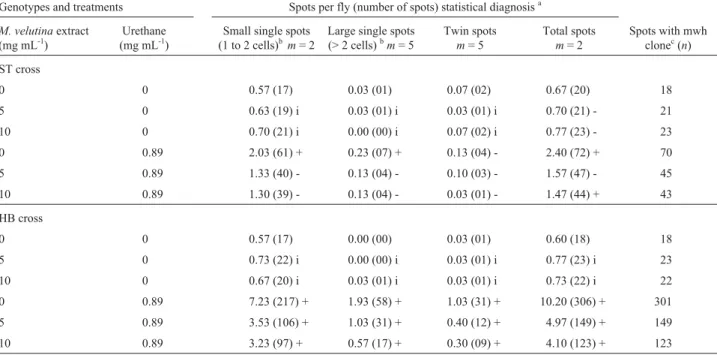
The mean total spot frequencies per fly was 0.60 in the HB cross negative control group and 0.67 in the ST cross negative control group, both values being within the normally observed range. However, the frequencies of spontaneous spots normally observed in HB crosses are usually slightly higher than those observed in the ST cross (Graf and van Schaik, 1992; Lehmannet al., 2000; Cunha et al., 2001). Compared to the negative control group, ure-thane induced a statistically significant number of small single spots and large single spots in the ST cross and in all three categories of spots with the HB cross and demon-strated a high bioactivation effect, with 2.4 spots per fly in the ST crossvs.10.2 spots per fly in the HB cross. After chronic treatment of the ST cross larvae with different con-centrations of extract only there were no statistically signif-icant differences in any of the three categories of spot frequencies in the adult flies (Table 2).However, for ST cross larvae grown on media supplemented with 10 mg mL-1of extract plus 0.89 mg mL-1of urethane there was a statistically significant reduction in the total spots recorded on adult flies as compared to the number of spots seen on adult flies the larvae of which had been exposed to urethane only. For the HB cross, for both concentrations of M velutinaextract there was a significant reduction in all three categories of urethane-induced spots and in the total ure-thane-induced spots seen on adult flies.
The spot size distributions for the negative control group, the extract only group, the urethane only group and the extract plus urethane group are presented in Figures 1A
and 1B, which clearly show that the extract was not geno-toxic under our experimental conditions. In contrast, after chronic treatment with urethane alone or urethane plus ex-tract there was a predominance of small single spots, with the frequency of larger spots decreasing with increasing spot size. These data are in line with those reported with bleomycin, diethylnitrosamine and procarbazine (Graf et al., 1984), cyclophosphamide (Spanó et al., 2001) and a phytotherapeutic extract fromStryphnodendron adstringes (Mart.) Coville (Sousaet al., 2003).
Discussion
Evaluation of micronucleus induction is the primary in vivotest in a battery of genotoxicity tests and is recom-mended by regulatory agencies around the globe as part of product safety assessment. The assay, when performed cor-rectly, detects both clastogenic and aneugenic effects (Krishna and Hayashi, 2000). Micronuclei in young eryth-rocytes arise primarily from acentric fragments or chromo-somes that are unable to migrate and follow the mitotic spindle during cell division in erythropoietic blast cells (Salamone and Heddle, 1983; Ouaneset al., 2003), an crease in the MNPCE frequency being an indication of in-duced chromosome damage (Krishna and Hayashi, 2000). In our experiments, we found that the MNPCE frequency in the groups treated with the three different extract concen-trations was not significantly different from the frequency seen in the negative control group. Cyclophosphamide has been widely used as a positive control in rodent micro-nucleus assay (Krishna and Hayashi, 2000) and, in our ex-periments, showed a statistically significant induction of
Extract (mg per 100 g bw) M1 M2 M3 M4 M5
Without cyclophosphamide
0 08 08 08 07 09 40 4.0±0.71
10 11 15 08 09 11 54 5.4±2.68
20 01 00 03 02 01 07 0.7±1.14
40 03 09 04 03 04 23 2.3±2.51
With cyclophosphamide†
0 46 59 45 44 56 250 25.0±6.96*
10 15 11 22 10 09 67 6.7±5.32**
20 08 02 08 03 05 26 2.6±2.77**
40 09 12 11 11 08 51 5.1±1.64**
†
Equivalent to 0.2 mg per 100 g bw.
MNPCE. However, we also found that all concentrations of the extract prevented significant induction of MNPCE by cyclophosphamide.Taken together, these results indicate that the extract was not genotoxic under the conditions of this assay but does contain components that exert an anti-genotoxic effect on cyclophosphamide-induced DNA le-sions.
In the SMART the genotoxicity and antigenotoxicity of the extract was investigated in somatic cells of theD. melanogasterimaginal disk by feeding larvae derived from both ST and HB crosses on media containing different con-centrations of the extract. The HB cross results in constitu-tively increased cytochrome P-450-dependent enzyme activities and therefore make the wing spot test more sensi-tive for the detection of promutagens and procarcinogens (Graf and Singer, 1992; Graf and van Schaik, 1992). Ure-thane, a promutagen which can occur naturally in some fer-mented food (Ough, 1976), was used as positive control because it has a clear genotoxic potential in Drosophila, with a clear dose response and dependence on metabolic activation. The metabolic pathway most probably involves cytochrome P-450-dependent enzyme activities (Frölich and Würgler, 1990).
The SMART data showed that at the concentrations tested the extract did not induce somatic mutation and re-combination in theD. melanogasterST or HB crosses. As expected, when compared to the negative control group, urethane not only showed a statistically significant
induc-Table 2- TheD. melanogasterwing spot somatic mutation and recombination test (SMART) results showing the number of flies and frequency of spots observed in the marker-heterozygous (MH) progeny of the standard (ST) and the high bioactivation (HB)D. melanogastercrosses after chronic treatment of larvae (n = 30 per treatment) with different concentrations ofMandevilla velutinacrude extract (CE) either alone or supplemented with 0.89 mg mL-1of urethane. Marker-heterozygous flies (mwh/flr3) were evaluated.
Genotypes and treatments Spots per fly (number of spots) statistical diagnosisa
M. velutinaextract (mg mL-1)
Urethane (mg mL-1)
Small single spots (1 to 2 cells)bm= 2
Large single spots (> 2 cells)bm= 5
Twin spots m= 5
Total spots m= 2
Spots with mwh clonec(n)
ST cross
0 0 0.57 (17) 0.03 (01) 0.07 (02) 0.67 (20) 18
5 0 0.63 (19) i 0.03 (01) i 0.03 (01) i 0.70 (21) - 21
10 0 0.70 (21) i 0.00 (00) i 0.07 (02) i 0.77 (23) - 23
0 0.89 2.03 (61) + 0.23 (07) + 0.13 (04) - 2.40 (72) + 70
5 0.89 1.33 (40) - 0.13 (04) - 0.10 (03) - 1.57 (47) - 45
10 0.89 1.30 (39) - 0.13 (04) - 0.03 (01) - 1.47 (44) + 43
HB cross
0 0 0.57 (17) 0.00 (00) 0.03 (01) 0.60 (18) 18
5 0 0.73 (22) i 0.00 (00) i 0.03 (01) i 0.77 (23) i 23
10 0 0.67 (20) i 0.03 (01) i 0.03 (01) i 0.73 (22) i 22
0 0.89 7.23 (217) + 1.93 (58) + 1.03 (31) + 10.20 (306) + 301
5 0.89 3.53 (106) + 1.03 (31) + 0.40 (12) + 4.97 (149) + 149
10 0.89 3.23 (97) + 0.57 (17) + 0.30 (09) + 4.10 (123) + 123
aStatistical diagnoses according to Frei and Würgler [1988]: +, positive; w+, weakly positive; -, negative; i, inconclusive; Multiplication factor:m.
p < 0.05.bIncluding rareflr3single spots.cConsideringmwhclones frommwhsingle and twin spots.
Figure 1- Size distributions for single spots after chronic treatments with 5 mg mL-1and 10 mg mL-1ofMandevilla velutinacrude extract (CE) and
originating from larvae in the group fed on media supple-mented with 10 mg mL-1of extract plus urethane. Further-more, with the HB cross there was a statistically significant reduction in all three categories of spots and in total spots for flies emerging from larvae treated with both concentra-tions of extract plus urethane. These results indicate that the extract acts as an antigenotoxic on urethane-induced DNA lesions and suggests that components of the extract interact with cytochrome P-450 leading to a reduction in the forma-tion of the active urethane metabolite that provides geno-mic instability.
Our results are in line with those of Idaomaret al. (2002), who reported the results of aD. melanogasterwing spot test which showed that essential oils extracted from the medicinal plants Helichrysum italicum, Ledum groenlandicum and Ravensara aromatica produced an antimutagenic effect against urethane and that this could be explained by the interaction of constituents of these plants with the cytochrome P-450 activation system leading to a reduction of the formation of the active urethane metabo-lite. The effect could also be attributed to certain molecules that are involved in these oils. El Hamsset al.(2003) de-scribed the modulating action of bell pepper (Capsicum annuum) and black pepper (Piper nigrum) on the effects of the alkylating agent methyl methanesulfonate and the pro-mutagen urethane when investigated using a D. melanogasterwing spot test. These authors concluding that suppression of metabolic activation or interaction with the active groups of mutagens could be mechanisms by which these spices exert their antimutagenic activity. Kurodaet al. (1992) has suggested that in co-treatments an anti-mutagen can act as a desanti-mutagen which can chemically or enzymatically inactivate a mutagen or inhibit the metabolic activation of promutagen.
It has been reported that M. velutinacontains bio-active triterpenoids (Calixto and Yunes, 1991; Maraschin et al., 2000) which include velutinol A (Yuneset al., 1993) and the active terpenes MV 8608, MV 8612 and MV 8613 (Brito and Brito, 1993), while Bentoet al.(2003) has also reported a pentasaccharide derivative of velutinol A called velutinoside A. Nevertheless, there is still a lack of infor-mation on the genotoxic and antigenotoxic properties of compounds extracted fromM. velutina.
The in vivo rodent micronucleus assay has been widely used to detect genotoxicity (Krishna and Hayashi, 2000; Villaniet al., 2007; Doppalapudiet al., 2007) and the somatic mutation and recombination tests in D.
did not induce chromosome breakage and was non-aneu-genic since it did not affect spindle fiber function, while the D. melanogasterSMART data showed that the extract did not induce somatic mutation or recombination. Further-more, the results show that the extract has antigenotoxic ef-fects on cyclophosphamide-induced lesions in mice and urethane-induced DNA lesions inD. melanogaster. In ad-dition, the SMART results indicate that the action of the ex-tract may involve the interaction of constituents of the extract with cytochrome P-450, but, however, the exact mechanisms are not well understood due to the limited data reported in the literature with respect to the inhibitory ef-fects of these constituents. The results of our study show that the in vivo rodent micronucleus test and the D. melanogasterSMART are versatile and sensitivein vivo eukaryotic systems for the determination of non-genotoxic and antigenotoxic activity ofM. velutinaextract. However, further studies are needed to elucidate the precise mecha-nisms involved in the antigenotoxic activity ofM. velutina extract, with such studies including not only the investiga-tion of different concentrainvestiga-tions ofM. velutinacrude extract but also the main constituents of the extract.
Acknowledgments
This work was partially supported by the Brazilian agencies CNPq, CAPES, FAPEMIG and UFU.
References
Appezzato-da-Glória B and Estelita MEM (2000) The develop-mental anatomy of the subterrranean system inMandevilla illustris (Vell.) Woodson and M. vellutina(Mart. ex Sta-delm.) Woodson (Apocynaceae). Rev Brasil Bot 23:27-35. Bento ES, Calixto JB, Hawkes GE, Pizzolatti MG, SantAna AEG
and Yunes RA (1996) The structure of velutinol A is (15R,16R,20S)-14,16:15,20:16,21-triepoxy-15,16-seco-14
β,17 α-pregn-5-ene-3β,15-diol. A combined quantitative Overhauser effect and molecular modelling study. J Chem Soc, Perkin Trans 2:1359-1366.
Bento ES, Sant’Ana AEG, Hawkes GE, Calixto JB and Yunes RA (2003) The structure of velutinoside A: A pregnane pen-tasaccharide from Mandevilla velutina. Tetrahedron Lett 44:8335-8337.
Biondo R, Pereira AMS, Marcussi S, Pereira PS, França SC and Soares AM (2003) Inhibition of enzymatic and pharmaco-logical activities of some snake venoms and toxins by
Brito ARMS and Brito AAS (1993) Forty years of Brazilian me-dicinal plant research. J Ethnopharmacol 39:53-67. Calixto JB and Yunes RA (1986) Effect of a crude extract of
Mandevilla velutinaon contractions induced by bradykinin and des-Arg-9-bradykinin in isolated vessels of the rabbit. Br J Pharmacol 88:937-941.
Calixto JB and Yunes RA (1990) Blockade of kinin-induced re-sponses of the guinea-pig isolated urinary bladder by the ex-tract ofMandevilla velutina. Gen Pharmacol 21:285-290. Calixto JB and Yunes RA (1991) Natural bradykinin antagonists.
Mem Inst Oswaldo Cruz 86:195-202.
Calixto JB, Nicolau M and Yunes RA (1985) A selective antago-nist of bradykinin action from a crude extract ofMandevilla velutina. Effect on isolated rat uterine smooth-muscle. Braz J Med Biol Res 18:A728-A728.
Calixto JB, Nicolau M, Pizzolatti MG and Yunes RA (1987) Kinin antagonist activity of compounds from Mandevilla velutinain the rat isolated uterus. Br J Pharmacol 91:199-204.
Calixto JB, Zanini JC, Cruz AB, Yunes RA and Medeiros YS (1991) Extract and compounds obtained fromMandevilla velutinainhibit arachidonic acid-induced ear edema in mice, but not rat stomach contraction. Prostaglandins 41:515-526. Campos MM, Leal PC, Yunes RA and Calixto JB (2006)
Non-peptide antagonists for kinin B-1 receptors: Never insights into their therapeutic potential for the management of in-flammation and pain. Trends Pharmacol Sci 27:646-651. Cunha KS, Reguly ML, Graf U and Andrade HHR (2001)
Taxa-nes: The genetic toxicity of paclitaxel and docetaxel in so-matic cells of Drosophila melanogaster. Mutagenesis 16:79-84.
Dapkus D and Merrell DJ (1977) Chromosomal analysis of DDT-resistance in a long-term selected population ofDrosophila melanogaster. Genetics 87:685-697.
Doppalapudi RS, Riccio ES, Rausch LL, Shimon JA, Lee PS, Mortelmans KE, Kapetanovic IM, Crowell JA and Mirsalis JC (2007) Evaluation of chemopreventive agents for geno-toxic activity. Mutat Res 629:148-160.
El Hamss R, Idaomar M, Alonso-Moraga A and Muñoz-Serrano A (2003) Antimutagenic properties of bell and black pep-pers. Food Chem Toxicol 41:41-47.
Fragiorge EJ, Spanó MA and Antunes LMG (2007) Modulatory effects of the antioxidant ascorbic acid on the direct geno-toxicity of doxorubicin in somatic cells of Drosophila melanogaster. Genet Mol Biol 30:449-455.
Frei H and Würgler FE (1988) Statistical methods to decide whether mutagenicity test data from Drosophila assays indi-cate a positive, negative or inconclusive result. Mutat Res 203:297-308.
Frei H and Würgler FE (1995) Optimal experimental design and sample size for the statistical evaluation of data from so-matic mutation and recombination tests (SMART) in Droso-phila. Mutat Res 334:247-258.
Frölich A and Würgler FE (1990) Genotoxicity of ethil carbamate in the Drosophila wing spot test: Dependence on geno-type-controlled metabolic capacity. Mutat Res 244:201-208. Graf U and Singer D (1992) Genotoxicity testing of promutagens in the wing somatic mutation and recombination test in
Drosophila melanogaster. Rev Int Contam Ambient 8:15-27.
Graf U and van Schaik N (1992) Improved high bioactivation cross for the wing somatic mutation and recombination test inDrosophila melanogaster. Mutat Res 271:59-67. Graf U, Abraham SK, Guzman-Rincón J and Würgler FE (1998)
Antigenotoxicity studies in Drosophila melanogaster. Mutat Res 402:203-209.
Graf U, Frei H, Kägi A, Katz AJ and Würgler FE (1989) Thirty compounds tested in the Drosophila wing spot test. Mutat Res 222:359-373.
Graf U, Würgler FE, Katz AJ, Frei H, Juon H, Hall CB and Kale PG (1984) Somatic mutation and recombination test in
Drosophila melanogaster.Environ Mutagen 6:153-188. Guzmán-Rincón J and Graf U (1995)Drosophila melanogaster
somatic mutation and recombination test as a biomonitor. In: Butterworth FM, Corkum LD and Guzmán-Rincón J (eds) Biomonitors and Biomarkers as Indicators of Environmental Change. Plenum Press, New York, pp 169-181.
Hällström I and Blanck A (1985) Genetic regulation of the cyto-chrome P-450 system inDrosophila melanogaster. I Chro-mosomal determination of some cytochrome P-450-depen-dent reactions. Chem Biol Interact 56:157-171.
Henriques MGMO, Fernandes PD, Weg VB, Yunes RA, Cordeiro RSB and Calixto JB (1991) Inhibition of rat paw edema and pleurisy by the extract from Mandevilla velutina. Agents Actions 33:272-278.
Hirschmann GS and Dearias AR (1990) A Survey of medici-nal-plants of Minas-Gerais, Brazil. J Etnopharmacol 29:159-172.
Idaomar M, El Hamss R, Bakkali F, Mezzouga N, Zhiri A, Baudoux D, Muñoz-Serrano A, Liemans V and Alonso-Mo-raga A (2002) Genotoxicity and antigenotoxicity of some essential oils evaluated by wing spot test of Drosophila melanogaster.Mutat Res 513:61-68.
Kastenbaum MA and Bowman KO (1970) Tables for determining the statistical significance of mutation frequencies. Mutat Res 9:527-549.
Krishna G and Hayashi M (2000)In vivorodent micronucleus as-say: Protocol, conduct and data interpretation. Mutat Res 455:155-166.
Kuroda Y, Jain AK, Tezuka H and Kada T (1992) Antimuta-genicity in cultured mammalian cells. Mutat Res 267:201-209.
Lehmann M, Graf U, Reguly ML and Andrade HHR (2000) Inter-ference of tannic acid on the genotoxicity of mitomycin C, methylmethanesulfonate, and nitrogen mustard in somatic cells of Drosophila melanogaster. Environ Mol Mutagen 36:195-200.
Lindsley DL and Zimm GG (1992) The Genome ofDrosophila melanogaster. Academic Press, San Diego, 1133 pp. Maraschin M, Carobrez SG, Persike D, Peixoto ML, Ferreira AG,
Ferracin R, Verpoorte R and Fontana JD (2000) Cell wall polysaccharides from Mandevilla velutina (Apocynaceae) cultured cells: Extraction and chemical structure. Carbohyd Polym 41:55-60.
Maraschin M, Sugui JA, Wood KV, Bonham C, Lancas FM, Araujo PS, Yunes RA, Verpoorte R and Fontana JD (2001) Supercritical CO2 extraction of velutinol A from
Mandevilla velutina (Apocynaceae) cultured cells and MALDI-TOF MS analysis. Biotechnol Lett 23:77-82. Mattos WM, Campos MM, Fernandes ES, Richetti GP, Niero R,
ef-Neves PC, ef-Neves MC, Cruz AB, Sant’ana AE, Yunes RA and Calixto JB (1993) Differential effects of Mandevilla velutinacompounds on paw oedema induced by phospho-lipase-A2 and phospholipase-C. Eur J Pharmacol 243:213-219.
Ouanes Z, Abid S, Ayed I, Anane R, Mobio T, Creppy EE and Bacha H (2003) Induction of micronuclei by Zearalenone in Vero monkey kidney cells and in bone marrow cells of mice: Protective effect of vitamin E. Mutat Res 538:63-70. Ough CS (1976) Ethylcarbamate in fermented beverages and
foods. I. Naturally occurring ethylcarbamate. J Agric Food Chem 24:323-328.
Pantaleão SM, Alcântara AV, Alves JPH, Pavanin LA, Graf U, Rezende AAA, Valadares BLB, Fragiorge EJ, Souza NC, Guterrez ZR,et al.(2007) Assessing the impact of pollution on the Japaratuba River in Brazil using the Drosophila wing spot test. Environ Mol Mutagen 48:96-105.
Salamone MF and Heddle JA (1983) The bone marrow micro-nucleus assay: Rationale for a revised protocol. In: de Serres FJ (ed) Chemical Mutagens: Principles and Methods for their Detection, v. 8. Plenum Press, New York, pp 111-149. Saner C, Weibel B, Würgler FE and Sengstag C (1996)
Metabo-lism of promutagens catalyzed byDrosophila melanogaster
CYP6A2 enzyme in Saccharomyces cerevisiae. Environ Mol Mutagen 27:46-58.
Santos ARS, Trentin AP, Ferreira J, Yunes RA and Calixto JB (2003) Mechanisms involved in the antinociception caused by compound MV8612 isolated fromMandevilla velutinain mice. Brain Res 961:269-276.
Schmid W (1975) The micronucleus test. Mutat Res 31:9-15.
genotoxicity of a phytotherapeutic extract from
Stryphnodendron adstringens (Mart.) Coville in somatic and germ cells ofDrosophila melanogaster.Environ Mol Mutagen 41:293-299.
Spanó MA, Frei H, Wurgler FE and Graf U (2001) Recom-binagenic activity of four compounds in the standard and high bioactivation crosses of Drosophila melanogasterin the wing spot test. Mutagenesis 16:385-394.
Téllez MGO, Rodríguez HB, Olivares GQ, Sortibrán ANC, Cetto AA and Rodríguez-Arnaiz R (2007) A Phytotherapeutic ex-tract ofEquisetum myriochaetumis not genotoxic either in thein vivowing somatic test of Drosophila or in thein vitro
human micronucleus test. J Ethnopharmacol 111:182-189. Villani P, Cordelli E, Leopardi P, Siniscalchi E, Veschetti E,
Fresegna AM and Crebelli R (2007) Evaluation of geno-toxicity of oral exposure to tetravalent vanadium in vivo. Toxicol Lett 170:11-18.
Yunes RA, Pizzolatti MG, Santana AEG, Hawkes GE and Calixto JB (1993) The structure of velutinol-A, an anti-inflamma-tory compound with a novel pregnane skeleton. Phytochem Anal 4:76-81.
Zanini JC, Medeiros YS, Cruz AB, Yunes RRA and Calixto JB (1992) Action of compounds fromMandevilla velutinaon cróton oil-induced ear edema in mice - A comparative study with steroidal and nonsteroidal anti-inflammatory drugs. Phytother Res 6:1-5.
Associate Editor: Carlos F.M. Menck