EVALUATION OF A NESTED-PCR FOR MYCOBACTERIUM TUBERCULOSIS DETECTION IN BLOOD AND URINE SAMPLES
Heidi Lacerda Alves da Cruz1, Rosana de Albuquerque Montenegro1, Juliana Falcão de Araújo Lima1, Diogo da Rocha
Poroca², Juliana Figueirêdo da Costa Lima1, Lílian Maria LapaMontenegro1, Sergio Crovella²,³*, Haiana Charifker
Schindler1
¹Departamento de Imunologia, Centro de Pesquisas Aggeu Magalhães, Fundação Oswaldo Cruz, Recife, PE, Brasil; ²Departamento de Genética, Universidade Federal de Pernambuco, Recife, PE, Brasil; ³Genetic Service IRCCS Burlo Garofolo,
University of Trieste, Italy.
Submitted: January 05, 2010; Returned to authors for corrections: March 14, 2010; Approved: November 04, 2010.
ABSTRACT
The polymerase chain reaction (PCR) and its variations, such as the nested-PCR, have been described as promising techniques for rapid diagnosis of tuberculosis (TB). With the aim of evaluating the usefulness of a nested-PCR method on samples of blood and urine of patients suspected of tuberculosis we analyzed 192 clinical samples, using as a molecular target the insertion element IS6110 specific of M. tuberculosis genome. Nested-PCR method showed higher sensitivity in patients with extrapulmonary tuberculosis (47.8% and 52% in blood and urine) when compared to patients with the pulmonary form of the disease (sensitivity of 29% and 26.9% in blood and urine), regardless of the type of biological sample used. The nested-PCR is a rapid technique that, even if not showing a good sensitivity, should be considered as a helpful tool especially in the extrapulmonary cases or in cases where confirmatory diagnosis is quite difficult to be achieved by routine methods. The performance of PCR-based techniques should be considered and tested in future works on other types of biological specimens besides sputum, like blood and urine, readily obtainable in most cases. The improving of M. tuberculosis nested-PCR detection in TB affected patients will give the possibility of an earlier detection of bacilli thus interrupting the transmission chain of the disease.
Key words: Mycobacterium tuberculosis, nested-PCR, blood, urine, molecular diagnosis, IS6110 insertion sequence.
INTRODUCTION
Among all the infectious diseases affecting humans, tuberculosis (TB) remains one of the most lethal (32). In
Brazil, 80.000 cases are registered annually with incidence rate of 37,1/100,000 inhabitants, according to data from the Ministry of Health. The country occupies the 19th place among the 22 nations, responsible for 80% of all cases of TB
worldwide, with a mortality rate of 3.07 per 100,000 inhabitants (17). In 2005, 9.7% of reported cases occurred in children and adolescents. The incidence rate varies from 14 to 70 cases per 100,000 inhabitants, according to the different national regions. The States with the highest reported cases that exceeds the average rate of incidence in Brazil are Amazonas, Rio de Janeiro, Pernambuco, Pará, Ceará and Rio Grande do Sul, respectively, which corresponds to 63% of the cases of Brazil. In 2004, 4,583 new cases of TB were registered in the State of Pernambuco, Northeast Brazil, with an incidence of 55.6 and 28.7/100000 inhabitants for all forms of TB and bacillary forms respectively (28).
One of the priorities for the disease control consists in an early diagnosis and appropriate treatment (5), resulting in the cure of patients and intervention in the transmission chain (8). The standard laboratory procedures for diagnosing the disease, based on microscopic examination of acid-fast bacilli, have low sensitivity, and mycobacterial culture requires about eight weeks to release the results (9, 11).
Molecular tests based on fragments amplification of genomic sequences of M. tuberculosis have been shown to be a useful tool, able to detect the bacillus in biological samples. The Polymerase Chain Reaction (PCR) is currently considered a rapid and sensitive method for detection of M. tuberculosis, able to detect less than 10 bacilli per mL in different biological specimens (1, 2, 10, 24). Although conventional PCR is very useful for the detection of M. tuberculosis, a procedure that associates two PCRs (nested-PCR) combines greater sensitivity and specificity (18).
The cost of PCR in tuberculosis diagnostic routine is reasonable, considering that the more rapidly a diagnosis in paucibacillary patients is performed, the sooner a specific treatment can be provided, thereby reducing the bacilli sources and, with them, the risk of infection by M. tuberculosis. The development and standardization of the test make the cost more affordable, considering that this technology should be reserved for reference centers in the investigation of cases that demand
difficult diagnoses (paucibacillary form/negative smear) and the disease severity (7, 12).
The purpose of this study is to evaluate the applicability of the nested-PCR technique as a tool to assist TB diagnosis, starting from clinical samples simple to collect, such as blood and urine of patients with difficulty to expectorate or whose sputum smear was negative, with the aim of determining the sensitivity and specificity of the technique, and assessing its importance in the context of clinical and laboratory criteria used in routine diagnostic elucidation of the disease.
PATIENTS AND METHODS
Population of Study
We analyzed blood and urine samples from 96 patients (median age 32.2 years ± 30.03, with 53 males and 43 females) suspected of TB, admitted in the period from October 2006 to May 2008 at the following outpatient clinic or public institutions of reference for diagnosis of tuberculosis located at Recife (Pernambuco, Brazil): Instituto de Medicina Integral Professor Fernando Figueira - IMIP, Hospital of the Universidade Federal de Pernambuco - HC / UFPE; Hospital Barão de Lucena, and other general hospitals of the Unified Health System (SUS) which sought the Research Center Aggeu Magalhães - CPqAM / FIOCRUZ.
The criteria for patients selection were based on the presence of clinical symptoms leading to suspect TB, such as prolonged cough, fever without apparent cause, weight loss and night sweat. In order to evaluate the performance of nested-PCR we used as gold standard the criteria employed by the health services in the diagnostic definition of TB such as the combination of epidemiological, clinical and laboratory data (chest X-ray, tuberculin test, sputum smear or culture) and patient history. Patients who started anti-TB treatment, immune-compromised or under corticosteroids-based medication were excluded from the study.
according to the criteria described above, while 38 were characterized by clinical symptoms not suggestive of TB. Among the TB patients, 33 had pulmonary TB and 25 had extrapulmonary TB. The extrapulmonary forms included 11 cases of pleural TB, 3 ganglionary, 2 renal, 2 osteoarticular, 2 miliary, 2 cutaneous, 1 meningoencefalic, 1 tuberculous peritonitis and 1 pericardial. We also enrolled 60 healthy subjects (median age of 20.4 years ±14.0, with 37 female and 23 males), without clinical symptoms of tuberculosis, using them as controls.
Collection of Clinical Samples and Laboratory Processing We collected 4.0 mL of peripheral blood from each patient using tubes with EDTA and 10.0 mL of urine samples obtained in three consecutive mornings. The biological specimens were kept under refrigeration at 4 °C for up to 24 hours, until DNA extraction. Urine samples were first decontaminated according to the protocol of Sechi et al. (27) for the possible elimination of other bacteria present in the sample. Afterward the urines were centrifuged for 20 min at 4500rpm. Next the supernatant was removed, the pellet resuspended in 2 mL of 2% NaOH and incubated for 15 min at 37 °C. Then 40 mL of PBS were added to inhibit the NaOH and, after that, the samples were centrifuged at 4500rpm for 20 min. Subsequently, the pellets were resuspended in 1 mL of PBS and stored at -20 °C for subsequent DNA extraction.
DNA Extraction
DNA extraction followed the protocol proposed by Rossetti et al. (25) with some modifications. An aliquot of 500 l of each sample collected was centrifuged at 13000rpm for 10 min. and washed 3X with Tris-EDTA (TE) 1X. The pellet was resuspended in 100 l of TE 1X and thermoblock heated at 100ºC for 10 min. After centrifugation, the supernatant was transferred to a new eppendorf tube and was added 5 l of resin (Sephaglas BandPrep Kit, Amersham Pharmacia Biotech) and 6M of sodium iodide 2:1 to it, then the
eppendorf was stirred manually for 5 minutes and incubated at room temperature for 5 min. The tubes were centrifuged at 13000rpm for 1 min, the supernatant discarded, and 200 l of 70% ethanol was added, and then the tubes were centrifuged for 1 min. The sediment was dried at room temperature for 60 min, resuspended with 40 l of TE 1X and incubated in a water bath at 50 °C for 10 min. After centrifugation for 1 min., the supernatant was transferred to a new tube and stored at -20 °C until the samples were processed. A negative control using TE was included during DNA extraction and it was manipulated as a biological sample to assess any type of contamination among the samples.
Amplification of genomic DNA of M. tuberculosis
The IS6110 insertion sequence of M. tuberculosis (GenBank NP_215310.1) was the target used for nested-PCR. In the first reaction a DNA fragment of 409 bp was amplified using a pair of external primers: TJ5
(5'-CCGCAAAGTGTGGCTAAC-3') and TJ3
(5’-ATCCCCTATCCGTATGGT G-3'); in the second reaction, the first PCR amplicon served as template for the nested amplification of a 316 bp fragment using a pair of internal primers: OLI5 (5'-AACGGCTGATGACCAAAC-3') and STAN3 (5’-GTCGAGTACGCCTTCTTGTT-3'), all described by Ritis et al. (23).
Amplification reactions have been run on a MasterCycler Gradient (Eppendorf) thermal-cycler. The first amplification reaction mix consisted of 50mM KCl, 10mM Tris-HCl, pH 8.3, MgCl2 2.5 mM, dNTP (200 µM each), 30 pmol of each
oligonucleotide TJ5 and TJ3 and 2.5 unit of Taq DNA polymerase (Gibco BRL) in a final volume of 50 µL. Denaturation process was carried out at 94 °C for 30s,
annealing at 68 °C for 1 min. and extention to 72 °C for 1 min.,
the primers OLI5 and STAN3. PCR products were analyzed by electrophoresis in a 2% agarose gel, stained with ethidium bromide, visualized in ultraviolet translumination and photographed by a polaroid documentation system (Kodak MP4 + System).
For each amplification, a positive control containing DNA extracted from the reference strain M. tuberculosis H37Rv to verify if there were no complications in the amplification during the NPCR reaction, and a negative control containing Milli-Q water to exclude the possibility of contamination were included.
Ethical Considerations
All patients signed a written informed consent, agreeing to participate to the study, approved by the Ethics Committee in Research of Aggeu Magalhães Research Center - CPqAM/FIOCRUZ, Recife (CEP – Nº 55/05, approved on March 05, 2005). The necessary precautions to preserve the freedom of consent of the patients were observed.
Statistical Analysis
Each patient underwent a clinical-epidemiological questionnaire developed and standardized by the researchers’ team. Epi-Info epidemiology software (Version 6.02) was used for data record and statistical analysis, with confidence interval (CI) of 95%. P values less than 0.05 were considered statistically significant.
RESULTS
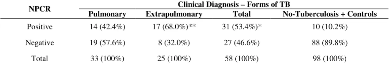
The results obtained by Nested-PCR (NPCR) are shown in Table 1. Among the 58 TB patients, 31 (53.4%) were NPCR
positive and 27 (46.6%) negative. Of the 38 patients without clinical symptoms suggestive of TB, 10 (26%) were identified as positive by NPCR. Comparing the results of NPCR the sensitivity, specificity, positive and negative predictive values were respectively 53.4% (CI: 40,0-66,5), 89.8% (CI: 81,6-94,7), 75.6% (CI: 59,4-87,1) and 76.5% (CI: 67,5-83,7). The NPCR was negative in all samples of control group.
Table 1 shows the correlation between the form of TB and the results of NPCR. In patients with the pulmonary form, NPCR was positive in 42.4% of cases, while in patients with the extra-pulmonary form, NPCR was positive in 68% of cases.
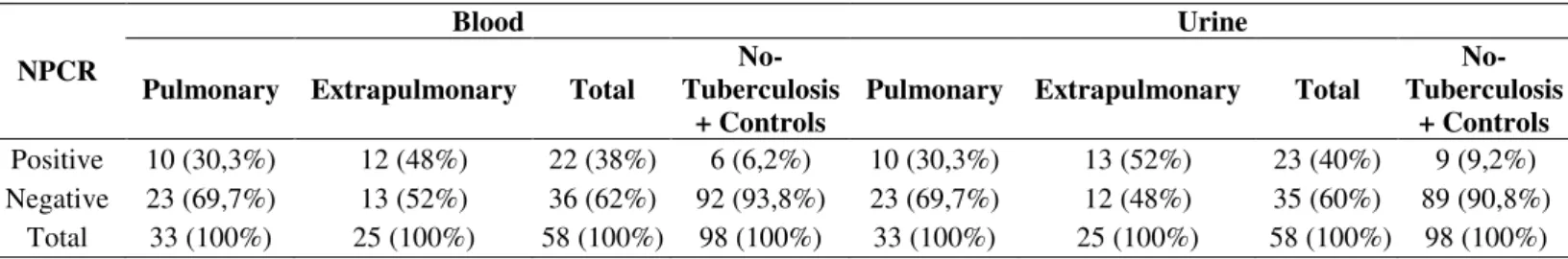
One hundred ninety two biological samples (96 whole blood and 96 urine) were collected from 96 patients suspected of TB. In this group, NPCR was positive in 53.4% (31/58) of TB cases regardless of the type of biological sample analyzed. However when considering the sensitivity and specificity of NPCR according to the type of biological sample analyzed, considering as gold standard the final clinical diagnosis, the test showed a sensitivity of 38% (22/58) (p<0.046) and 40% (23/58) (p<0.028) respectively, and specificity of 93.8% (92/98) and 90.8% (89/98), in blood and urine respectively (Table 2).
Comparing the results of NPCR for detection of M. tuberculosis in blood and urine in the different form of tuberculosis, we observed that in the group of patients with pulmonary TB, the NPCR was positive in 30.3% (10/33) in blood and urine samples. In the group of extrapulmonary TB, the NPCR showed higher positivity in both blood 48% (12/25) and urine 52% (13/25) when compared to the previous group. However, no significant differences between the samples and within the groups were observed (Table 2).
Table 1. Comparison between the results of NPCR for IS6110 and the clinical form of TB. Clinical Diagnosis – Forms of TB NPCR
Pulmonary Extrapulmonary Total No-Tuberculosis + Controls
Positive 14 (42.4%) 17 (68.0%)** 31 (53.4%)* 10 (10.2%)
Negative 19 (57.6%) 8 (32.0%) 27 (46.6%) 88 (89.8%)
Total 33 (100%) 25 (100%) 58 (100%) 98 (100%)
Sensitivity: 53.4% Specificity: 89.8% PPV: 75.6% PPN: 76.5%
*p <0,001
Table 2. Results of NPCR for IS6110 in relation the type of sample analyzed and the clinical form of TB.
Blood Urine
NPCR
Pulmonary Extrapulmonary Total
No-Tuberculosis
+ Controls
Pulmonary Extrapulmonary Total
No-Tuberculosis
+ Controls Positive 10 (30,3%) 12 (48%) 22 (38%) 6 (6,2%) 10 (30,3%) 13 (52%) 23 (40%) 9 (9,2%) Negative 23 (69,7%) 13 (52%) 36 (62%) 92 (93,8%) 23 (69,7%) 12 (48%) 35 (60%) 89 (90,8%)
Total 33 (100%) 25 (100%) 58 (100%) 98 (100%) 33 (100%) 25 (100%) 58 (100%) 98 (100%) Sensitivity: 38% Specificity: 93.8% Sensitivity: 40% Specificity: 90.8%
DISCUSSION
Tuberculosis is a serious and persistent problem of public health, especially in developing countries, and is responsible for high rate of morbidity and mortality (14). Since the need to obtain an early, rapid and sensitive diagnosis is significantly increasing, we considered a diagnostic method that may assist in advancing the diagnosis of paucibacillary forms of tuberculosis. The PCR and its variations, such as the nested-PCR, have been indicated as a promising molecular technique for a rapid diagnosis of tuberculosis (1, 10, 24). Miyazaki et al. (16) examined the potentiality of nested-PCR for tuberculosis diagnosis. The sensitivity of their nested-PCR was evaluated by using 10-fold bacterial dilutions. The bacterial count detection limit for the first PCR was 102 Colony Formation
Unit (CFU) for M. tuberculosis, but the sensitivity increased approximately 1,000-fold to 0.1 CFU, following the second PCR.
The results of NPCR coincided with the final clinical diagnosis in 61.5% (59/96) of cases (Table 1). From the 27 NPCRs false-negatives, in the group of TB patients, 41% of them were from patients under 15 years of age. Therefore, a negative test does not rule-out the diagnosis of TB. Considering that children and adolescents in general have paucibacillary form, probably the negative result of the technique could be associated with a number of bacilli circulating insufficient to be detected by NPCR.
According to Bach (3), clinical samples often contain
inhibitory substances that could interfere with the PCR reaction, making necessary to adopt procedures able to remove all inhibitors simultaneously. However, according to Böddinghaus et al. (4), until now, no single procedure has been demonstrated ideal for blocking these inhibitors, among them the hemoglobin, polysaccharides present in sputum, some reagents and components of swabs. According to the authors, false-negative results can be detected through the inclusion of a control-positive reaction. In our study, we used the reference strain H37Rv M. tuberculosis as positive control and a reagent blank (no target DNA) as negative control.
a latent TB co-associated with other pathologies.
Moreover the NPCR can detect the presence of DNA of M. tuberculosis since it amplifies genetic material derived from dormant/nonviable bacilli. Cases of patients with infection by mycobacteria are not diagnosed as soon as needed so that non-treated infected individuals continue to spread the bacilli. The importance of molecular methods is evident, as tools for increase diagnostic efficiency in health services (16). On the other hand, PCR might present a high rate of contamination, as described by Kox et al. (15), in fact, for the NPCR, where two amplifications are carried out, there is a greater possibility of contamination at the opening of the tubes between the first and second reaction. In our study we added negative controls in all extractions of DNA and in the reactions of NPCR with the aim to control the risk of false-positive results.
When considering a new diagnostic method, the performance of the gold standard is a crucial parameter. Due to the difficulty of isolating the M. tuberculosis in paucibacillary samples, the use of routine diagnosis is based on indirect indications, such as epidemiological criteria , clinical, laboratory data (in the Mantoux tuberculin test and chest X-ray), evolution and response to the treatment. The lack of a diagnostic gold standard remains a serious issue for evaluating new diagnostic, especially in HIV-infected persons and in paucibacillary disease (e.g., extrapulmonary TB and pediatric disease).
In pediatric patients, although there are few studies in the literature regarding the evaluation of molecular tests for the identification of TB in children, most of them used the above mentioned “indirect” criteria as the gold standard, and this may lead to cases with diagnostic inaccuracy (21, 29). In addition, children with TB can not produce sputum and obtaining material for culture often requires invasive procedures that cannot be easily repeated.Because of the paucibacillary nature of specimensand the irregular distribution of bacilli that tend to clump together, the sensitivity of smear microscopy is very low. Nevertheless, the interpretation of the results of NPCR
should be used and interpreted in conjunction with conventional and clinical data.
An extension of this research is being developed with larger number of subjects with smear and/or positive culture in different biological specimens, to better assess the magnitude of the above limitations.
In relation to the clinical form of TB, the highest rate of NPCR detection and correlation with clinical diagnosis were observed in the extrapulmonary form, including patients younger than 15 years of age, probably due to increased circulation of the bacillus in body fluids and tissues. Although our findings are preliminary and obtained on a low number of individuals, also in the case of paucibacillary form, the positivity of the NPCR was found both in blood and in urine even in subjects with the pulmonary form of the disease.
The application of PCR in biological samples has been developed and tested over many years. However, the standardization of PCR as a diagnostic tool to be implemented in routine laboratories requires careful processing of samples to avoid complications, such as contamination and production of nonspecific or false positive/negative that may interfere with the test results (5). In the literature, only sputum, blood and cerebrospinal fluid (CSF) have been shown to represent good substrates for PCR analysis (20). Although the low number of patients analyzed and the use of multiple parameters (as indicated above) as gold standard it can be observed that the sensitivity and specificity of NPCR for M. tuberculosis detection in blood and urine samples, considered together, showed a trend for better performance, even in the pulmonary and extrapulmonary tuberculosis, when compared with the results obtained on blood or urine for samples analyzed separately.
35.5% respectively. However, the sensitivity of PCR varies widely when compared to other studies, where results can vary between 13% and 97.4% (25, 26, 30).
Regarding the use of NPCR in urine samples to our knowledge little information is available in the literature. In our study, we found a sensitivity of 40% (23/58) and specificity of 90.8% (89/98). The variation in sensitivity of the test within blood and urine samples has been already shown in several studies. Kafwabulula et al. (13) found 55% sensitivity when evaluating patients with pulmonary TB, but Sechi et al. (27) and Torrea et al. (31) showed that the sensitivity of the NPCR in TB patients with pulmonary TB ranged from 23% to 40%.
Kafwabulula et al. (13) reported that variation in sensitivity in urine may be due to different factors. Patients with pulmonary tuberculosis usually do not present circulating mycobacterial DNA that filters through to the urine. We can also hypothesize that M. tuberculosis or its fragments are intermittently excreted in the urine of patients and it may therefore be prudent to collect urine in at least three consecutive days or more samples over 24 hours. The DNA present in urine can be from: whole mycobacteria circulating in the blood and filtered into the urine, the presence of fragments of mycobacteria, free DNA or dormant mycobacteria existing in the kidneys, which become active after the onset of the disease in the lungs. However, false-negative results can occur due to the collection of a few samples from each patient or the non-homogeneous distribution of mycobacteria in the urine (13, 31).
Rebollo et al. (22) showed that PCR detection of mycobacteria is more sensitive in HIV-positive patients than in immunocompetent patients with tuberculosis, since the mycobacterial burden is greater in these cases and more fragments of DNA are available. However, the authors found sensitivity lower than our study (16%).
The variation in sensitivity is one of the obstacles that prevent the full deployment and standardization of this technique in the laboratories of clinical analysis centers. This
variation is directly related to many factors such as different protocols employed, procedures for decontamination of samples and variations in the composition of lysis buffer (22). PCR has been used with different biological samples as a tool for TB diagnosis. However, the collection of some of these specimens requires invasive and laborious process (broncho-alveolar fluid, gastric lavage, biopsy). The use of blood and urine samples is a simple, convenient and not invasive procedure, avoiding the use of aggressive collection techniques (6). It should be noted that other biological specimens, especially in children, adolescents and paucibacillary forms of the disease should be considered in the standardization of PCR-based systems as auxiliary tools in confirming the clinical diagnosis.
Furthermore the choice of target IS6110 favored the performance of the nested-PCR due to multiple copies of the target in the genome of M. tuberculosis. In fact, in the study of Ogusku et al. (19), the PCR assay for IS6110 region has shown better results than the 16S rDNA target, which is present in a single copy in the genome of M. tuberculosis. The NPCR protocol presented in this study is suitable for TB diagnosis in underdeveloped countries because is simple, has limited costs and needs few equipment commonly available in a basic molecular biology laboratory.
ACKNOWLEDGMENTS
The authors thank the doctors and health professionals from HC-UFPE that collaborated with the research. We thank the staff of Laboratory Imunoepidemiologia of CPqAM/FIOCRUZ that assisted in carrying out the techniques. This work was supported by FACEPE and PDTIS.
REFERENCES
1. Ahmed, N.; Mohanty, A.K.; Mukhopadhyay, U.; Batish, V.K.; Grover, S. (1998). PCR-based rapid detection of Mycobacterium tuberculosis in
2. Assis, N.C.S.; Lopes, M.L.; Cardoso, N.C.; Costa, M.M.; Sousa, C.O.; Lima, K.V.B. (2007). Diagnóstico molecular da tuberculose pulmonar. J. Bras. Patol. Med. Lab.,43(1): 1-7.
3. Bach, A.H. (2003). Application of molecular techniques in the development of test for diagnosis of tuberculosis in paucibacillary and
genotypic characterization of Mycobacterium tuberculosis. Recife,
Brazil, 110p. (M.Sc. Dissertation. Universidade Federal de Pernambuco. UFPE).
4. Böddinghaus, B.; Wichelhaus, T.A.; Brade, V.; Bittner, T. (2001). Removal of PCR inhibitors by silica membranes: evaluating the Amplicor
Mycobacterium tuberculosis kit. J. Clin. Microbiol., 39 (10): 3750–3752. 5. Bollela, V.R.; Sato, D.B.; Fonseca, B.A.L. (1999). Problems in the
standardization of polymerase chain reaction for diagnosis of pulmonary tuberculosis. Rev. Saúde Pública., 33(3): 281-286.
6. Butcher, P.D.; Hutchinson, N.A.; Doran, T.J.; Dale, J.W. (1996). The application of molecular techniques to the diagnosis and epidemiology of mycobacterial diseases in: Mycobacterial disease - old problems, new solutions. J. Appl. Bacteriol., 81: 53-71.
7. Coberly, J.S.; Chaisson, R.E. (2001). Tuberculosis. In: Nelson, K.E.; Williams, C.M.; Graham, M.H. Infectious Disease Epidemiology – Theory and Practice. Marylan, Aspen.
8. Ferreira, A.A.A.; Queiroz, K.C.S.; Torres, K.P.; Ferreira, M.Â.F.; Accioly, H.; Alves, M.S.C.F. (2005). Factors associated with pulmonary tuberculosis and smear: a contribution to diagnosis in public health services. Rev. Bras. Epidemiol., 8(2):142-149.
9. Hargreaves, N.J.; Kadzakumanja, O.; Phiri, S.; Nyangulu, D.S.; Salaniponi, F.M.L.; Harries, A.D.; Squire, S.B. (2001). What causes smear negative pulmonary tuberculosis in Malawi, an area of high HIV sero prevalence? Int. J. Tuberc. Lung. Dis.,5(2): 113-22.
10. Honoré-Bouakline, S.; Vincensini, J.P.; Giacuzzo, V.; Lagrange, P.H.; Hermann, J.L. (2003). Rapid diagnosis of extrapulmonary tuberculosis by PCR: impact of sample preparation and DNA extraction. J. Clin. Microbiol. 41(6): 2323–2329.
11. Hudson, C.P.; Wood, R.; Maartens, G. (2000). Diagnosing HIV-associated tuberculosis: reducing costs and diagnostic delay. Int. J. Tuberc. Lung. Dis., 4(3): 240-245.
12. Huggett, J.F.; Mchugh, T.D.; Zumla, A. (2003). Tuberculosis: amplification-based clinical diagnostic techniques. Int. J. Biochem.Cell. Biol., 35(10): 1407–1412.
13. Kafwabulula, M.; Ahmed, K.; Nagatake, T.; Gotoh, J.; Mitarai, S.; Oizumi, K.; Zumla, A. (2002). Evaluation of PCR-based methods for the diagnosis of tuberculosis by identification of mycobacterial DNA in urine samples. Int. J. Tuberc. Lung. Dis., 6(8): 732–737.
14. Khan, M.A.; Mirza, S.H.; Abbasi, S.A.; Butt, T.; Anwar, M. (2006). Peripheral blood-based polymerase chain reaction in diagnosis of pulmonary tuberculosis. J. Ayub Med. Coll. Abbottabad., 18(2): 25-28.
15. Kox, L.F.F.; Rhienthong, D.; Medo-Miranda, A.; Udomsantisuk, N.; Ellis, K.; Leeuwen, J.; Van Heusden, S.; Van Kujper, S.; Kolk, H.J. (1994). A more reliable PCR for detection of Mycobacterium tuberculosis in clinical samples. J. Clin. Microbiol., 32(3): 672–678. 16. Marchi, A.M.; Juttel, I.D.; Kawacubo, E.M.; Dalmarco, E.M.; Blatt, S.L.;
Cordova, C.M.M. (2008). Evaluation of methods for detection and identification of Mycobacterium species in patients suspected of having pulmonary tuberculosis. Braz. J. Microbiol., 39(4): 613-618.
17. Ministry of Health. 2009. Health Surveillance Secretariat. National Program for Tuberculosis Control. Available at: http://www.cve.saude.sp.gov.br/htm/tb/eventos/forum/04TB09_Brasil_D Barreira.pdf. Accessed 26 Mar 2010.
18. Miyazaki, Y.; Koga, H.; Kohno, S.; Kaku, M. (1993). Nested polymerase chain reaction for detection of Mycobacterium tuberculosis in clinical samples. J. Clin. Microbiol., 31(8): 2228-2232.
19. Ogusku, M.M.; Sadahiro, A.; Hirata, M.H.; Hirata, R.D.C.; Zaitz, C.; Salem, J.I. (2003). PCR in the diagnosis of cutaneous tuberculosis. Braz. J. Microbiol., 34:165-170.
20. Ogusku, M.M.; Salem, J.I. (2004). Análise de diferentes primers utilizados na PCR visando o diagnóstico da tuberculose no estado do amazonas. J. Bras. Pneumol., 30 (4): 433-439.
21. Portillo-Gómez, L.; Morris, S.L.; Panduro, A. (2000). Rapid and efficient detection of extrapulmonary Mycobacterium tuberculosis by PCR
analysis. Int. J. Tuberc. Lung. Dis., 4(4): 361-370.
22. Rebollo, M.J.; Garrido, R.S.J.; Folgueira, D.; Palenque, E.; Díaz-Pedroch, C.; Lumbreras, C.; Aguado, J.M. (2006). Blood and urine samples as useful sources for direct detection of tuberculosis by polymerase chain reaction. Diagn. Microbiol. Infect. Dis., 56(2): 141-146.
23. Ritis, K.; Tzoanopoulos, D.; Speletas, M.; Papadopoulos, E.; Arvanitidis, K.; Kartali, S.; Sideras, P. (2000). Amplification of IS6110 sequence of
M. tuberculosis complex in HIV negative patients with Fever of Unknown Origin (FUO) and evidence of extrapulmonary disease. J. Intern. Med., 248(5):415-424.
24. Rodriguez, J.C.; Fuentes, E.; Royo, G. (1997). Comparison of two different PCR detection methods. Application to the diagnosis of pulmonary tuberculosis. APMIS Acta Pathol. Microbiol. Immunol. Scand., 105(8): 612-616.
25. Rossetti, M.L.R.B.; Jardim, S.; Rodrigues, V.D.F.S.; Moura, A.R.; Oliveira, H.; Zaha, A. (1997). Improvement of Mycobacterium tuberculosis detection in clinical samples using DNA purified by glass
matrix. J. Microbiol. Methods., 28(2): 139-146.
26. Schuger, N.W.; Condos, R.; Lewis, S.; Rom, W.N. (1995). Amplification of DNA of Mycobacterium tuberculosis from peripheral blood of patients
with pulmonary tuberculosis. Lancet. 344 (8917): 232-23.
28. Mycobacterium tuberculosis by PCR analysis of urine and other clinical
samples from AIDS and non-HIV-infected patients. Mol. Cell. Probes., 11(4): 281–285.
29. SINAN. (2006). National System of Health Surveillance: progress report: California/ Ministry of Health Secretariat of Health Surveillance - 2. ed. - Brasília: Ministry of Health.
30. Smith, K.C.; Starke, J.R.; Eisenach, K.; Ong, L.T.; Denby, M. (1996). Detection of Mycobacterium tuberculosis in clinical specimens from
children using a polymerase chain reaction. Pediatrics., 97(2): 155-60. 31. Taci, N.; Yurdakul, A.S.; Ceyhan, I.; Berktas, M.B.; Ögretenso, Y.M.
(2003). Detection of Mycobacterium tuberculosis DNA from peripheral
blood in patients with HIV-seronegative and new cases of smear-positive pulmonary tuberculosis by polymerase chain reaction. Respir. Med.,
97(6): 676-681.
32. Torrea, G.; Van De Perre, P.; Ouedraogo, M.; Zougba, A.; Sawadogo, A.; Dingtoumda, B.; Diallo, B.; Defer, M.C.; Sombié, I.; Zanetti, S.; Sechi, L.A. (2005). PCR-based detection of the Mycobacterium tuberculosis complex in urine of HIV-infected and uninfected pulmonary and extrapulmonary tuberculosis patients in Burkina Faso. J. Med. Microbiol., 54(Pt 1): 39-44.
33. World Health Organization. (2007). Global Tuberculosis Control. Brazil. WHO Report.
34. Zambardi, G.; Druetta, A.; Roure, C.; Fouqué, B.; Girardo, P.; Chypre, C.; Marchand, J.; Freney, J.; Fleurette, J. (1995). Rapid diagnosis of