Cyto m e galo virus infe ctio n in re nal
transplant re cipie nts diagno se d by
ne ste d-PCR
1Unidade Multidepartamental de Pesquisa em Virologia,
Faculdade de Medicina de Ribeirão Preto, Universidade de São Paulo, Ribeirão Preto, SP, Brasil
2Instituto de Investigaciones en Ciencias de la Salud,
Universidad Nacional de Asunción, Asunción, Paraguay V.H. Aquino1,2 and
L.T.M. Figueiredo1
Abstract
A prospective study of cytomegalovirus (CMV) infection was carried out on 34 renal transplant recipients managed at a General Hospital in Ribeirão Preto, SP, Brazil. Serologic tests showed that all patients were infected with CMV before renal transplantation. Two nested-PCR techniques with primers that recognize sequences of the glyco-protein B (gB) and H (gH) genes were used for CMV detection in blood and urine samples during the post-transplantation period. CMV was detected more frequently in blood samples than in urine samples (P<0.001). Thirty-three patients had CMV detected at least once in blood and/or urine samples. Seven of these patients (21.2%) were diagnosed as having symptomatic CMV infection and showed a worse clinical outcome, with a higher death rate (P = 0.03). No association between CMV viremia and graft rejection was observed. Nested-PCR was not useful to identify patients at risk for symptomatic CMV infection since only 21.2% of the patients with CMV infection were symptomatic.
Co rre spo nde nce
L.T.M. Figueiredo
Unidade Multidepartamental de Pesquisa em Virologia, FMRP, USP Av. Bandeirantes, 3900 14049-900 Ribeirão Preto, SP
Brasil
Fax: + 55-16-633-6631
Research supported by FAPESP (No. 94/6001-6).
Received O ctober 15, 1999 Accepted November 8, 2000
Ke y wo rds
·Cytomegalovirus
·Polymerase chain reaction
·Kidney transplantation
Intro ductio n
Cytomegalovirus (CMV) is one of the most common causative agents of infections that affect renal transplant recipients. In coun-tries where diagnosis of active infection and treatment of symptomatic cases are not a routine the patients usually show a worse outcome. The incidence of symptomatic CMV infection during the post-transplant period ranges from 20 to 60% (1,2). Diagno-sis of CMV infection in renal transplant re-cipients should be carried out by detection of the virus. The polymerase chain reaction (PCR) is a highly sensitive technique that
may detect CMV earlier than cell culture or antigenemia determination (3-5). In this study, we used two nested-PCR techniques, recog-nized as highly sensitive (6), for CMV detec-tion in clinical specimens. The purpose of this study was to examine the relationship of CMV infection with clinical aspects of renal transplant recipients.
Mate rial and Me tho ds
Study po pulatio n
Preto General Hospital (RPGH) of São Paulo University between July 1996 and October 1997 were prospectively enrolled in this study.
Immunosuppressive therapy was started on the day of transplantation with oral ad-ministration of cyclosporine, prednisolone, and azathioprine. Cyclosporine administra-tion was started at a dose of 8 mg kg-1
day-1
, and was reduced to 1 mg kg-1
day-1
weekly until a maintenance dose of 4 mg kg-1 day-1
was reached. Prednisolone administration was started at a dose of 1 mg kg-1
day-1
for 10 days, and was reduced to a dose of 0.75 mg kg-1
day-1
for 20 days, 0.5 mg kg-1
day-1
for 30 days, and finally a maintenance dose of 0.25 mg kg-1 day-1. Azathioprine was always
ad-ministered at a dose of 2 mg kg-1
day-1
. The medical team diagnosed graft rejec-tion based on the criteria of Hibberd et al. (7).
Whole blood with EDTA as an antico-agulant and urine samples were obtained from patients admitted to the hospital before transplantation and weekly after transplan-tation. Thereafter, blood and urine samples were collected one to three times a month from outpatients. The patients were observed for at least 3 months except in cases of graft loss or death. A total of 343 blood samples, with an average of 10 per recipient (range 3-20), and 282 urine samples, with an average of 8 (range 1-13), were obtained from the 34 renal transplant recipients for analysis. A lung biopsy was also obtained from one patient.
Clinical and laboratory data were ob-tained by examination of the patients records at the RPGH. The patients were divided into two groups, those having symptomatic CMV infection and those having asymptomatic CMV infection, according to the method of Van der Berg et al. (8), with some modifica-tions. Briefly, the symptomatic CMV-in-fected patients were characterized by the presence of viral DNA detected in at least 2 consecutive samples of peripheral blood
leu-kocytes (PBLs) or urine, and unexplained fever (>37.5oC) for at least 3 days, in
combi-nation with at least one of the following features: arthralgia, leukopenia (<3 x 10⁹
/l), thrombocytopenia (150 x 109/l), liver
en-zyme elevation (ALT >50 U/l), pneumonitis or gastrointestinal ulceration without other causes. Asymptomatic CMV-infected pa-tients had viral DNA detected in at least 2 consecutive samples of PBLs or urine, with-out presenting the signs, symptoms, or labo-ratory abnormalities stated above. Graft re-jection associated with detection of CMV viremia was also analyzed in all patients.
The authors of this paper performed the diagnostic test for CMV infection and the medical team looking after the patients was responsible for treatment.
Se ro lo gy
Anti-CMV IgG and IgM were detected in serum by indirect immunofluorescence fol-lowing protocols described by Reynolds et al. (9), using anti-human IgG or IgM immu-noglobulin conjugated to fluorescein iso-thiocyanate (Biomerieux, Lyon, France).
Sample pre paratio n and D NA purificatio n
For PBL separation, 3 to 5 ml of EDTA-treated whole blood samples was processed immediately after collection. Briefly, 1 ml of 1% dextran was added to each blood sample and the mixture was incubated at 37o
C for 30 min. Ten milliliters of PBS was added to the supernatant fluids and centrifuged at 300 g
samples were resuspended in 200 µl of wa-ter.
Ne ste d-PCR
To reduce the risk of false-positive re-sults, each step of the nested-PCR was car-ried out at different locations with different pipettes, and using tips with filters (Gibco, Gaithersburg, MD, USA). Two nested-PCRs were carried out using 2 primer sets that recognize part of the glycoprotein B (gB) and H (gH) genes. The external primers (gB1604 5GAAACGCGCGGCAATCGG3 and gB1319 5TGGAACTGGAACGTTT GGC3; gH172 5TGGTGTTTTTCACG CAGGAA3 and gH203 5CCACCTGGAT CACGCCGCTG3) were those selected by Chou and Dennison (10) and Chou (11), and the internal primers (gBn1 5GCGCCGTT GATCCACACACC3 and gBn2 5TACG CCCAGCTGCAGTTCAC3; gHn1 5GCG TGAGGGTCCAGCGCTTC3 and gHn2 5CCTCACTGTCTTCACCGTCT3) were selected from the sequence of the CMV laboratory strain AD169 (12). The reaction mixture of the first round of amplification contained 5 µl DNA sample, 50 mM KCl, 10 mM Tris-HCl, pH 9, 3 mM MgCl2, 50 µM
each of the dNTPs, and 0.3 µM of primers gB1604 and gB1319 or primers gH172 and gH203. The mixture was incubated at 95oC
for 3 min in an automated thermal sequencer (Techne, Cambridge, UK), the temperature was reduced to 80o
C and 1 U of Taq DNA polymerase (Gibco) was added (hot start); the final reaction volume was 50 µl. The amplification of CMV DNA was carried out with 15 cycles of 60 s at 94o
C, 120 s at 65o
C, and 120 s at 72oC, followed by 30 cycles of
60 s at 94o
C, 90 s at 55o
C, and 120 s at 72o
C, including a final extension of 3 min at 72o
C. As a template in the second round of ampli-fication 2 µl from the first round was used, including the same components, except for 0.3 µM of the internal primers gBn1 and gBn2 or gHn1 and gHn2. Amplification was
carried out beginning with the hot start procedure, followed by 30 cycles of 60 s at 95o
C and 60 s at 65o
C, with a final extension of 3 min at 72o
C. Amplification products (95 bp for gB primers and 80 bp for gH primers) were visualized after electrophore-sis on 2% agarose gel stained with ethidium bromide.
A PCR with primers for ß-globin gene amplification was carried out in blood samples to detect possible false-negative re-sults, which were not included in the study (13).
Ne ste d-PCR se nsitivity
A laboratory strain of CMV (AD169) was used to determine the sensitivity of nested-PCR. Ten microliters of decimal di-lutions of this virus was added in quadrupli-cate to a 96-well microplate which contained a monolayer of human fibroblasts. The fi-broblast monolayer was observed daily for the cytopathic effect, characterized by the presence of giant rounded cells with increased cytoplasmic granules. The tissue culture in-fective dose 50 (TCID50) of the virus was
calculated by the Reed and Muench method (14). Two microliters of the same dilutions as used above was tested by nested-PCR using both gB and gH primer sets.
Statistical analysis
Re sults
All patients had anti-CMV IgG but not IgM before transplantation, indicating that all of them were infected with CMV before transplantation.
Both nested-PCRs used for CMV detec-tion were highly sensitive as compared to the observation of cytopathic effects in tissue culture. The CMV AD169 strain titer was 103.5
TCID50/ml. Nested-PCR with the gB
and gH primer sets was up to 10,000-fold
and 3,162-fold more sensitive than observa-tion of the cytopathic effect, respectively.
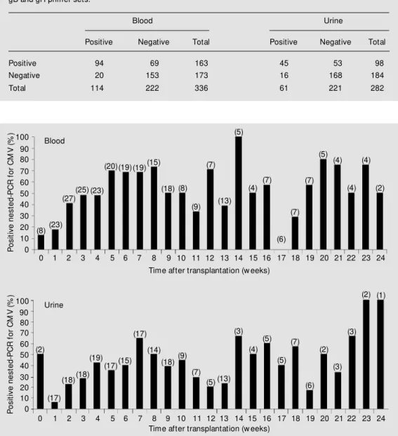
The results of nested-PCR using the gB and gH primer sets in blood and urine samples are shown in Table 1. CMV was detected more frequently in blood samples than in urine samples (P<0.001). Nested-PCR with the gB primer set detected a larger number of positive samples than nested-PCR with the gH primer set (P<0.001).
CMV was detected at least twice in urine or blood samples after renal transplantation
Table 1 - Positive and negative tests for CM V detected in blood and urine samples by nested-PCR using the gB and gH primer sets.
Blood Urine
Positive Negative Total Positive Negative Total
Positive 94 69 163 45 53 98
Negative 20 153 173 16 168 184
Total 114 222 336 61 221 282
P o s it iv e n e s te d -P C R f o r C M V ( % )100 24 23 22 21 20 19 (8)(23) 18 17 16 15 14 13 12 11 10 9 8 7 6 5 4 3 2 1 0
Time after transplantation (w eeks) 90 80 70 60 50 40 30 10 0 20 (27) (25) (23) (20)(19)(19)(15) (18) (8) (9) (7) (13) (5) (4) (7) (7) (7) (5) (4) (4) (2) (4) (6) P o s it iv e n e s te d -P C R f o r C M V ( % ) 100 90 80 70 60 50 40 30 10 0 20 24 23 22 21 20 19 18 17 16 15 14 13 12 11 10 9 8 6 5 4 3 2 1 0 7
Time after transplantation (w eeks) (2) (17) (18)(18) (19) (17)(15) (17) (14) (18)(9) (7) (5) (13) (3) (4) (5) (5) (7) (2) (6) (3) (3) (2) (1) Figure 1 - Percentage of patients
w ith CM V detected in blood and urine by nested-PCR w ith gB or gH primer sets. The number of patients studied is show n in pa-rentheses.
Blood
Asymptomatic
Graft rejection
Graft loss Death
Symptomatic
Thrombocytopenia
Fever
LE increase
Pneumonitis
Hepatomegaly
Splenomegaly
Gl ulceration
Graft rejection
Graft loss
Death
0 2 4 6 8 10 12 14 16 18 20 22 24 26 28 30 32
in 32 patients (94.1%). CMV was detected only in a lung biopsy during 13 months of follow-up in one patient. One patient with CMV detected once in urine died 4 weeks after transplantation. Figure 1 shows the per-centage of patients with CMV detected in blood and urine during a follow-up of 24 weeks after renal transplantation.
CMV was detected before transplanta-tion and within 1 week after the procedure in blood samples from 5 (16.1%) of 31 patients and in urine samples from 2 (10.5%) of 19 patients. The percentage of patients with CMV detected in blood and urine increased from the second week after transplantation, reaching a maximal level between the 5th and 8th week in blood (70-75%) and be-tween the 4th and 10th week in urine (43-65%).
The number of positive results observed during the period of 0 to 7 days after trans-plantation was compared with the number of positive results observed during the subse-quent periods, as shown in Table 2. A sig-nificant increase in CMV detection was ob-served after the first week of transplantation.
Of the 32 patients with active CMV in-fection, 6 (18.7%) were classified as having symptomatic CMV infection and 26 (78.8%) as having asymptomatic CMV infection. The clinical outcome and laboratory data for these patients are shown in Figure 2.
Symptoms related to CMV infection ap-peared between the 2nd and 5th week after renal transplantation in 4 (66.6%) patients. Two patients developed symptoms between the 14th and 15th week. Arthralgia and leu-kopenia were not observed in any patient.
Figure 2 - Clinical and laboratory aspects that may be related to the CM V infection observed in the 33 renal transplant recipients w ith CM V detected in blood and/ or urine. The percentages refer to the total number of sympto-m at ic and asysympto-m pt osympto-m at ic pa-tients. LE: Liver enzymes; GI: gastrointestinal.
Table 2 - Comparison of CM V detection in blood samples collected during the first w eek of trans-plantation w ith the subsequent periods.
All comparisons w ere carried out w ith the period of 0 to 1 w eek. * Statistically significant (Fisher’s exact test).
Weeks Number of samples P value
Total Positive
0-1 31 5
2-4 75 34 0.005*
5-8 73 51 <0.001*
9-12 42 21 0.002*
Five (83.3%) patients had CMV viremia and 3 (50%) had viruria at the time of appearance of symptoms. Three of these patients also had viremia 1 to 2 weeks before the onset of symptoms.
Fourteen patients developed graft rejec-tion and 13 of them showed CMV viremia at least once after renal transplantation. Eight (57.1%) of the latter patients showed CMV viremia within one week of transplantation or before graft rejection and 5 (35.7%) showed CMV viremia only after graft rejec-tion. These data show no association be-tween time of CMV viremia detection and graft rejection.
Graft rejection occurred in 4 (66.6%) of 6 symptomatic patients and in 9 (34.7%) of 26 asymptomatic ones, and graft loss occurred in 3 (50%) of 6 symptomatic patients and in 4 (15.4%) of 26 asymptomatic ones. These differences in frequency were not statisti-cally significant (P>0.05). However, the symptomatic patients showed a significant increase in death rate compared to the asymp-tomatic ones (P = 0.03).
D iscussio n
All patients were infected with CMV before renal transplantation as shown by the detection of anti-CMV IgG in all of them. The high frequency of CMV infection ob-served agrees with previous data indicating a prevalence of 90 to 100% CMV anti-bodies in Brazilian populations (15,16).
The PCR technique has shown higher sensitivity and earlier ability for CMV detec-tion as compared to antigenemia and viral isolation in cell culture (3-5). In the present study we used two highly sensitive nested-PCRs for CMV detection, one with the gB primer set and the other with the gH primer set. Nested-PCR with these primers detected 10,000- and 3,162-fold fewer viruses than the observation of cytopathic effect in cell culture, respectively.
Nested-PCR using gB primers detected
more positive samples than nested-PCR us-ing gH primers (P<0.001). This result agrees with our sensitivity test mentioned above. However, nested-PCR with gB primers did not detect 10.9% of positive blood samples and 14% of positive urine samples that were detected by using gH primers. Therefore, we believe that both gB and gH primers should be used for diagnosis, perhaps in a multiplex fashion.
CMV was detected more frequently in blood than in urine samples (P<0.001). This result is different from that obtained by Hokeberg et al. (17), who found a higher proportion of CMV viruria in kidney trans-plant recipients. The presence of PCR in-hibitors in urine samples may account for a negative result; however, in our study we used a column containing an anion-exchange resin for CMV DNA purification that elimi-nated possible PCR inhibitors. However, it was not possible to carry out the PCR for ß-globin in urine samples due to the low sensi-tivity of this test and to the low cellular DNA concentration in these samples.
CMV was detected at least twice after transplantation in 32 (94.1%) patients. The high prevalence of CMV DNA detected in this study is in agreement with previous reports showing that, depending on the sero-logic status of donor/recipient and type of immunosuppression, 60 to 100% of the pa-tients shed CMV (18-20).
transplan-tation since all patients were previously in-fected with CMV. Other evidence that our nested-PCR detected active CMV infection came from the observation of 4 patients who developed graft rejection. CMV was detected in these patients only after the onset of graft rejection and probably as a consequence of the vigorous immunosuppression (corticoids and OKT3) administered to control graft rejection, which led the latent virus to reacti-vate. However, it is possible that our nested-PCR detected latent infection in the lungs of one patient. This patient, not included among the CMV active infection cases, never pre-sented viremia or viruria and remained asymptomatic during follow-up.
CMV was detected within the first week of transplantation in 16.1 and 10.5% of the patients when blood and urine samples were analyzed, respectively (Figure 1). The num-ber of patients with CMV viremia and viruria increased in the second week after trans-plantation and reached the highest level (70-75%) between the 5th and 8th week in blood samples and (43-65%) between the 4th and 10th week in urine samples. Thus, CMV could be detected intermittently for a long period of time. The presence of CMV vire-mia and viruria within the first week of renal transplantation is in agreement with data reported by Rowley et al. (22). Bitsch et al. (3) and Barber et al. (5) also reported the detection of CMV DNA by PCR for long periods of time after kidney transplan-tation.
Six (18.7%) patients developed sympto-matic CMV infection, a frequency also ob-served in previous studies indicating the pres-ence of symptomatic infection in 20 to 60% of patients after transplantation (1,2,23). Rubin and Colvin (24) found that the time between the 4th and 12th week after renal transplantation is critical for symptomatic CMV infection. In this study, symptomatic CMV infection appeared between the 2nd and 5th week after renal transplantation in 4 (66.6%) patients. Two patients also
devel-oped symptomatic CMV infection between the 14th and 15th week after transplantation. Hokeberg et al. (17) detected arthralgia and thrombocytopenia in 66% and 7% of pa-tients with CMV disease, respectively, whereas in the present study no patient had arthralgia, and all of them had thrombocy-topenia. Leukopenia, commonly found in this kind of patient, was also not observed. CMV viremia, reported as a risk factor for symptomatic infection (25,26), was found in 3 patients (42.9%) starting 1 to 3 weeks before the onset of symptoms and persisted during the disease. Two other cases pre-sented CMV viremia only at the onset of symptoms.
Symptomatic CMV-infected patients showed a higher risk of death compared to asymptomatic ones (P = 0.03), in agreement with other reports (27-29).
CMV infection has been associated with decreased graft survival; however, the mechanisms by which the virus induces re-jection are not well known (30). Graft rejec-tion of the cellular type, which may be asso-ciated with CMV infection (31), was ob-served in 10 patients, but the association of diagnosis of CMV infection with graft rejec-tion was not detected in any patient.
The use of ganciclovir was not investi-gated because only 2 of the 6 symptomatic patients were treated.
Ackno wle dgm e nts
We are grateful to the medical team of the
Renal Transplantation Unit of Hospital das Clínicas, Faculdade de Medicina de Ribeirão Preto, USP, for collaboration during the study.
Re fe re nce s
1. Fryd DS, Peterson PK & Ferguson R (1980). Cytomegalovirus as a risk factor in renal transplantation. Transplantation, 30: 436-439.
2. Singh N, Dummer JS, Kusne S, Breinig M K, Armstrong JA, M akow ka L, Starzl TE & Ho M (1988). Infections w ith cytomega-lovirus and other herpesviruses in 121 liver transplant recipients: transmission by donated organ and the effects of OKT3 antibodies. Journal of Infectious Diseases, 158: 124-131.
3. Bitsch A, Kirchner H, Dennin R, Hoyer J, Frics L, Steinhoff J, Sack K & Bein G (1993). The long persistence of CM V DNA in the blood of renal transplant patients after recovery from CM V infection. Trans-plantation, 56: 108-113.
4. Gerna G, Zipeto D, Parea M , Revello M G, Silini E, Percivalle E, Zavattoni M , Grossi P & M ilanesi G (1991). M onitoring of hu-man cytomegalovirus infections and gan-ciclovir treatment in heart transplant re-cipients by determination of viremia, anti-genemia, and DNAemia. Journal of Infec-tious Diseases, 164: 488-498.
5. Barber L, Egan JJ, Lomax J, Yonan N, Deiraniya AK, Turner AJ, Woodcock AA & Fox AJ (1996). Comparative study of three PCR assays w ith antigenaemia and serol-ogy for the diagnosis of HCM V infection in thoracic transplant recipients. Journal of M edical Virology, 49: 137-144. 6. Albert J & Fenyo EM (1990). Simple,
sen-sitive, and specific detection of human immunodeficiency virus type 1 in clinical specimens by polymerase chain reaction w ith nested primers. Journal of Clinical M icrobiology, 28: 1560-1564.
7. Hibberd PL, Tolkoff-Rubin NE, Cosimi AB, Schooley RT, Isaacson D, Doran M , Delvecchio A, Delmonico FL, Auchincloss Jr H & Rubin RH (1992). Symptomatic cytomegalovirus disease in the cytome-galovirus ant ibody seroposit ive renal transplant recipient treated w ith OKT3. Transplantation, 53: 68-72.
8. Van der Berg AP, Van der Bij W, Van Son WJ, Anema J, Van der Giessen M , Schirm J, Tegzess AM & The TH (1989). Cytome-galovirus antigenemia as a useful marker of symptomatic cytomegalovirus infection after renal transplantation: a report of 130
consecutive patients. Transplantation, 48: 991-995.
9. Reynolds DW, Stagno S & Alford CA (1979). Laboratory diagnosis of cytome-galovirus infections. In: Lennette E & Schmidt NJ (Editors), Diagnostic Proce-dures for Viral, Rickettsial and Chlamydial Infections. 5t h edn. Am erican Public Health Association., Inc., New York. 10. Chou S & Dennison K (1991). Analysis of
interstrain variation in cytomegalovirus glycoprotein B sequences encoding neu-tralization-related epitopes. Journal of In-fectious Diseases, 163: 1229-1234. 11. Chou S (1992). M olecular epidemiology
of envelope glycoprotein H of human cy-tomegalovirus. Journal of Infectious Dis-eases, 166: 604-607.
12. Cranage M P, Kouzarides T, Bamkier AT Satchw ell S, Weston K, Tomlinson P, Barrell B, Hart H, Bell SE & M imson AC (1986). Identification of the human cy-tomegalovirus glycoprotein B gene and induction of neutralizing antibodies via its expression in recombinant vaccinia virus. EM BO Journal, 5: 3057-3063.
13. Vahey M T, W ong M T & M ichael NL (1995). A standard PCR protocol: rapid isolation of DNA and PCR assay for ß-globin. In: Dieffenbach CW & Dveksler GS (Editors), PCR Primer. A Laboratory M anual. Cold Spring Harbor Laboratory Press, Cold Spring Harbor, USA. 14. Reed LJ & M uench H (1938). A simple
method of estimating fifty per cent end-points. American Journal of Hygiene, 27: 493-497.
15. Suassuna JH, Leite LL & Villela LH (1995). Prevalence of cytomegalovirus infection in different patient groups of an urban university in Brazil. Revista da Sociedade Brasileira de M edicina Tropical, 28: 105-108.
16. Costa SCB, M iranda SRP, Alves G, Rossi CL, Figueiredo LTM & Costa FF (1999). Detection of cytomegalovirus infections by PCR in renal transplant patients. Brazil-ian Journal of M edical and Biological Re-search, 32: 953-959.
17. Hokeberg I, Eriksson BM , Sw eygberg-Wirgart B, Tufvesson G, Olding-Stenkvist E & Grillner L (1995). Diagnostic markers and risk factors of cytomegalovirus
infec-tion and disease in renal allograft recipi-ents. Scandinavian Journal of Infectious Diseases, 27: 435-440.
18. Pass RF, Long WK, Whitley RJ, Soong SJ, Diethelm AG, Reynolds DW & Alford Jr CA (1978). Productive infection w ith cy-tomegalovirus and herpes simplex virus in renal transplant recipients: role of source of kidney. Journal of Infectious Diseases, 137: 556-563.
19. Peterson PK, Balfour HH, M arker SC, Fryd DS, How ard RJ & Simmons RL (1980). Cytomegalovirus disease in renal allograft recipients: a prospective study of the clini-cal features, risk factors and impact on renal transplantation. M edicine, 59283-59300.
20. Pollard AB (1988). Cytomegalovirus infec-tions in renal, heart, heart-lung and liver transplantation. Pediatric Infectious Dis-ease Journal, 7: 97-102.
21. Taylor-Wiedeman J, Sissons JGP, Bory-siew icz LK & Sinclair JH (1991). M ono-cytes are a major site of persistence of hum an cyt om egalovirus in peripheral blood mononuclear cells. Journal of Gen-eral Virology, 72: 2059-2064.
22. Row ley AR, Wolinsky SM , Sambol SP, Barkholt L, Ehrnst A & Andersson JP (1991). Rapid detection of cytomegalovi-rus DNA and RNA in blood of renal trans-plant patients by in vitro enzymatic ampli-fication. Transplantation, 51: 1028-1033. 23. Smiley M L, Wlodaver CG, Grossman RA,
Barker CF, Perloff LJ, Tustin NB, Starr SE, Plotkin AS & Friedman HM (1985). The role of pretransplant immunity in protection from cytomegalovirus disease follow -ing renal transplantation. Transplantation, 40: 157-161.
24. Rubin RH & Colvin RB (1981). Effects of antithymocyte globulin on cytomegalovi-rus infection in renal transplant recipients. Transplantation, 31: 143-145.
25. Glenn J (1981). Cytomegalovirus infec-tions follow ing renal transplantation. Re-view s of Infectious Diseases, 3: 1157-1178.
human-leuko-cyte interferon in renal transplantation: ef-fects on cytomegalovirus and herpes sim-plex virus infections. New England Jour-nal of M edicine, 300: 1345-1349. 27. Harmon JB, Sibley RK, Peterson P &
Ferguson R (1982). Cytomegalovirus vire-mia and renal allograft morphology: are there distinct pathologic features? Labo-ratory Investigation, 46: 35 (Abstract). 28. Herrera GA, Alexander RW, Cooly CF,
Luke RG, Kelly DR, Curtis JJ & Gockerman JP (1986). Cytomegalovirus glomerulopa-thy: a controversial lesion. Kidney Interna-tional, 29: 725-733.
29. M aryniak R, First RM & Weiss M A (1985). Transplant glomerulopathy: evolution of morphologically distinct changes. Kidney
International, 27: 799-806.
30. Britt WJ & Alford CA (1996). Cytomega-lovirus. In: Fields BM , Knipe DM , How ley PM , Chanock RM , M elnik JL, M onath TP, Roizman B & Straus SE (Editors), Fields Virology. Lippincott-Raven Publishers, Philadelphia.
31. Colvin RB & Fang LST (1994). Interstitial nephritis. In: Tisher CC & Brenner BM (Editors), Renal Pathology w ith Clinical and Functional Correlations. 2nd edn. J.B. Lippincott Company, Philadelphia. 32. Delgado R, Lumbreras C, Alba C, Pedraza
M A, Otero JR, Gomez R, M oreno E, Noriega AR & Paya CV (1992). Low pre-dictive value of polymerase chain reaction for diagnosis of cytomegalovirus disease
in liver transplant recipients. Journal of Clinical M icrobiology, 30: 1876-1878. 33. Gerdes JC, Spees EK, Fitting K, Hiraki J,
Sheehan M , Duda D, Jarvi T, Roehl C & Robertson AD (1993). Prospective study utilizing a quantitative polymerase chain reaction for detection of cytomegalovirus DNA in the blood of renal transplant pa-tients. Transplantation Proceedings, 25: 1411-1413.