AbstrAct
Erythema elevatum diutinumis a rare neutrophilic der-matosis with vasculitis, which presents as persistent, symmetrical, purple or brownish papules and nodules, mainly in the extensor surface of the limbs.
We describe a case of erythema elevatum diutinum and polyarthritis as initial manifestations of Crohn’s disea se associated spondyloarthritis.
A 51-year-old man, from São Tomé e Príncipe, with previous history of treated tuberculosis and chronic hepatitis B infection, was admitted due to 4 months history of polyarthritis, hyperpigmented papules on the extensor surfaces, occasional episodes of bloody mu-cous diarrhea and significant weight loss. Histology of the skin showed myeloperoxidase positive neutrophilic granulocytes with moderate karyorrhexis, consistent with erythema elevatum diutinum. Colonoscopy showed erosions in sigmoid and rectum.
Diagnosis of erythema elevatum diutinumsecondary to Crohn’s disease with associated peripheral spondy-loarthritis was assumed. The patient was treated with prednisolone, sulphasalazine, metronidazole, azathio-prine and tenofovir with good clinical response. As erythema elevatum diutinum can be secondary to mul-tiple systemic diseases, including rheumatic diseases and inflammatory bowel disease, being aware and re -cognizing this entity can be of great importance for rheumatologists.
Keywords: Erythema elevatum diutinum; Crohn’s disea -se; Spondyloarthritis; Cutaneous vasculitis.
IntroductIon
Erythema elevatum diutinum (EED) is a rare neutrophilic dermatosis, first described by Hutchinson in 18881,
that has been considered a variant of leukocytoclastic vasculitis2. It has no racial predilection and it can
occur in any age, though it is most common between the fourth and sixth decades of life3.
EED often presents as persistent, symmetrical, firm, tender, red or purple papules and nodules that may coa lesce to form larger nodules or plaques. The exten-sor aspects of the extremities, near joints, are the pre-ferred location for skin lesions4.
The cause of EED is unknown, but it has been hypothesized that EED is mediated by immune comple -xes deposition in the perivascular dermis that induce an inflammatory response which in turn damages the ves-sel walls, resulting in fibrosis5. It has been described as
associated with neoplasms6, infections (Streptococcus,
hepatitis B (HBV) and C (HCV) virus, human immuno -deficiency virus (HIV))7, rheumatic diseases
(rheuma-toid arthritis (RA), lupus erythematosus)8,
inflamma-tory bowel diseases (ulcerative colitis, Crohn’s disease, celiac disease)9and other conditions.
The differential diagnosis of EED comprehends mainly Sweet syndrome, pyoderma gangrenosum, granuloma annulare, Kaposi’s sarcoma3. Skin biopsies
are the mainstay of the diagnosis, allowing differential diagnosis to be excluded3,4.
First-line treatment is to target underlying systemic conditions3. Other treatment options are limited, with
dapsone being the most commonly used agent10.
We describe a case of EED and polyarthritis as ini-1. Reumatologia, Centro Hospitalar Lisboa Norte; Unidade de
Investigação em Reumatologia, Instituto de Medicina Molecular 2. Dermatologia, Centro Hospitalar Lisboa Norte
3. Reumatologia, Centro Hospitalar Lisboa Norte; EpiDoC, CEDOC, Nova medical school
4. Reumatologia, Centro Hospitalar Lisboa Norte 5. Dermatopathologie Friedrichshafen, Friedrichshafen
Erythema elevatum diutinum in
Crohn’s disease-associated spondyloarthritis –
a rare vasculitis, an unusual association
Gonçalves MJ1, Romão VC1, Soares-de-Almeida L2, Canhão H3, Romeu JC4,
Kutzner H5, Pereira-da-Silva JA4
tial manifestations of Crohn’s disease, to raise aware-ness to this rare form of leucocytoclastic vasculitis, as an accurate diagnosis can prompt the investigation of underlying conditions.
cAsE rEport
A 51-year old black man was admitted to the Rheuma-tology Department, on July 2013, for diagnostic in-vestigation of arthralgia, cutaneous lesions and weight loss.
The patient had lived in São Tomé e Príncipe until December 2006. He had a previous history of pul-monary tuberculosis (TB) infection treated with mul-tiple antibacillary drugs. He was then submitted to left inferior lobectomy and his condition improved, only with mild cough persisting after surgery. In 2009, an incidental diagnosis of chronic HBV infection was per-formed. At the time, hepatic enzymes were normal.
On admission, the patient reported a four months history of symmetric, additive, inflammatory arthral-gia in small joints of the hand, wrists, elbows, knees and ankles, with good response to nonsteroidal anti--inflammatory drugs (NSAIDs). One month after the
beginning of these symptoms, non-pruritic cutaneous lesions on the arms and feet appeared. At the same time, the patient recalled having occasional episodes of bloody mucous diarrhea and had lost 16% of total body weight.
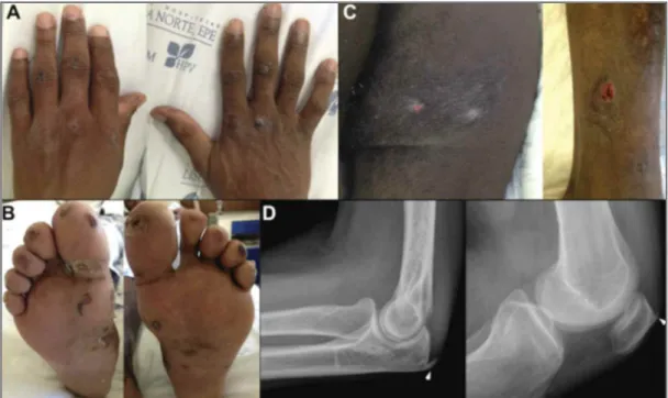
His physical exam was remarkable for hyperpig-mented papules on the extensor surfaces of the metacarpophalangeal (MCP) and proximal interpha-langeal (PIP) joints (Figure 1A), and purpuric lesions with ulcerations on the soles (Figure 1B), elbows and digital pulps, legs and buttocks (Figure 1C), symmet-rical polyarthritis (PIPs, MCPs, wrists, knees and an-kles), muscle wasting, a systolic murmur on the aortic focus and hepatosplenomegaly. The neurological exam was normal.
His laboratory workup was noteworthy for elevat-ed erythrocyte selevat-edimentation rate (ESR, 120mm/1sth,
314 100 000 UI/mL). He had positive anticitrullina -ted protein antibodies (ACPA, titre 40.2U/L, N<20) and equivocal anti-double stranded DNA antibodies (anti-dsDNA, titre 221.6, N<200). Rheumatoid factor (RF, N<16 UI/mL), neutrophil cytoplasmic anti-bodies (c-ANCA, p-ANCA, both N<20 UI/ml), antin-uclear antibodies (ANA, N< 1/160), human leukocyte antigen (HLA) B27 and serum cryoglobulins were neg-ative. Angiotensin converting enzyme was also within normal limits. A myelogram was performed, showing an eosinophil increase (12% of total cell population, N<6%), hypocellularity, with shift of the myeloid/ery-throid ratio in favor of the granulocytic series.
A thorough investigation was conducted to exclude an infectious cause. Bacteriological and mycobacterio -logical exams of blood, urine and stool were negative. Parasitological exam of the stool was also negative. Broncofibroscopy showed tracheomalacia and bron-choalveolar lavage was unremarkable, with negative bacteriological and mycobacteriological exams.
Multiple cutaneous biopsies of the skin lesions, in various locations, were performed. Gram, periodic acidSchiff (PAS) and Grocott staining, as well as ba -cteriological and mycoba-cteriological exams, including
Mycobacterium leprae, were negative. Immunohisto-chemistry (anti-leishmania, anti-mycobacterium tuber-culosis) was also negative.
Transesophageal echocardiography excluded the presence of vegetations and determined mild aortic in-sufficiency. A whole-body computerized tomography showed no images suggesting tumoral lesions or sources of occult infection. A liver biopsy was per-formed, showing mild to a moderate mononucleated inflammatory infiltrate, portal and acinar fibrosis, fo-cal hepatocellular necrosis, interface hepatitis and sig-nificant staining for HBV markers. HBV infection treat-ment was initiated promptly with tenofovir (300mg/day).
Articular ultrasound confirmed polyarticular syno -vitis, no erosions found. Radiographs (including the axial skeleton) showed no structural damage. Fine en-thesophytes were found bilaterally on the insertion of the tricipital and quadricipital tendons (Figure 1D).
On electromyography (EMG), low sensitive action potentials were found in the right ulnar, peroneal and sural nerves, with no changes in the motor compo-nent. Sural nerve biopsy revealed asymmetric inflam-matory infiltrate in both nerve and vessels, suggesting vasculitic neuropathy.
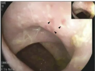
Colonoscopy showed erosions in sigmoid and re
-ctum, which histologically corresponded to undeter-mined inflammatory bowel disease (IBD) findings (moderate diffuse lamina proprialymphoplasmacytic infiltrate with lymphoid aggregates, basal plasmacyto-sis, criptitis, erosions and one non-necrotizing granu-loma), with mild activity (Figure 2). Duodenum biopsies exhibited an intense lymphoplasmocytic infiltrate, with epithelial regeneration and some areas of atrophy.
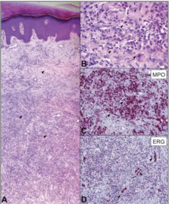
The anatomopathological study of skin lesions showed, on the medium and reticular dermis, a high number of myeloperoxidase (MPO) positive perivas-cular neutrophilic granulocytes and some eosinophils with massive karyorrhexis, but without blood ex-travasation or fibrinoid deposits associated. Advanced lesions showed slight dermal fibrosis. Nuclear endo -thelial marker – ERG and CD31 staining demons trated the presence of many capillary vessels (Figure 3).
After clinical correlation, these findings established the diagnosis of erythema elevatum diutinum (EED), assumed to be associated to Crohn’s disease with pe-ripheral spondyloarthritis. The patient was treated with prednisolone (PDN) 1mg/Kg/day, sulphasalazine 2g/day and metronidazole 1000mg/day. A good clinical response followed and the patient became asym -ptomatic within 16 weeks, with significant improve-ment of the cutaneous lesions (regression of the lesions to cicatricial changes) and weight gain. Steroids were then tapered until 0.5mg/Kg/day of PDN. At this dose, the patient experienced muco-bloody diarrhea and
FIGurE 2. Colonoscopy showed erosions in sigmoid and rectum, which histologically corresponded to undetermined inflammatory bowel disease (IBD) findings (moderate diffuse
monoarthritis of the tibiotalar joint. Azathioprine was added with improved disease control and no relapse of skin or joint involvement, and allowing further steroid tapering. Steroids were discontinued after a year with-out disease relapse. Antiviral treatment for hepatitis B was effective, with significant decrease of viral load after 12 months (3723 UI/mL, log103.57).
dIscussIon
Erythema elevatum diutinumis an inflammatory der-matosis, characterized by a neutrophilic infiltrate of the dermis in the absence of cutaneous infection. In the present case, we found an intense infiltrate of poly-morphonuclear cells (MPO+) with moderate karyor-rhexis and with older lesions showing fibrosis. The ab-sence of blood extravasation and the preab-sence of late-stage lesions showing fibrosis is characteristic of
clas-sic EED11and allow the distinction among other forms
of leukocytoclastic vasculitis. Other neutrophillic dermatoses (such as Sweet syndrome) are rarely associa -ted with vasculitis, and some are associa-ted with granu lomatous inflammation (e.g granuloma annulare), absent in EED3.
The clinical presentation of EED in our patient showed typical aspects (painless, non pruritic brow -nish and purpuric nodules and papules in extensor surfaces of the limbs) and more atypical features, such as the presence of ulcerations in elbows, buttocks and legs3,4.
EED was reported to be associated with numerous pathologies, including rheumatic diseases, viral infec-tions and inflammatory bowel disease. In the present case, the initial clinical picture with symmetric addi-tive polyarthritis, elevated CRP and ESR and low-titer ACPA positivity could point to RA12. However, the
presence of mucobloody diarrhea along with endos -copic and histologic documentation of intestinal in-flammation, affecting both duodenum and colon con-curred to Crohn’s disease (although with only a single granuloma identified in the colon tissue sample). Moreover, intestinal, cutaneous and articular manifes-tations occurred almost simultaneously. Also, enthe-sopathy, as observed in our patient, is a common fea-ture of spondyloarthritis and no bone erosions were found in joint radiography or ultrasound. Overall, a diagnosis of Crohn’s disease with associated polyar -thritis was assumed as more likely. Immunosuppres-sive treatment was started with global improvement. Corticosteroid tapering was followed by disease re-lapse (abdominal pain, diarrhea and monoarthritis of the ankle), a clinical picture that is actually more charac teristic of Crohn’s disease with associated spon -dyloarthritis.
Thus, both polyarthritis and EED seem less likely to be associated chronic infection with HBV, serological-ly detected several years before. In addition, as men-tioned above, the relapse of the disease occurred when a decrease in viral load had already been documented. Nevertheless HBV infection could also be a possible trigger for EED. Although in this case HBV infection was diagnosed long ago, and apparently then there were no systemic manifestations of the hepatic disease. Peripheral sensitive neuropathy diagnosed in this patient was subclinical, the onset being impossible to date. EMG was previous to treatment with metronida-zole, a recognized neurotoxic drug. Active chronic hepatitis B could cause peripheral neuropathy by vas-FIGurE 3. Erythema elevatum diutinumpresenting with the
cular involvement (vasculitis) or direct immune-me-diated damage13. In accordance, our patient’s nerve
biopsy revealed an intense infiltrate of T lymphocytes in both vessel and nerve. However, it is difficult to ex-clude other etiology for the neuropathy, such as Crohn’s disease itself. Peripheral neuropathy has also been reported in association with Crohn’s disease al-though it occurs more commonly in ulcerative coli-tis14.
Mild peripheral eosinophilia and central nonclo nal eosinophilia was documented. However, eosino -philic infiltration in all the tissues sampled was never found and other common manifestations of idiopa thic hypereosinophilic syndrome were absent. Reactive forms of eosinophilia are far more common than pri-mary forms and include inflammatory bowel disease15.
EED lesions almost disappeared after initiation of sulphasalazine, highdose prednisolone and metro -nidazole. Dapsone remains the treatment of choice for EED10. This option has limitations since dapsone is a
suppressive rather than curative therapy, and with-drawal usually results in prompt and severe recurrence of disease10. As in other neutrophilic dermatoses,
sul-fapyridine has showed similar effects as dapsone. Sul-phasalazine, being a compound derived from sul-fapyridine, may be in part responsible for the cuta-neous improvement. Also, treatment of the underlying condition is associated with an improvement of the EED lesions.
To the best of our knowledge, the association be-tween EED (per se, an uncommon vasculitis) and Crohn’s disease has been previously reported in only 3 cases in literature9, 16, 17.
This case illustrates a complex investigation that has led to an unexpected diagnosis. As erythema elevatum diutinumcan be associated with multiple diseases, in-cluding rheumatic diseases and IBD, being aware and recognizing this entity can be of great importance for rheumatologists.
corrEspondEncE to
Maria João Gonçalves
Serv. Reumatologia, Hospital de Santa Maria Av Prof Egas Moniz, Lisboa, Portugal E-mail: mjoaomgoncalves@gmail.com
rEFErEncEs
1. Hutchinson J. On two remarkable cases of symmetrical purple congestion of the skin in patches, with induration. Br J Der-matol1888; 1:10–15.
2. Yiannias JA, el-Azhary RA, Gibson LE., Erythema elevatum diu-tinum: a clinical and histopathologic study of 13 patients, J Am
Acad Dermatol. 1992; 26:38-44.
3. Momen SE, Jorizzo J, Al-Niaimi F. Erythema elevatum diuti-num: a review of presentation and treatment. J Eur Acad Der-matol Venereol. 2014; 28 (12): 1594-1602.
4. Gibson LE, El-Azhary RA , Erythema Elevatum Diutinum., Cli-nics Dermatology 2000;18:295–299.
5. Gibson LE, Su WP, Cutaneous vasculitis, Rheum Dis Clin North Am. 1995; 21:1097-1113.
6. Hatzitolios A, Tzellos TG, Savopoulos C et al. Erythema eleva-tum diutinum with rare distribution as a first clinical sign of non-Hodgkin’s lymphoma: a novel association? J Dermatol. 2008; 35:297-300.
7. Kim H. Erythema elevatum diutinum in an HIV-positive pa-tient. J Drugs Dermatol. 2003; 2:411-412.
8. Woody CM, Lane JE, Davis LS. Erythema elevatum diutinum in the setting of connective tissue disease and chronic bacterial infection, J Clin Rheumatol. 2005; 11:98-104.
9. Walker KD, Badame AJ. Erythema elevatum diutinum in a pa-tient with Crohn’s disease. J Am Acad Dermatol. 1990; 22:948-952.
10. Seneschal J, Guillet S, Ezzedine K, et al, Alternative Procedure to Allow Continuation of Dapsone Therapy despite Serious Ad-verse Reaction in a Case of Dapsone-Sensitive Erythema Ele-vatum Diutinum, Dermatology. 2012; 224:115-119. 11. Crowson AN, Mihm MC Jr, Magro CM. Cutaneous vasculitis:
a review. J Cutan Pathol. 2003; 30:161-173.
12. Biliavska I, Stamm TA, Martinez-Avila J et al; Application of the 2010 ACR/EULAR classification criteria in patients with very early inflammatory arthritis: analysis of sensitivity, specificity and predictive values in the SAVE study cohort, Ann Rheum Dis. 2013; 72:1335-1341.
13. Verma R, Lalla R, Babu S. Mononeuritis multiplex and painful ulcers as the initial manifestation of hepatitis B infection. BMJ Case Rep. 2013,2013: bcr2013009666 (published online). 14. A Wills, CJ Hovell, Neurological complications of enteric
di-sease, Gut 1996, 39: 501-504.
15. Butt NM, Lambert J, Ali S, et al; British Committee for Stan-dards in Haematology. Guideline for the investigation and ma-nagement of eosinophilia. Br J Haematol. 2017 doi: 10.1111/ bjh.14488. [Epub ahead of print]
16. Orteu CH, McGregor JM, Whittaker SJ et al, Erythema Eleva-tum Diutinum and Crohn Disease: A Common Pathogenic Role for Measles Virus? Arch Dermatol. 1996; 132:1523-1525. 17. Elsner J, Kiehl P, Kapp A, Weiss J. [Erythema elevatum and