Coinfection of
Leishmania (Viannia)
braziliensis
and
Streptococcus pneumoniae
in
Multiple Cutaneous Lesions
Paulo R. Cortes1,2, Laura S. Chiapello1, David Dib2, Monica V. Herrero3, Carmen T. Nuncira2, Carlos De Petris2, Jose Echenique1*
1Departamento de Bioquímica Clínica (CIBICI-CONICET), Facultad de Ciencias Químicas, Universidad Nacional de Córdoba, Córdoba, Argentina,2Hospital Pediátrico del Niño Jesús, Córdoba, Argentina,
3Hospital Córdoba, Córdoba, Argentina
*jeche@fcq.unc.edu.ar
Case Presentation
A 12-year-old girl was seen by a physician in Frias (Santiago del Estero Province, northern Argentina region) due to a lesion in her left leg that presented a painless papule, which later became ulcerative. The mother referred that the first lesion appeared after an insect bite in the left leg of her daughter, but she did not remember any febrile episode. She reported no other symptoms or past medical history associated with skin ulcerations, diabetes, recurrent ear infections, or chronic diseases. In a few days, new lesions with similar characteristics appeared on her arms and legs. The first clinical impressions were ulcers caused by group A beta-hemo-lytic streptococci or staphylococci. No bacteriological cultures or other microbiological studies were performed to determine the putative cause of those lesions. Nevertheless, she received an empirical antibiotic treatment, such as topical rifampicin (3 applications/day of a 100 mg/ml solution) concomitantly with penicillin G benzathine (1,200,000 UI, one dose, i.m.) cephalexin (500 mg orally every 12 h for 14 days) and ciprofloxacin (500 mg orally every 12 h for 7 days), which were administered sequentially due to clinical worsening of the skin lesions. After 2 weeks, the physicians could not observe any lesion improvement after these treatments, and judged by the evolution of lesions, they suspected aLeishmaniainfection and interrupted anti-biotic treatment. The patient was transferred to the Hospital Pediatrico del Niño Jesus (chil-dren’s hospital, Cordoba, Argentina) for an accurate diagnosis.
Diagnosis
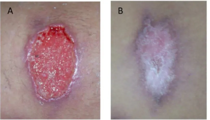
On admission, the patient presented multiple skin ulcers on the left leg and in both arms, and she was hospitalized several days for diagnosis and treatment. The clinical examination showed oval ulcers with an average diameter of 3 x 5 cm, hard edges, erythematous halos, painful to the palpation, granular aspect, and covered with a yellowish secretion (Fig 1A).
These ulcers were located on the inner face of the left leg (n= 2), the inner face of the right thigh (n= 1), and the inner face of the right arm (n= 3). Histopathological analysis of lesion biopsies noted abundant fibrin deposition, neutrophil granulocytes, erythrocytes, and necrotic tissues. Neoformed blood vessels, endothelial cell proliferation, and young connective tissues were also observed in subjacent samples. Deep and peripheral samples of lesions showed inflammatory infiltrates constituted by lymphocytes and plasma cells. All these findings were compatible with a nonspecific chronic inflammation. Initial laboratory studies revealed a OPEN ACCESS
Citation:Cortes PR, Chiapello LS, Dib D, Herrero MV, Nuncira CT, De Petris C, et al. (2016) Coinfection ofLeishmania (Viannia) braziliensisand
Streptococcus pneumoniaein Multiple Cutaneous Lesions. PLoS Negl Trop Dis 10(3): e0004388. doi:10.1371/journal.pntd.0004388
Editor:Genevieve Milon, Institut Pasteur, FRANCE
Published:March 10, 2016
Copyright:© 2016 Cortes et al. This is an open access article distributed under the terms of the Creative Commons Attribution License, which permits unrestricted use, distribution, and reproduction in any medium, provided the original author and source are credited.
Funding:This study was supported by National Council of Scientific and Technological Research (CONICET); National Agency of Scientific and Technological Promotion (ANPCYT; FONCYT PICT, Prestamo BID) and the Scientific and Technological Secretary of the National University of Cordoba (SECYT-UNC). Nubia Yandar has a fellowship from ANPCYT. Jose Echenique is a member of the Research Career of CONICET. The funders had no role in study design, data collection and analysis, decision to publish, or preparation of the manuscript.
normal number of white blood cell count (8,700/μl with 69% granulocytes and 26%
lympho-cytes) and increased erythrocyte sedimentation rate (ESR [40 mm/h]), hemoglobin 139 g/liter, hematocrit at 42 liter/liter and normal blood glucose (95 mg/dL) and urea (20 mg/dL) levels. Once biological samples from lesions were obtained for microbiological tests, the patient received an empirical treatment with rifampicin/trimethoprim (300/80 mg orally every 12 h for 15 days), topical treatments of skin lesions with fusidic acid cream (2%), and povidone/ iodine solutions every 12 h.
To determine a putativeLeishmaniainfection, Giemsa-stained thin smears of dermal scrap-ings were analyzed, which revealed amastigotes inside macrophages, consistent with leishmani-asis (Fig 2).
Furthermore, sterile biopsy specimens for culture were remitted to the Instituto Nacional de Parasitología“Fatala-Chaben.”Immediately, an antiprotozoal treatment with meglumine anti-moniate (Glucantime, 20 mg/kg/day i.m. and 1 mg/kg/day intralesion for 1 month) was
Fig 1. Skin lesion caused byLeishmania (Viannia) braziliensisand coinfected withStreptococcus pneumoniae.(A) Image of an untreated ulcer when the patient was admitted at the hospital. (B) Image of the same ulcer shown in panel A after the treatment with antibiotics (rifampicin/trimethoprim) and meglumine antimoniate (Glucantime).
doi:10.1371/journal.pntd.0004388.g001
Fig 2. Microscopic identification ofLeishmaniaamastigote forms.Biological samples obtained from ulcer were stained by Giemsa technique. (A) image of a peripheral lesion biopsy; (B) image of a deep lesion biopsy. The amastigote forms are indicating by arrows.
administered. Then,Leishmaniapromastigotes were identified after 10 days of culture [1], and the amastigotes isolated from lesions were identified asLeishmania (Viannia) braziliensisby molecular tests, which is one of the most commonLeishmaniaspecies circulating in Santiago del Estero, Argentina [2].
To detect a possible bacterial coinfection, blood cultures were taken before initiation of empirical antibiotic treatment, but the results were negative. However, the purulent material obtained from lesions was cultured in blood agar plates, and alpha-hemolytic colonies grew, showing a clear mucoid phenotype. Unexpectedly, bacterial strains recovered from four lesions were identified asStreptococcus pneumoniaeby classical tests, such as optochin susceptibility and bile solubility. This was confirmed by PCR amplification of specific pneumococcal genes, such aslytA,psaA,ply, andsodA, as described [3,4]. In addition, thesodAgene was partially amplified and sequenced (Macrogen Inc. Seoul, Korea) [3]. The DNA sequences were analyzed in the GenBank database and they showed 99.99% of homology with theS.pneumoniae sodA
gene, confirming the identification of this pathogen. The pneumococcal strains were also sero-typed by the Quellung reaction [5] at the Instituto Nacional de Enfermedades Infecciosas (“Carlos Malbran”), and they were identified as serotype 3. To determine the putative origin of these isolates, they were also analyzed by BOX-PCR using a unique BOXA1R primer and fol-lowing the protocol described [3]. BOX-PCR is a molecular technique that amplifies, by PCR, a DNA-repetitive element named BOX, which is used for epidemiological studies ofS. pneumo-niaeThe BOX patterns showed identical profile (Fig 3), suggesting a clonal relationship between the isolated strains.
The antibiotic susceptibility tests for the pneumococcal strains were carried out using agar diffusion methodology and E-tests forβ-lactams antibiotics following the guidelines of the Clin-ical and Laboratory Standards Institute [6]. The isolates were susceptible to penicillin (Minimal Inhibitory Concentration [MIC]: 0.006 mg/L) and cefotaxime (MIC:<0.006 mg/L) determined by E-test, and to erythromycin, rifampicin, trimethoprim-sulphamethoxazole, clindamycin, chloramphenicol, ofloxacin, and only resistant to tetracycline as determined by disk diffusion,
Fig 3. BOX-PCR DNA profiles ofS.pneumoniaeisolates.Four pneumococcal strains (lines 2–5) isolated from different ulcers showing genetic identity between them and the same clonal origin. DNA from R6 pneumococcal strain was used as control (line 1). MM, molecular DNA marker (phageλDNA digested with HindIII/EcoRI).
indicating that the empirical antibiotic treatment administered on admission (rifampicin/tri-methoprim) was appropriate. After that, the patient was discharged. A month later, when she came back to the hospital for a follow-up appointment, the ulcer lesions were completely healed (Fig 1B), and she did not present any side effects from the medication. In this manuscript, the patient has given written informed consent for publication of her case details.
Case Discussion
Leishmaniasis is an infectious disease caused by a protozoan microorganism transmitted to humans via the bite of a female sand fly. It is disseminated in five continents and causes more than 30,000 deaths every year. The World Health Organization (WHO) estimates that approxi-mately 350 million people are at the risk of contracting this disease [2]. Different species of
Leishmaniaare responsible for cutaneous and visceral diseases that are endemic to tropical and subtropical regions in 98 countries. Cutaneous leishmaniasis is the most common disease that is caused by allLeishmaniaspecies that are pathogenic to humans and is considered endemic in northern Argentina [1]. WHO also estimates that 0.7–1.2 million new cases of cutaneous leishmaniasis occur annually [7].
It is known thatL.(V.) braziliensisnormally express virulence factors to disrupt the natural skin barrier to establish cutaneous leishmaniasis [8], and these lesions predispose to coinfec-tions of bacterial pathogens (Table 1) [9–17].
Here, we report the first case of coinfection withLeishmania and S.pneumoniae. This bacte-rial pathogen resides normally in the human upper respiratory tract, but it is also the causal agent of infections in children and adults such as otitis, sinusitis, pneumonia, septicemia, men-ingitis, and it is also isolated from skin [18]. Pneumococcal skin infections are more frequently found in immunocompetent patients associated with HIV, lupus erythematosus, diabetes, burns, cancer, and alcoholism. Listed by order of relevance, the clinical sources of these infec-tions were surgical wounds, burns, cellulitis, pyomyositis, fasciitis, and abscess infecinfec-tions [18].
Table 1. Bacterial species that coinfect skin ulcers caused by cutaneous leishmaniasis.
Bacterial species References
Bacillusspp. [16]
Bacteroides fragilis [15]
Enterobacterspp. [14,15,16,17]
Enterococcusspp. [9,10,14]
Escherichia coli [9,11,15,16]
Klebsiellaspp. [9,11,14,15,16,17]
Micrococcusspp. [14]
Morganella morganii [14]
Mycobacterium ulcerans [13]
Peptostreptococcusspp. [15]
Prevotellaspp. [15]
Proteusspp. [9,11,15,16,17]
Pseudomonas aeruginosa [9,12,14,15,16]
Staphylococcusspp. [11,14,16]
Staph.aureus [9,10,11,14,16,17]
S.agalactiae [15]
S.pneumoniae This work
S.pyogenes [9,14,15,16]
Based on these clinical features, we cannot discard thatS.pneumoniaecontributed to the infec-tious process of lesions caused byLeishmania.
Regarding the origin of bacterial infection, the pneumococcal strains isolated from different
Leishmaniaskin lesions appear to have the same source of contagion. We think that the pneu-mococcal isolates that infected skin ulcers came from the patient`s nasopharynx, but we don’t have evidence to discern whether these strains were part of the patient’s microbiota or were acquired in the community. We cannot rule out the possibility that the patient was a nasal car-rier of the pneumococcal isolates found in the skin lesions, and this fact is related to pneumo-coccal survival after the empirical antibiotic therapy. The patient was initially treated with cephalexin and ciprofloxacin, because the lesions appeared to be caused byS.pyogenesor
Staph. spp. However, these antibiotics are not indicated for pneumococcal infections because they are less active against this pathogen. She was also treated with penicillin G that is effective for the treatment of pneumococcal infections. Nevertheless, it was reported that penicillin con-centrations in saliva were much lower than the corresponding serum concon-centrations during penicillin therapy, indicating that decolonization is not possible with this antibiotic, and thatS.
pneumoniaeis able to survive in nasopharynx under these conditions [19]. For these reasons, we think that the patient possibly was a nasal carrier after the antibiotic treatments. These bac-terial isolates were identified as serotype 3, which was underrepresented in pneumococcal car-riage studies [20], but it was one of the most frequent serotypes isolated from pneumococcal invasive disease in Argentina [21]. There is also the possibility that the patient had acquired pneumococcal strains in the community when antibiotics were interrupted.
Concerning the way by whichS.pneumoniaecoinfected these ulcerative lesions caused orig-inally byLeishmaniaon the arms and legs by the nasopharynx, we propose that this bacterial pathogen was disseminated through contaminated respiratory tract secretions like saliva when the patient exhaled or coughed onto skin lesions. Once the first skin lesion was coinfected with
S.pneumoniae, the other ulcerative lesions could have been coinfected by scratching. On the other hand, although the patient did not report clinical symptoms compatible with septicemia or pneumonia before or after her hospitalization, there is a possibility that this strain had been disseminated by a hematogenous spread from the first coinfected lesion. In this sense, Kalima et al. [22] reported that 30/34 cases of pneumococcal infections, where skin was the primary site of infection, had bacteraemia. Thirty percent of these cases did not present underlying pre-disposing conditions. In addition, these coinfected lesions also represented a risk factor for the invasive disease development caused byS.pneumoniae.
After a precise microbial diagnosis, the coinfected lesions were successfully treated with antibacterial (rifampicin/trimethoprim) and antiprotozoal agents (meglumine antimoniate). We think that antiprotozoal treatment was essential for skin healing, but a concomitant anti-bacterial therapy helped to heal the ulcerative lesions.
In view of the pathogenic potential ofS.pneumoniaeand a high carriage rate in children, we expect to find more cases in skin infections; however, this kind of coinfection has never been reported before. We propose thatS.pneumoniaeinfections are being underestimated in skin lesions of patients fromLeishmaniaendemic areas, probably by a misidentification with other gram-positive cocci that may coinfect these ulcers (seeTable 1).
Acknowledgments
We would like to thank Alex Saka for revising this manuscript. Consent of publication was obtained from the patient.
References
1. Segura EL, Juan N, Piquin AL, Cuba Cuba CA et al. (2000) Molecular and biologic characterization of Leishmania parasites implicated in an epidemic outbreak in northwestern Argentina. Parasitol Res 86: 504–8. PMID:10894479
2. Cuba CA, Torno CO, Ledesma O, Visciarelli E, et al. (1996) Human cutaneous leishmaniasis caused by Leishmania (Viannia) braziliensis in Santiago del Estero, Argentina: identification of parasites by monoclonal antibodies and isoenzymes. Rev Inst Med Trop 38: 413–21.
3. Cortes PR, Orio AG, Pinas GE, Echenique J. (2008) Characterization of in vitro-generated and clinical optochin-resistant strains ofStreptococcus pneumoniaeisolated from Argentina. J Clin Microbiol 46: 1930–4. doi:10.1128/JCM.02318-07PMID:18417665
4. Albarracin Orio AG, Cortes PR, Pinas GE, Echenique JR. (2008) A new serotype 14 variant of the pneumococcal Spain9V-3 international clone detected in the central region of Argentina. J Med Micro-biol 57: 992–9. doi:10.1099/jmm.0.2008/000505-0PMID:18628501
5. Habib M, Porter BD, Satzke C. (2014) Capsular serotyping of Streptococcus pneumoniae using the Quellung reaction. J Vis Exp: e51208. doi:10.3791/51208PMID:24637727
6. CLSI. (2015) Performance Standards for Antimicrobial Susceptibility Testing; Twenty-Fifth Informa-tional Supplement. CLSI document M100-S25. Wayne, PA: Clinical and Laboratory Standards Institute;
7. Alvar J, Velez ID, Bern C, Herrero M, Desjeux P, et al. (2012). Leishmaniasis Worldwide and Global Estimates of Its Incidence. PLoS ONE 7: e35671. doi:10.1371/journal.pone.0035671PMID: 22693548
8. Silva-Almeida M, Pereira BA, Ribeiro-Guimaraes ML, Alves CR (2012) Proteinases as virulence factors
inLeishmania spp. infection in mammals. Parasit Vectors 5: 160. doi:10.1186/1756-3305-5-160
PMID:22871236
9. Edrissian GH, Mohammadi M, Kanani A, Afshar A, et al. (1990) Bacterial infections in suspected cuta-neous leishmaniasis lesions. B World Health Organ 68: 473.
10. Shirazi MH, Ranjbar R, Asgari V, Mohebali M, Hamidian M. (2007) Study of bacterial infections among the patients with suspected cutaneous leishmaniasis. Pak J Biol Sci 10: 4555–8. PMID:19093532
Key Learning Points
• Cutaneous leishmaniasis is the most common clinical disease caused by all the Leish-maniaspecies that are pathogenic to humans.
• Other bacterial coinfections ofLeishmanialesions have been described, but this is the first report of coinfection withLeishmaniaandS.pneumoniae.
• S.pneumoniaeis also able to cause skin infections. For that reason, the pneumococcal diagnosis could be underestimated inLeishmanialesions coinfected with bacterial pathogens, particularly in endemic areas.
• An accurate microbiological diagnosis ofLeishmaniacoinfections is essential for a cor-rect antimicrobial treatment of skin infections.
11. Ziaie H, Sadeghian G. (2008). Isolation of bacteria causing secondary bacterial infection in the lesions of cutaneous leishmaniasis. Indian J Dermatol 53: 129–31. doi:10.4103/0019-5154.43217PMID: 19882011
12. Van Der Vliet D, Le Guern AS, Freitag S, Gounod N (2006). Pseudomonas aeruginosa otochondritis complicating localized cutaneous leishmaniasis: prevention of mutilation by early antibiotic therapy. Am J Trop Med Hyg 75: 270–2. PMID:16896131
13. Mougin B, Avenel-Audran M, Hasseine L, Martin L, et al. (2011) A cutaneous ulcer resulting from
Myco-bacterium ulcerans-Leishmania braziliensiscoinfection in South America. Am J Trop Med Hyg 85:
897–9. doi:10.4269/ajtmh.2011.11-0126PMID:22049045
14. Isaac-Márquez AP, Lezama-Dávila CM. (2003) Detection of Pathogenic Bacteria in Skin Lesions of Patients with Chiclero’s Ulcer. Reluctant Response to Antimonial Treatment. Mem Inst Oswaldo Cruz, Rio de Janeiro, Vol. 98(8): 1093–1095.
15. Fontes CO, Carvalho MA, Jacques R and Farias LM (2005). Identification and antimicrobial susceptibil-ity of microorganisms recovered from cutaneous lesions of human American tegumentary leishmania-sis in Minas Gerais. J Med Microb 54, 1071–1076.
16. Doudi M, Setorki M, Narimani M. (2012) Bacterial superinfection in zoonotic cutaneous Leishmaniasis. Med Sci Monit, 18(9): BR356–361 PMID:22936185
17. Sadeghian G, Ziaei H, Shirani Bidabadi L, and Zolfaghari Baghbaderani A. (2011). Decreased effect of glucantime in cutaneous leishmaniasis complicated with secondary bacterial infection. Indian J Derma-tol. 56(1): 37–39. doi:10.4103/0019-5154.77549PMID:21572789
18. Garcia-Lechuz JM, Cuevas O, Castellares C, Perez-Fernandez C, et al. (2007)Streptococcus
pneu-moniaeskin and soft tissue infections: characterization of causative strains and clinical illness. Eur J
Clin Microbiol 26: 247–53.
19. Strömberg A, Friberg U, Cars O (1987). Concentrations of phenoxy-methylpenicillin and cefadroxil in tonsillar tissue and tonsillar surface fluid. Eur J Clin Microbiol. 6:525–9. PMID:3125047
20. Gentile A, Prieto N, Rodriguez M, Gagetti P et al (2009). First national study of prevalence for nasopha-ryngeal carriage of Streptococcus pneumoniae among non-vaccinated children attending daycare cen-ters in Argentina. Available:http://antimicrobianos.com.ar
21. SIREVA Group (Sistema de Redes de Vigilancia de los Agentes Bacterianos Responsables de Neu-monía y Meningitis) (2014). Pan American Health Organization Available:http://antimicrobianos.com. ar/category/resistencia/sireva.