A
BNORMAL
HEMOGLOBINS AND THALASSEMIAS IN
COSTA RICA, OTHER COUNTRIES OF
CENTRAL AMERICA, AND PANAMA1
German. E Sbetq2 Mar&q AZtafuZ.Za,J GuiZZermo Sancho, 4 and M&mz SaZgadoos
I
N’I‘RODUCTION
The hematologic heritage of the people of Costa Rica-and of Central America in general-is derived from the genetic traits of the native Amerindians and of the African, Asian, and European immigrants who settled there. In turn, these genetic traits tend to reflect the var- ied ecologic conditions of these peoples’ historic backgrounds. Among other things, some of the genetic markers in- volved are characterized by a balanced polymorphism-this being especially true of the genes for hemoglobins S and C , both clearly of African extraction, that are considered deleterious to prolifera- tion of malarial parasites, especially Phs- medium fdct~arum.
The influence of Asian immi- grations in this picture has not been properly established. But it is well known that Black and Caucasian populations
’ This work will also be published in Spanish in the Bo-
emigrated to Central America from vari- ous parts of Africa, the Antilles, and Eu- rope; that native Amerindians made contributions to the present populations that vary greatly from country to country; that there have been differences, which persist, in the degree of racial mixing in the different countries; and that the se- lective pressure exerted by malaria has also been highly variable in the different regions. All of these factors are responsi- ble for the different frequencies of ab- normal hemoglobins and thalassemias found today in Central America and throughout tropical America (1). G
4 Physician. Resales Hospital, San Salvador, El Salvador. ’ Instructor. School of Microbiology, National Autono-
mous University of Honduras, Tegucigalpa, Honduras. 6 An exhaustive search of the existing Central American literature on abnormal hemoglobins and thalassemias was made in the course of preparing this review. Among the disappointments encountered, our failure u. -.
letin de /a Oficina Sanitah Panamericana, vol. p04, I -1 i 1988.
* Professor, Center for Research on Abnormal Hemoglo- bins and Related Disorders (Centro de InvestigaciBn en Hemoglobinas Anormales y Trastornos Afines- CIHATA), University of Costa Rica, San Juan de Dios Hospital, San Jose, Costa Rica.
3 Physician, Hematology Service, Central Hospital, So- cial Security, Panama City, Panama.
AB
NORMAL
HEMOGLOBINS
The treatment and control of malnutrition and various infectious dis- eases illustrate the advances achieved in medicine over the last two decades. As medicine has increased its knowledge of these illnesses, other inborn conditions capable of producing disease in human beings (especially children) have taken on greater importance. Among these we should note what are termed innate er- rors of metabolism, previously consid- ered untreatable medical or research curiosities.
In times past, individuals af- flicted with these hereditary ailments were obviously subject to infections and malnutrition; and it is logical to think that being more susceptible, they proba- bly tended to die in their first years of life, and even when recognized did not constitute a large or significant group within the population. At present- thanks to better clinical control, im- proved knowledge, and more accessible laboratory techniques-a steadily grow- ing proportion of the medical problems related to innate metabolic errors are be- ing recognized; a certain amount of ge- netic counseling is being provided for af- fected families; and some treatment methods are being used.
Among the hemoglobino- pathies, the problems of abnormal he- moglobins S and C and related disorders (such as GGPD deficiency) are of special interest to the countries of the Americas in general because of their prevalence and damaging effects. In 1971 an ac- count by Arends (2) sought to report the
prevalence of abnormal hemoglobins and related disorders in the countries of Latin America. Since then there has been growing interest in learning about the hemoglobinopathies in our countries- about both their epidemiologic aspects and their genetic, clinical, and anthropo- logic contexts.
Hemoglobins S and C
These hemoglobins are found in Central America among Black and mixed populations of clearly African ex- traction. No hemoglobin (Hb) polymor- phism has been found in the small purely indigenous groups studied in Costa Rica (3, 4) and Panama (3).
In Costa Rica, a significant difference has been noted between the frequency of the Hb C marker among Black subjects in the Atlantic Coast prov- ince of Lirndn (2.40%) and subjects of mixed racial origins in the northern Pa- cific region of the province of Guanacaste
(0.29%) (6, 7). The frequency of the Hb S marker is nearly the same in both regions (8.2 % and 8.8 % , respectively).
Superficially, at least, popula- tion movements suggest an explanation for this difference (8). Specifically, the Blacks living in the northern Pacific re- 0 gion of Costa Rica appear to have come from parts of Africa where the prevalence 8 of Hb C is relatively low; in contrast, the g Jamaican immigrants who became agri- 5
cultural workers along Costa Rica’s At-
lantic Coast may have come originally 3 from parts of West Africa such as Ghana 8 where the prevalence of Hb C is rela- 2 tively high. 2 Prevalences of Hb S and Hb C l
problems in other parts of Central Amer-
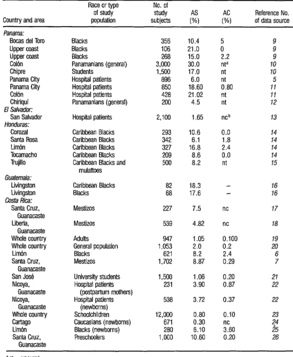
TABLE 1. Frequencies of heterozygous Hb S and Hb C markers (AS and AC) found by various investigators in a wide range of Central American populations.
Country and area
Race or type No. of of study study
population subjects of data source Reference No.
Panama:
Bocas del Tore Upper coast Upper coast Colbn Chipre Panama Cii Panama Cii Colon Chiriqui
El Salvador:
San Salvador
Honduras:
Corozal Santa Rosa Limon Tocamacho Trujillo
Guatemala:
Livingston Livingston
Costa Rica:
Santa Cruz, Guanacaste Liberia,
Guanacaste Whole country Whole country
Limbn Santa Cruz,
Guanacaste San Jose Nicoya,
Guanacaste Nicoya,
Guanacaste Whole country
4
2 Cartago
-4 Limbn -3
-8
Santa Cruz, Guanacaste
Blacks 356 10.4 5 9
Blacks 106 21 .o 0 9
Blacks 268 15.0 2.2 9
Panamanians (general) 3,000 30.0 nta IO
Students 1,500 17.0 nt 10
Hospital patients 896 6.0 nt 5 Hospiil patients 850 18.60 0.80 11 Hospital patients 428 21.02 nt II Panamanians (general) 200 4.5 nt 12 Hospital patients 2,100 1.65
Caribbean Blacks Caribbean Blacks Caribbean Blacks Caribbean Blacks Caribbean Blacks and
mulattoes
293 10.6 342 6.1 327 16.8 209 8.6 500 8.2
ncb 0.0 1.8 2 nt
13 14 14 14 14 15 Caribbean Blacks 82 18.3 16
Blacks 68 17.6 16 Mestizos 227 7.5 nc 17 Mestizos 539 4.82 nc 18 Adults 947 1.05 0.100 19 General population 1,053 2.0 0.2 20 Blacks 621 8.2 2.4 6 Mestizos 1,702 8.87 0.29 7 University students
Hospital patients (postpartum mothers) Hospital patients
(newborns) Schoolchildren Caucasians (newborns) Blacks (newborns) Preschoolers
1,500 1.06 0.20 231 3.90 0.87 538 3.72 0.37 12,000 0.80
671 0.30 280 6.10 1,000 10.60
0.10 ;c60 0.20
21 22 22 ;i 2% 26 +
American populations to detect Hb S and Hb C. Some of the apparent varia- tions in these frequencies could arise from different laboratory techniques em- ployed by different research groups, and also from the fact that many investigators employed relatively imprecise tests of sickling induced with 2% sodium meta- bisulfite (27) to demonstrate the pres- ence of Hb S. In addition, use of pheno- typically subjective criteria for racial classification could have caused substan- tial variation in the results listed in Table l-including results of the survey of
12,000 Costa Rican schoolchildren (23),
more detailed data from which are shown in Table 2.
Aside from exceptional condi- tions of hypoxia, the heterozygous he- moglobin S condition (oAfiS) is benign and by itself causes no significant public health problem (28). But it appears to have no positive adaptive value when the selective pressure of malaria is absent. And since sickle cell disease caused by the homotygous condition (flsps) and by OS in combination with other genes for abnormal hemoglobin makes this gene a social liability, over the generations in places where malaria is absent its preva- lence can be expected to decline (29). Of course, this decline is so slow as to be all but irrelevant for most health purposes.
Therefore, it is important to know the prevalences of this and other genes for abnormal hemoglobins in affected popu- lations, so that health authorities can be alerted to watch for associated illness, so that physicians can have an indication of the relative significance of sickle-cell and related diseases among the hemolytic anemias of a given population, and so that appropriate countermeasures can be taken (1).
In this context it is worth not- ing the program for education and ge- netic counseling on sickle-cell disease that is now under way in Panama. Under this program, a commission of Social Se- curity offrcials and community members seek to improve the approach to this public health problem.
The problem is serious in Pan- ama because the prevalence of the Hb S gene is very high. As a result, the disease places major demands on health re- sources and imposes a heavy economic burden on the patients’ families and the national economy-quite aside from the physical and psychological toll it takes on the patients themselves and the signifi-
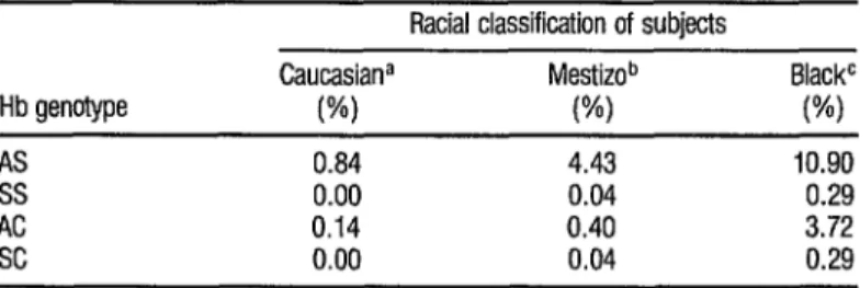
TABLE 2. Frequencies of Hb S and Hb G markers among 12,tUJO Costa Rican schoolchildren classified as belonging to diirerd racial groups.
8
Racial classification of subjects
& Caucasiana Mestizob BlackC q Hb genotype (%I VW (%) l
*
AS 0.84 4.43 10.90 u
ss 0.00 0.04 0.29 D AC 0.14 0.40 3.72
SC 0.00 0.04 0.29
% 2
a N = 6.911 b N = 4,740
cant social consequences for the patients and their relatives (11). The dimensions of the problem among populations of mainly African extraction are indicated by the relatively high percentages of pa- tients with the homozygous genotype among groups of hospital patients stud- ied in the provinces of Panama and Co- lon (12). These percentages are shown in Table 3.
In Costa Rica, the first case of homozygous hemoglobin C disease (in- volving the Hb CC genotype) ever de- tected in the country was found in a boy
12 years of age in 1985 (3O), The boy, who was Black, had marked splenome- galy, an infectious process, and an impor- tant anemic picture-possibly because he was experiencing a hemolytic crisis mediated by infection.
Sickling Syndromes
Sound information is lacking on the natural history and clinical picture of sickle-cell disease in the Central Amer- ican countries. However, studies of hos- pitalized patients have been performed in Cuba (1) and Jamaica (31), and a co- hort study begun in 1973 in Jamaica is seeking to define the clinical and hema- tologic behavior from birth of children with sickle-cell disease. Generally speak-
ing, most of these studies have not fo- cused on the benign forms of sickle-cell anemia, since these can only be identi- fied through systematic studies covering a large part of the population.
Overall, the limited clinical experience to date in Costa Rica suggests that sickle-cell anemia shows the same broad variation in degrees of clinical se- verity that is found in other countries. Thus, we observe patients who are se- verely affected and others with benign manifestations.
In 1961, Elizondo (32) showed the Central American Medical Confer- ence of that year a case of priapism caused by sickle-cell disease in a Mestizo subject with a Panamanian father and a Costa Rican mother. In 1965, Elizondo and Solano (33) reported a heterozygous hemoglobin S and hemoglobin C (SC) case in a subject of mixed racial origins from Guanacaste who exhibited a long- standing untreated chorioretinitis, neu- rologic alterations, and splenomegaly.
Zomer, in an unpublished re- port (34), has reviewed the files of 20 pa- tients with sickle-cell disease at the San Juan de Dios Hospital in San Jose, Costa Rica, from 1967 to 1974. The ages of “adult” subjects at their first admission to this hospital ranged from 13 to 45 years, the average being 21 years. All of these patients came from parts of the country known to have an Hb S prob- lem. In all, the 29 study patients had been hospitalized 114 times, for a total
5
TABLE 3..g disease (Hb SS) reported by Frequenciis of heterozygous Hb AS subjects and subjects with sickle cell studies of hospiil patients in two provinces of Panama PJ
Q in 1981 (12). 23
2 No. of Hb AS Hb SS
Province subjects (%I (%I Panam@ 850 18.6
Col6n 428 21.0
of 2,419 days, the average stay being 21 days. This latter figure contrasts with the overall average length of stay at this hos- pital, which was 11.5 days during the
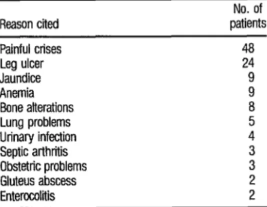
study period. The reasons for hospital- ization of these patients and the main complications observed are shown in Table 4.
Since 1978 we have been rry- ing to correctly identify the hemoglobin genotypes of patients with the sickling syndrome. At times these genotypes have not been identified correctly for lack of adequate analytic and genetic cri- teria. However, it is known that electro- phoretic testing based on the positions of the Hb A, Hb F, and Hb S bands can serve to distinguish between various sick- ling syndromes including SS disease (@fls); Hb S--P0 thalassemia (psfi*h”l “); Hb S--S@’ thalassemia (/3s(S/3)*do); SS combined with alpha thalassemia (for ex- ample: (Y--/• fisfls); Hb S combined with the hereditary persistence of (pan- cellular) fetal hemoglobin found in Black populations @Is, HPFH); SS com- bined with hereditary persistence of (het- erocellular) fetal hemoglobin (fisPs,
TABLE 4. Reasons cited fur 114 hospitalizations and fur
vadous compkations observed in 29 sickle cell anemia patfents over 13 years of age who were admitted to the San Juan de Dios hospital in San Jo&, Costa Rica, in the 1967-1974 period (32).
Reason cited patients No. of Painful crises
Leg ulcer Jaundice Anemia Bone alterations Lung problems Urinary infection Septic arthritis Obstetric problems Gluteus abscess Enterocoliiis
48
24 9 9
a
5 4
; 2 2
HEW); and Hb S combined with abnor- mal beta chain hemoglobins such as Hb Korle Bu (/3S/3KodeBu).7 This last doubly heterozygous condition has been found in a Black Costa Rican patient, now quite old, who for many years was diagnosed as having “atypical sickle-cell disease.”
A careful analytical examina- tion that includes electrophoresis of he- moglobin under both alkaline and acidic conditions, a quantitative solubility test, measurement of A2 and F hemoglobins,* staining of Hb F in a blood smear, and examination of the patient’s family his- tory will generally permit determination of the patient’s true genotype. Routine hematologic and clinical studies in such cases make it possible to advise the clinic of the existence of a disorder related to classic sickle-cell disease. This diagnostic
’ The Greek letters alpha (cc), beta (6). gamma (y), and delta (6) refer to different kinds of nolvneptide chains in hemoglobin molecules. The prim&l-type of hemo- globin found in the normal adult-known as hemoglo- bin A or Hb A-consists of two (Y and two ~3 polypep- tide chains, each chain being folded around a pocket containing a heme molecule. Thalassemias, caused by partially functional or nortfimctional genes for produc- tion of the alpha or beta chains, are termed 01 or fl tha- lassemias. Since each person possesses two separate genes for production of the (Y chain, the normal (Y glo- q bin genotype can be designated as orculorcu. Varying de- 8 grees of thalassemia are associated with partially or completely nonfunctional genotypes designated as o-l 3 ~01, cc-/o--, NY/--, or --/--. Alternativelv, the geno- zi types or phenotypes of subjects with thalassemia c&i be Q written usine + and o superscrims. with CY+ and B+ des- . -. 2 ignating p&al function and on and p” indicating non- function.
In this system of annotation. P indicates normal w production’of the /3 chain found in hemoglobin A, OS indicates presence of the sickle cell gene. and 16 BP in- dicates nonfunction of the genes r~spomible‘for’pto- ducing both the 0 and 6 chains. To designate genes in- volved with thalassemia, the letters “thal” (e.g., 131ha’ ._ “) .
may be used (3). 4;
s A, hemoelobin. nom-tally a minor constituent of adult +r e hemoglobin, contains two delta (8) polypeptide chains QJ instead of two beta chains; Hb F (fetal hemoglobin)
contains two gamma(r) chains instead of the fl chains. ii 2
process is relatively easy when the hemo- globin level exceeds 9.0 gl dl, when mi- crocytosis is evident, when splenomegaly persists in an adult, and when moderate clinical symptoms are present. The level of reticulocytes and the percentage of ir- reversibly sickled cells are also important factors.
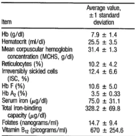
On studying cases of sickle- cell disease in 14 individuals with mixed racial backgrounds (36), we found the hematologic values indicated in Table 5. The family study carried out in connec- tion with this work led to detection of four people with the homozygous SS genotype combined with alpha thalasse- mia whose cases met the criteria cited for this double condition (37). These four subjects yielded the average hematologic values shown in Table 6. In addition, 38 relatives of the homozygous SS patients were found to possess the heterozygous Hb S gene. These 38 subjects were classi-
TABLE 5. Hematologic values found for 14 subjects with sicklecell disease in Costa Rica.
Item
Average value, + 1 standard deviation Hb (g/dl) 7.9 I!I 1.4 Hematocrit (ml/dl) 25.5 f 3.5 Mean corpuscular hemoglobin 31.4 f 1.3
concentration (MCHS, g/dl)
Reticulocytes (%) 10.2 + 4.2 Irreversibly (ISC, %) sickled cells 12.4 + 6.6 2
2 Hb F (%) 10.6 + 5.0 i Hb A2 (%) 3.5 2 0.33 2 2 Tdal iron (pg/dl) Serum 75.0 + 31.1 iron-binding 328.2 + 69.8 .g =wcity Wdl)
u
Q Folates (nanograms/ml) 14.7 f 9.4 *
2 Vitamin B12 (picograms/ml) 670 + 254.6
48
TABLE 6. Hematologic values found for four subjects with the homozygous sickle-cell genotype and alpha tha- lassemia in Costa Rica.
Item Average value Hb (g/dl)
Hematocrit (ml/dl)
Mean corpuscular hemoglobin concentration (MCHC, g/dl) Reticulocytes (%)
Irreversibly sickled cells (ISC, %) Hb A2 (%)
HB F (%)
9.55 29.1 30.4
a.0
11.0 3.1 14.6
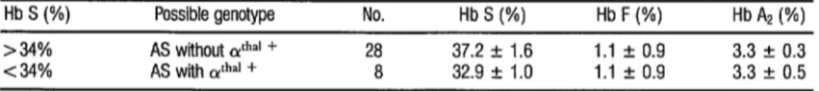
fied according to whether Hb S ac- counted for more or less than 34% of their total hemoglobin, as shown in Ta- ble 7. (The table only includes 36 sub- jects because two with iron deficiency were dropped from the “less than 34% ” group. )
In general, it is known that a significant subgroup of the Black popu- lation with the homozygous sickle cell trait (psDs) also has ofhal (38, 39). Many of these cases are confused with fisflthal O. This coexistence of fisfls and othal (espe- cially oyfhalO) gives rise to a hematologic picture characterized by microcytosis, hy- pochromia, high levels of Hb F, and higher figures for total Hb than is ob- served in classic sickle-cell disease. A slight relative increase of Hb A2 (39) has also been described in these cases.
TABLE 7. Concentratfons of Hb S, Hb 6 and Hb A2 as a percentage of total hemoglobin (plus or minus one standard deviation) in 36 subjjcts heterozygous for Hb S. The subjects were divided info two groups for analytical purposes, those with over 34% Hb S and those with less than that percentage (30, 33).
HbS(%) Possible genotype No. HbS(%) HbF(%) HbA*(%) >34% AS withoutdha'+ 28 37.2 + 1.6 1.1 f 0.9 3.3 f 0.3 <34% ASwithc@'+ 8 32.9 + 1.0 1.1 f 0.9 3.3 + 0.5
tends to be more conspicuous with oF o than with CYST + (41).
In this regard, eight of the 36 Hb S (AS) cases listed in Table 7 also ap- peared to have CYST + , a logical finding since a large percentage of the people where the study subjects resided (in the province of Puntarenas) are of African extraction, and since the (Y*~ + gene is
very prevalent among Blacks (42). While environmental factors such as temperature and altitude may di- minish the clinical picture of sickle-cell disease, socioeconomic factors are of pri- mary importance, since improvement of the clinical picture and life expectancy of sickle-cell disease patients depends on general improvement of living stan- dards (I).
It should also be noted that we have observed Costa Rican disease cases caused by the psfiM combination (both flthal + and pthal “). These cases have had variable clinical pictures that were sometimes indistinguishable from SS disease and sometimes slight or moder- ate, independent of whether they in- volved /3*d + or @*d O. Thus the progno- sis for such a case cannot be arrived at merely on the basis of these hemoglobin patterns.9 We have also confirmed the usual and mild clinical picture of the dis- ease caused by the combined presence of
‘) There are a good many molecular variations within the general thalassemia cy+, cP, PC, and p” categories; many of these molecular variations are not yet well un- derstood.
hemoglobins S and C, together with the serious ocular complication involving proliferative retinitis and vitreous hem- orrhage (43).
Uncommon Abnormal
Hemoglobins
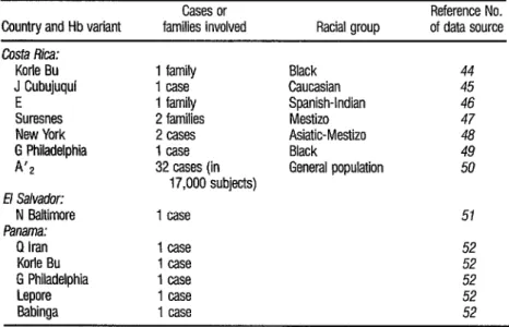
Several abnormal hemoglo- bins have been detected in Central America. These abnormalities involve the alpha, beta, and delta chains as well as combinations of portions of those chains such as that found in Hb Lepore. Table 8 lists the electrophoretic mutants reported as of 1984. The list is modest, partly for lack of systematic studies in the Central American countries except for surveys conducted in Costa Rica (23). (To support efforts that are needed in this re- gard, the Costa Rican multidisciplinary group studying hemoglobinopathies, with PAHO support, has published an an-
alytical protocol applicable to developing countries for identifying Hb variants, thalassemia syndromes, and glucose-6- phosphate dehydrogenase (G6PD) defi- ciency-50. )
TABLE 8. Uncommon hemoglobin variants reported in Central America as of 1984.
Country and Hb variant families Cases involved or Racial group Reference of data source No.
Costa Rica:
Korle Bu J Cubujuqui E
Suresnes New York G Philadelphia
A’2
E/ Salvador:
N Baltimore
Panama:
Q Iran Korle Bu G Philadelphia Lepore Babinga
1 family 1 case 1 family 2 families 2 cases 1 case 32 cases (in
17,000 subjects) 1 case
1 case 52
1 case 52
1 case 52
1 case 52
1 case 52
Black 44 Caucasian 45 Spanish-Indian 46 Mestizo 47 Asiatic-Mestizo 48 Black 49 General population 50 51
ethnic groups (such as Hb Korle Bu and Hb G Philadelphia) have come from Af- rica (44, 49, J3). Some of these markers are specific to particular regions of Af- rica; hence, their presence in Central America tends to define the regions of origin of the Black populations among which they appear. For example, Hb Korle Bu, associated with populations in Ghana and the Ivory Coast, has been found in both Costa Rica and Panama (44, 53).
Hb Cubujuqui, an abnormal hemoglobin produced by substitution of serine for arginine at position 141 (helix HC 3) on the (Y polypeptide chain, has been found in one instance in Costa Rica. The clinically silent condition, which was identified in a Caucasian Costa Rican schoolboy, resulted from a spontaneous point mutation (45). Point mutations of this sort should give rise to slight polycythemia whenever the argi-
nine at position 141 of the Q! chain is compromised in stabilizing the desoxy form of the hemoglobin molecule.
Hemoglobin E has been found in Costa Rica in three members of a Cau- casian-Mongoloid Salvadoran family (46). Hb E is very frequent in Burma and Thailand, as well as in certain Mongoloid populations of India and Malaya; and it has been suggested that much of El Sal- vador’s present population has resulted from the interbreeding of an indigenous (Mongoloid) race with Blacks and Cauca- sians, giving rise to Mongoloid ( - 80%)
and Caucasian-Black ( - 20 % ) mixtures
(54). It has also been suggested that Hb E may have reached tropical America directly from Asia, or else via Africa (as- suming that people from Malaysia trav- eled to the East Coast of Africa and then to the Americas--I).
ally (47), the first having been described in France in a male Caucasian child (55). The Costa Rican involved was a healthy woman 23 years old with Hispanic-Indig- enous ancestors and a racially mixed ap- pearance. In the original report on Hb Suresnes, it was noted that the indicated substitution of histidine for arginine would mildly upset the oxy-deoxy bal- ance of the hemoglobin molecule, pro- ducing slight erythrocytosis.
Hb New York-produced by substitution of glutamic acid for valine at position 113 (helix G15) on the fi chain-was fast described in a Chinese- North American family (56). This hemo- globin has been reported in Costa Rica (48) in three members of two related families of Asian-Mestizo appearance. This apparent ancestry could explain Hb New York’s presence, since subsequent publications report it being found among Chinese subjects in Taiwan (57). Hemoglobin BZ, also known as hemoglobin A’ *, results from a muta- tion affecting the delta chain (causing substitutions of arginine for glycine at position 16, helix A13). A study carried out in Costa Rica (23) has confirmed the
existence of small amounts of Hb B2 in some subjects. Specifically, its prevalence among Black study subjects was found to be considerable (2.58 % ); its prevalence was lower (0.29%) among Mestizos, and still lower among Caucasians (0.07 %).
In Panama, the Babinga delta chain variant (produced by substitution of aspartic acid for glycine at position 136, helix H14) has been found, as have hemoglobins Q Iran and Lepore (52).
Panama has experienced tremendous Spanish-Black-Amerindian racial mixing and significant Arab, Jewish, and Asian immigration-including arrival of a large number of Hindu immigrants from India.
T
HALASSEMIAS
The indigenous peoples of Central America do not appear to have genes for thalassemia. That is, the few re- ports citing such genes indicate that they have resulted from relatively recent racial mixing, in the manner of the Hb S genes found among these Amerindians (58). It can thus be inferred that the genes for thalassemia in these countries have been introduced by immigrants from places where these hereditary defects are rela- tively common.
In this vein, it is plausible to think that the epidemiology of the tha- lassemias, like that of the abnormal he- moglobins, has undergone significant changes in Central America because of racial mixing, a process that in the past was sometimes limited or controlled by racial segregation. At present, given the modern trend toward ethnic dilution and lowering of social barriers, th.e situa- tion could continue to change signifi- cantly.
It is not generally possible to pinpoint the regions of origin of the thalassemias, partly because genes for thalassemia were widely distributed among various ancestral populations. In general, although there are marked vari- ations in their frequency, both alpha and beta thalassemias are found in African and Mediterranean countries (59, 60). In Spain, for example, pthal is present in 1.1% to 3.43 % of the population
(61, 62), although @’ in that country is much less common (6.3).
In most cases it is not possible to establish the racial origin of a specific thalassemia gene, partly because the clinical picture produced by each type of
thalassemia tends to be similar in differ- ent racial groups. Thus, even though beta thalassemia is usually moderate among Blacks, it sometimes shows the severe sorts of clinical pictures more com-
monly observed in Europeans.
At the molecular level, a great heterogeneity has been observed in both alpha and beta thalassemias, different genetic lesions having been demon- strated in different racial groups (64). In this vein, gene mapping has shown indi- vidual and demographic variations of each thalassemia variant studied in Black, Oriental, and Caucasian individ- uals. Since tropical America has a largely mixed population, it is expected that such genetic analyses will yield worth- while anthropologic information and shed considerable light on the heteroge- neity of existing thalassemia phenotypes and genotypes.
Alpha Thalassemias
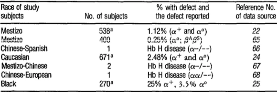
Costa Rica is the only Central American country where the presence of c@ has been reported (Table 9). Identi- fication of this marker has always been accomplished on the basis of hemato- logic criteria and family studies. In gen- eral, it seems clear that the prevalence of o@@’ in Costa Rica has arisen from a ra- cial/ethnic mixing of the population that has mediated the Caucasian (Euro-
pean) influence as well as the Black (Afri- can) and Asian (especially mainland Chi- nese) influences.
The different forms of alpha thalassemia generally result from the in- teraction of two basic alleles (oar), both of which produce the alpha polypeptide chain. ofhal + , involving partial nonfunc- tion, is associated with the (-a) haploid genotype; while ofhalo, involving com- plete nonfunction, is associated with the (--) haploid genotype. Studies of Asian newborns suggest that the o+ and o? genes occur frequently among the Chi- nese, Malaysian, and Thai peoples ((59). Different molecular lesions producing c~+ and o” phenotypes have also been de- tected in Mediterranean peoples, though with a lesser frequency (70).
The o? gene is not generally found in the Black race, a circumstance that may explain the absence of hemo- globin H disease among Blacks (71). (The presence of this disease in the Costa Rican and Panamanian populations is due to those populations’ European and Asian ancestries-65-67).
Thai experience has demon- strated that the presence of Bart’s hemo- globin among newborns provides the most practical basis for diagnosing o?’
TABLE 9. Cases of alpha thabssemia diagnosed in Costa Rica.
Race of study subjects Mestizo Mestizo
Chinese-Spanish Caucasian Mestizo-Chinese Chinese-European Black
a Umbilical cord samples.
No. of subjects
F138~ 400
da
2
1
270a
% with defect and Reference No. the defect reported of data source 1 .I 2% (or+ and &) 22
0.25% (01”; /3*@) 65
Hb H disease (a-/--) 66
2.48% (a’ and a”) 24
Hb H disease ((Y-A-) 67
Hb H disease (OK&-) 68
in Oriental populations (69). (Blacks who are heterozygous for Qua + do not always show increased levels of Ban’s he- moglobin-1).
In Costa Rica, as Table 9 indi- cates, three population surveys of alpha thalassemia have been conducted. The one involving 671 Caucasian subjects (newborns) evaluated the concentration of Ban’s hemoglobin semiquantitatively using starch-gel elecuophoresis. This study found the frequency of the silent (x+ phenotype to be 2.08%) and that of the o@’ o gene (severe thalassemia) to be
0.40% (24). Another survey, of umbili- cal cord blood samples from 270 Black newborns, yielded Ban’s hemoglobin values indicating that 25% of the new- borns had the (Y+ character while 3.5 % had 01~ (2,).
Beta Thalassemia
Aside from data obtained in Costa Rica, there is no precise informa- tion on either the presence or the fre- quency of the phal marker in Central America. This is partly because no ana- lytical methodology has been devised to permit detection of the flti marker. The available Costa Rican data on the mark- er’s presence, summarized in Table 10,
were obtained by means of a simple and practical analytical approach that has been described elsewhere (83).
Another important point here is that data on the prevalence of flthal in Central America tend not to be represen- tative of the ethnic groups comprising the population, either because only a few samples were analyzed or because sound criteria for adequately determining the racial composition of the study popula- tions were lacking.
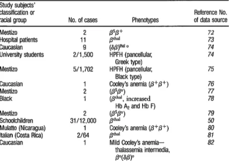
TABLE 10. Cases of beta thalassemia and hereditary persistence of fetal hemoglobin (HPFH) diag- nosed in Costa Rica. (Of indicates the beta thalassemia gene with some p-chain production; p indicates the beta thalassemia gene with no P-chain production.)
Study subjects’ classification or
racial group No. of cases Phenotypes of data source Reference No. Mestizo
Hospital patients Caucasian University students Mestizo
Caucasian Mestizo Black Mestizo Schoolchildren Mulatto (Nicaragua) Italian (Costa Rica) Caucasian
2 11 2/I ,950o 5/l ,702
1 2 1 31/122000
1 2164
1
@Lvn o
HPFH (pancellular, Greek type) HPFH (pancellular,
Black type) CyleT’s anemia (p+p+) $!I )
, mcreased ’ Hb A2 and Hb F) (Pp”)
pd
Coeley’s anemia (p+p+) Pd”l
Mild Cooley’s anemia- thalassemia intermedia, p”@PP
72 73 74 74 75 76 77 78 79 ii 81 82
Regarding actual cases of beta thalassemia, isolated cases have been re- ported in El Salvador and Panama. Also, the previously mentioned study of
12,000 Costa Rican schoolchildren
showed 0.26% to have the disorder-a finding illustrating the relative impor- tance of this gene in a fundamentally Caucasian population.
Since the eradication of ma- laria in Costa Rica, it has not been possi- ble to corroborate the controversial find- ing of high hemoglobin A2 values in connection with that pathology (84).
The doubly heterozygous cooft$~ s psflthal-i.e., Pspthal +, , or 0 (6 /3)*h” “-has also been re- ported in Costa Rica (72, 77, 79). How- ever, it is not possible to identify the het- erozygous /3+ and p” states with routine or conventional hematologic methods unless one uses an appropriate genetic approach. The most solid evidence for specific primary action by @+ and /3 genes is therefore obtained from hema- tologic and biochemical studies of dou- bly heterozygous states that involve an- other structural variant (such as ps) of the beta chain. For this reason, the distinc- tion between /3+ and p” should only be made on the basis of an appropriate framework of genetic information-as was done, for example, in an article re- lating detection of the p” gene in con- junction with hemoglobin S to the 2 observed concentrations of hemoglobins 2 F and S (79).
i
2 In addition, homozygous beta Q thalassemia (known as thalassemia major .g or Cooley’s anemia) has been reported in
9,
a ;i Costa Rica (76, 80). The first reported 2
case was diagnosed in a Caucasian Costa Rican subject and the second was found 2
in a Black girl from Nicaragua. (This lat- ter diagnosis provided the basis for our conclusion that this pathology existed in
54 Nicaragua.) Subsequently, a case of mild Cooley’s anemia (thalassemia inter-
media) with one delta chain gene involved+*” 0(8/3)*~ ~-was diagnosed in a Caucasian girl (82).
It is well known that the eth- nic distribution of thalassemia major var- ies from country to country, although in general the disorder is found most often in Caucasians, among whom it produces a clinical picture similar to that observed in Mediterranean populations (1). It is generally accepted that this disease is less severe among Blacks, a conclusion sup- porting the theoretical existence of a Black /3++ gene distinct from the Medi- terranean /3+ gene (64).
Finally, within this context, it should be noted that both Black and Greek varieties of hereditary persistence of fetal hemoglobin (HPFH) have been reported in Costa Rica (21, 75).
GL
UCOSE-6-PHOSPHATE-
DEHYDROGENASE
POLYMORPHISM
IN COSTA RICA
The role of glucose-&phos- phate-dehydrogenase (GGPD) deficiency in hemolysis, especially among Blacks, has been under study in Costa Rica for over 15 years (6). This hemolytic phe-
nomenon has also been detected in El Salvador and Honduras (15, 51, 54). Re- garding Costa Ricans, the frequency de- tected in males by giving the methe- maglobin reduction test to 621 subjects
was 12.6 % (85); a lower frequency
dren showed 2.3 % of the boys (irrespec- tive of race) to be affected (23).
In 1974 a new GGPD variant called Gd San Jo& was reported in a male Costa Rican Caucasian subject (87). In 1982 another new mutant with strong clinical expression, designated Gd Puerto LimBn, was identified (13); and this was followed by identification of an- other pathogenic variant, Gd E Santa- maria, in 1983 (88). At present, electro- phoretic studies permit phenotypic characterization of all the GGPD variants studied in our laboratories, and also per- mit us to determine their degree of activity.
S
UMMARY
The prevalences of abnormal hemoglobins and thalassemias depend largely upon the hereditary racial compo- sition and geographic origins of the af- fected populations. For this reason, the highly varied immigration and racial pat- terns found in Central America and Pan- ama make it logical to expect prevalences that vary greatly from one country to an- other and even from one population to another within a given country. Unfortu- nately, literature on the problem is scanty, and so the review presented here draws heavily upon fairly extensive infor- mation available from Costa Rican stud- ies, supplemented wherever possible with data from Panama and other parts of Central America.
The sickle cell gene for hemo- globin S (Hb S) has not been found among purely indigenous groups studied in Costa Rica and Panama. However, noteworthy prevalences of Hb S have
been reported among Blacks and other population groups in Costa Rica, El Sal- vador, Guatemala, Honduras, and Pan- ama, with apparent levels of the hetero- zygous Hb S marker reaching as high as 30 % of one survey population in the lat- ter country. In addition, the presence of the hemoglobin C marker has been re- ported in Costa Rica, Honduras, and Panama; and various sickling syn- dromes-including heterozygous Hb S C, Hb S combined with Hb Korle Bu, homozygous Hb S with alpha thalasse- mia, heterozygous Hb S with alpha (a+) thalassemia, and Hb S with beta thalas- semia (both fl+ and /3”)-have been found in Costa Rica.
Regarding uncommon abnor- mal hemoglobins, the hemoglobins A’ *, E, G Philadelphia, Korle Bu, J Cubu- juqui, New York, and Suresnes have been found in Costa Rica; the hemoglo- bin N Baltimore has been found in El Salvador; and the hemoglobins Babinga, G Philadelphia, Korle Bu, Lepore, and Q Iran have been found in Panama.
Costa Rica is the only Central American country where precise infor- mation is available on the presence of c+’ and flthal markers. The CYST studies involved have detected (Y*~ marker prev- alences ranging from 1.12 % to 25 % of S the various study populations, as well as :: isolated cases of hemoglobin H disease. The pthal studies have detected various 2 z kinds of beta thalassemia genotypes-in-
eluding Cooley’s anemia @VP+), mild Cooley’s anemia (p”(Sfly), (Sfi)o, psfl+, PW, and beta thalassemia associated with increased concentrations of hemo- globins A2 and F.
RE
FERENCES
variants (Gd E Santamaria, Gd Puerto LimBn, and Gd San Jose) have been found initially in Costa Rica, and Costa Rican studies designed to detect associ- ated hemolysis have found frequencies ranging from 2.3 % to 12.6 % in various study populations. Such hemolysis has also been detected in El Salvador and Honduras.
Colombo, B., and G. Martinez. Hemo- globinopathies: II. Tropical America. C&z Haematoj (London) 10:‘730, 1981.
Arends, T. Hemoglobinopathies and Enzyme Deficiencies in Latin American Populations. In: The Ongoing Evohticm
of
Latin American Pojdations, C. C. Thomas, Springfield, 1971, pp. 509-559.Cordero, R., and B. Monge. Analisis san- guinea y parasitosc6pico en indigenas en Costa Rica. Revista M&&a de Costa Rica 4~43, 1972.
Sknz, G. E Unpublished data, 1976.
Calero, C. Drepanocitemia y anemia dre- oanocitica en el Istmo de Panama. con esnecial referencia a sus manifestaciones ciinicas ; inci- dencia por raza. Archives del Hospital’ Saato Tom& (Panama City) 1:27, 1946.
SLnz, G. E, G. Arroyo, J. Jim&ret, A. Gu- tierrez, M. Barrenechea, E. Brilla, and E. Va- lenciano. Investigacidn de hemoglobinas anormales en poblaci6n de raza negra costarri- cerise. Rev BiolTrop 19:251, 1971.
S&em, G. E, M. A. Alvarado, E Atmetlla, G. Arroyo, R. JimEnez, and E. Valenciano. Inves- tigaci6n de hemoglobinas anormales en pobla- cidn costarricense de1 Guanacaste. Acta M& dica Costarrtkense 16: 147, 1973.
Mel&dez, C., and Q. Duncan. El Negro en Costa Rica. Editorial Costa Rica, San Jose, 1976.
Ferrell, R. E., A. Ntitiez, T. Bertin, D. R. La- barthe, and W. J. Schull. The blacks of Pan- ama: Their genetic diversity as assessed by 15 inherited biochemical systems. Am J Phys An- thropd48:269, 1978.
10
11
12
13
14
15
16
17
18
19
20
21
Zspino, H., and A. Santamaria. Personal com- munication, 1980.
Altafulla, M. La enfermedad por celulas falci- formes en Panama. Rev&a M&&a de Panama’ 6.620 1981 ., .
Altafulla, M. Origen e incidencia de1 gene falciforme en la Repfiblica de Panama. Revista Mhz’ica de Panamri 6:233, 1981.
Elizondo, J., G. E Sknz, C. A. PBez, and M. RamBn. GGPD-Puerto Limdn: A new de& cient variant of glucose-6-phosphate dehydro- genase associated with congenital nonsphero- cytic hemolytic anemia. Hwn Genet 62:110, 1982.
Rucknagel, D. L. Personal communication, 1976.
Stefan, R. Estudio epidemiokjgico-clinico de la drepanocitosis en la poblaci6n gar’&na de Honduras. Thesis. National Autonomous University of Honduras Tegucigalpa, 1982.
Tejada, C., N. L. S. Gonzalez, and N. San- chez. El factor Diego y el gen de ctlulas falci- formes entre 10s Caribes de rata negra en Livingston, Guatemala. Rev CoC MZd 16:83, 1965.
Solano, L., M. Cabezas, and L. Elizondo. Es- tudio sobre drepanocitosis y hemoglobina “S” en Santa Cruz de Guanacaste. Acta M&&a Costamiense 9:59, 1966.
Solano, L.. and E Mainieri. Estudio sobre dre- panocitosis y hemoglobina S en Liberia, Guanacaste. Acta M&&a Costarricense 10:175, 1967.
Rivera, A., and G. E Sknt. Datos numericos y estadisticos minimos sobre la incidencia de hemoglobinas anormales en Costa Rica. Re- vista MZa’jca del Hospital National de Nifios (Costa Rica) 2:95, 1968.
Elizondo, J., and M. Zomer. Hemoglobinas anormales en la poblacidn asegurada constarri- cerise. Acta M&&a Costamkense 13:249,
1970.
22
23
24
25
26
27
28
29
30
31
32
S%enz, G. F., M. Alvarado, E. Alfaro, M. Barrenechea, J. Jimenez, and G. Montero. Diagndstico neonatal de hemoglobinopatlas. Sangre 22~339, 1977.
Saent, G. I?, J. EIizondo, G. Arroyo, E. VaIen- ciano, L. E Rojas, J. Jim6net, A. G. Montero, and J. E. %inchez. Hemo 1obinopatIa.s en 12.000 escolares. Acta Me zca Costarhense -3 23:89, 1980.
S?ienz, G. E, E. Blanco, J. Jimcnez, A. G. Montero, E. Valenciano, G. Arroyo, and J. E. Stichez. Diagndstico neonatal de aIfa-talase- mia en poblacidn cauchica. Revista Costar-r-i: tense de Ciencias MZdicas 2~97, 1980.
Sienz, G. E, M. Chaves, M. Barrenechea, S. Grant, J. Jimenez, and G. A. Montero. He- moglobinas anormales, alfa talasemia y deli- ciencia de la G6PD eritrocltica en reciin naci- dos de raza negra. Sangre 29:861, 1984.
Saenz, G., M. Chaves, J. Briceilo, E. Quin- tana, G. Arro
tero. Pohmo % rsmo de la hemoglobina y de la o, E. Valenciano, and A. Mon- G6PD eritrocitica en poblaci6n pre-escolar de Santa Cruz, Guanacaste. Rev&a Costarricense a’.. Ciench M&&as 6:126, 1985.
Schneider, R. G., et aI. Sickling test: Pi&& in performance and interpretation. JAMA 202:419, 1967.
World Health Organization. WHO Week& EpidemioCogical Record Geneva, vol. 5 5, 4 . July 1980.
Livingstone, E B. Aspects of the opulation dynamics of the abnormal hemog ohms and ‘I GBPD deficiency genes. Am J Hum Genet 16:435, 1964.
SLnz, R. I., G. E SBenz, and M. Chaves. En- fermedad homocieotica nor Hb C: Renorte de1 primer case cost&rice&. Rev&a M.&&a de/ Hospital Naciond de Nigos (Costa Rica) 19:25, 1984.
Serjeant, G. R. The Clinicd Features of Sick/e Cell Disease. North Holland Publishing, Am- sterdam, 1974.
Elizondo, J. Anemia de cilulas fakiformes. Reporte deun case de priapism0 por drepano- citosis. Estudio de una famiha costarricense. Presentation to the IX Central American Med- ical Congress (Costa Rica, 1961), pp. 162-171.
33
34
35
36
37
38
39
40
41
42
43
44
45
Elizondo, J., and L. Solano. Hemoglobino- patia SC. Acta M&&a Costarhense 8:15, 1965.
Zomer, M. Anemia drepanocitica: Experiencia en eI Hospital San Juan de Dios (1967-1974). Unpublished paper, 1975.
Serjeant, G. R. SickLe Cell Disease. Oxford University Press; Oxford, New York, and Tokyo; 1985.
SLnz, G. E, M. Chaves, E. Castro, M. Ra- m6n, M. Barboza, J. Jimenez, P. Montero, G. Arroyo, and E. Valenciano. Slndromes dre- panociticos en poblaci6n de la provincia de Puntarenas. Revha Costarriicense de Ciencias M&&as (Costa Rica) (in press).
Serjeant, G. R., K. P Mason, and B. E. Ser- jeant. Negro or-thalassaemia: Genetic studies in homotygous sickIe cell disease. J Med Genet 17:281, 1980.
Embury, S. H., A. M. Dozy, J, Miller, and J. R. Davis. Concurrent sickle cell anemia and cr-thalassemia. N EngC J Med 306:270, 1982.
SeweII, A., D. Millard, and G. Serjeant. The interaction of alpha thalassemia with SS dis- ease. In: G. J. Brewer (ed.). The Red Cell. Alan R. Liss, New York, 1978, pp. 93-102.
Honig, G. R., M. Koshy, G. Mason, and L. N. Vida. Sickle cell syndrome: II. The sickle cell anemia and cY-thaiassemia syndrome. J Pea&r 92~556, 1978.
Mimer, P E Thalassemias, Hemoglobino- pathies and Sickle Ceil Disease. In: V. E Fair- banks (ed.). Hematolbgy (vol; 2). John Wiley and Sons, 1983.
Higgs, D. R., L. Pressley, J. B. Clegg, D. LJ. Weatherall, and G. R. Serjeant. Alpha-thalas- semia in black populations. Johns Hopkins MedJ 146:300, 1980.
Saenz, G. E, V. Ramirez., and M. Chaves. En- fermedad por hemoglobma SC y patologia oc- ular: A prop&it0 de un case. Acta M&&a CostatriGense 3:249, 1982.
Elitondo, J., G. E SPenz, M. A. Alvarado, and M. Ramon. HaIIazgo de la hemoglobina Korle-Bu (aIfarbetaz 75 asp-asn) en Costa Rica. Sangre 1:54, 1976.
Saenz, G. E, M. Alvarado, J. Elizondo, G. Arroyo, and E AtmetIIa. Chemical character- ization of a new haemoglobin variant, Haemo- globin J Cubuju
46
47
48
49
50
51
52
53
54
55
2 2
Zenz, G. E, J. Elizondo, B. Colombo, M. Al- rarado, G. Arroyo, and E Atmetila. Hallatgo le la hemoglobina E (Betaz6s’“-‘“) en Costa Rica. Sangre 5~652, 1977.
Saenz, G. E, M. Alvarado, G. Arroyo, E. Al- fare, G. Montero, and J. Jim&z. Hemoglo- bin Suresnes in a Costa Rican woman of Span- ish-Indian ancestry. Hemoglobin 4:383, 1978.
SBenz, G. E, J. Elizondo, G. Arroyo, J. Ji- menez, and A. G. Montero. Finding of the hemoglobin New York in Costa Rica. Hemo- globin l:lOl, 1980.
SLnt, G. F. 1 J. Elizondo, G. Arroyo, E. Valen- ciano. 1. Timtnez. A. G. Montero. E. Sanchez, and S.“&ant. Hallazgo de la hemoglobina G Philadelphia (alfa 68 (E 17) Asn-Lis) en Costa Rica: Consideraciones bioquimico-genEticas. Sangre 2~224, 1981.
Saenz, G. I?, J. Elizondo, G. Arroyo, J. Ji- menez, A. G. Montero, and E. Valenciano. DiagnBstico de hemoglobinopatias y de tras- tornos afines: Enfoque poblacional de1 pro- blema. Bol Of Sanit Panam 2:127, 1981. Bloch, M., and H. Rivera. Hemoglobinas anormales v deficit de GGPD en El Salvador. Sangre 14:i21, 1969.
Castillo, Z., and M. Altafulla. Hemoglobino- patias en Panama. Revljta M&&a de Panamd, 1983.
Altafulla, M. Personal communication, 1983.
Bloch, M., G. Sancho, R. A. Cea, H. Rivera, A. N. Zelava. and E. Montes. La deficiencia de glucosa-6-fosfato dehidrogenasa en El Salva- dor. Rev&a del Institzzto de Investigaciones MZdicas (EISahador) 1:397, 1972.
Poyart, C., R. JSrishnamoorthy, E. Bursuax, G. Gacon. and D. Labie. Structural and func- tional studies of haemoglobin Suresnes or cxa 141 (HC3) Apg-His &, a new high-oxygen affinity mutant. FEBS Lett 69:103, 1976.
4
z. 56 Ranney, H. M., A. S. Jacobs, and R. L. Nagel.
9 Haemoglobin New York. Nature 213:876,
tu
.E 1967. -
-ci P,
2 57 Blackwell, R. Q., C. S. Liu, and C. L. Wang.
Q Hemoglobin New York in Taiwan. Am J Phyr
m Anthropd 34:329, 1971.
59 Silvestroni, E., and I. Bianco. Diffusione e fre- quenze della microcitemia e delle anemie mi- crocitiche nell’Italia Continentale e in Sicilia, Atti I1 problema sociale della microcitemia e de1 morbo di Cooley’. Institz&o Itahzno diMe- dicina So&e (Rome) 1: 5 1, 1962.
60 Stamatoyanno
lassaemia, g ucose-6-phosphate 1; dehydro- oulos, G., and P Fessas. Tha-
genase deficiency, sickling and malaria ende- micity in Greece: A study of five areas. Br Med J 1:875, 1964.
6 1 Cotrina de Luna, 1. L.. F. S. St.&et, I? Llorente Gutierrez, E Vill&-me~a, J. Rico,. and A. Pefia. Hemoglobinopat’las en la Provtncia de Gta- nada. Rev Chn Esp (Madrid) 157:373, 1980.
62
63
64
65
66
67
68
Pellicer, A.., and A. Casado. Frequencies of thalassaemra and GGPD deficiency in five provinces in Spain. Am J Hum Genet 22:298, 1970.
Rotman, C., J. Capdevila, S. Woessner, and I. Marti. Hemoglobinopatia H en una familia es- patiola. Rev C/in Esp (Madrid) 103:373, 1966.
Weatherall, D. J., and J. B. Clegg. The Tha- lassaemia Syndromes. Blackwell Scientific Publishers, Oxford, 1980.
Saenz, G. E, J. Elizondo, G. Arroyo, J. Ji- minez, and A. G. Montero. Sindromes dre- panociticos en Costa Rica: VI. Sindrome de heterocigosis doble Slalfa-talasemia. Sangre 24~205, 1979.
Sknz, G. F., J. Jim&nez, and L. Mora. En- fermedad por hemoglobina H en Costa Rica. sangre 3:333, 1979.
Saenz, G. F., J. Bricefio, A. G. Montero, J. Timenez. and M. Chaves. Enfermedad nor he- moglobma H: Estudio de una nueva familia.
Acta M&&a Costarricense 24 : 15 1, 198 1.
SCnz, G. E, M. Castillo, and L. Salazar. Tala- semia intermedia: A prop6sito de un case de alfa talasemia tipo enfermedad por Hb H. Acta Mgdica Costamiense 27~179, 1984.
69 Wasi, P , S. Na-Nakorn, and S. Pootrakul. The
58 Livingstone, F. B. Abnormal Hemoglobins in Human Populations. Aldine Publishers, Chi- cago, 1967.
JO
71
72
73
74
75
76
77
78
Kan, Y. W., A. M. Dozy, G. Stamato anno- poulos, M. G. Hadijminas, 2. Zachana es, M. d Furbetta, and A. Cao. Molecular basis of he- moglobin H disease in the Mediterranean pop- ulation. Br!ood 54:1434, 1979.
Dozy, A. M., Y. W. Kan: and S. H. Embury. Alpha globm gene orgamtation in blacks pre- cludes the severe form of alpha thalassaemia. Nature 280:605, 1979.
Zomer, M., and A. Rivera. Primer case de he- moglobinopatla S-talasemia en Costa Rica. Acta MZdica Costarrhense 10:71, 1967.
Zomer, M., J. Elizondo, and E. Quesada. Ana- lisis de 11 cases de beta talasemia en Costa Rica. Acta Mgdica Costarriense 2:129, 1973.
SLnz, G. E, M. A. Alvarado, and G. Arroyo. F(delta-beta) talasemia en Costa Rica. Acta MZdica CostarriGense 1:63, 1974.
Sknz, G. E, M. A. Alvarado, G. Arroyo, and G. Montero. Persistencia hereditaria de he- moglobina fetal (PHHbF) en Costa Rica. Re- v&a M&ka de1 Hospital National de Niiios (Costa Rica) 2:135, 1975.
Sknz, G. E, B. Monge, G. Arroyo, and M. A. Alvarado. Enfemredades de Coolev (Beta+ ?g- lasemia mayor) en Costa Rica. &z&e 1: 117, 1976.
Saent, G. E, G. Sanchez, and B. Monge. Sindromes dreuanociticos en Costa Rica: IV. Hemoglobina S/beta-delta talasemia (SIFta- lasemia). Acta M.Zdica Costarnkense 4:3, 1976.
S%enz, G. F., J. Elizondo, G. Arroyo, G. Mon- tero, and J. JimEnez. ‘Iklasemia A,F en raza negra costarricense: A prop&it0 de un case. Acta Mgdica ConstaniGense 4 : 373, 1977.
79
80
81
82
83
84
85
86
87
88
Saenz, G. E, J. Elizondo, and C. A. Paez. Ha- llazgo de1 gene p”-talasemico (supresor) en Costa Rica: V. Slndrome de heterocigosis doble SlbetaO-tal. Sangre 2:196, 1978.
SPenz, G. F., M. Navarette, L. Mora, J. Ji- minez, A. G. Montero, and G. Arroyo. Beta talasemia mayor (enfermedad de Cooley): Consideraciones sobre biologia molecular y es- tudio de una nueva familia. Acta MZdica Cos- tarrkense 2:143, 1981.
SLnz, G. F., M. Chaves, G. Arroyo, E. Valen- ciano, J. Jimtnez, A. G. Montero, J. E. S&n- chez, C. Vega, and P Contreras. Escrutinio de hemoglobinopatias y GGPD en una poblaci6n italiana radicada en Costa Rica. Revista Costa- rrikense de Ciencizs M&&as 2~185, 1982.
SBenz, G. F., J. M. Carrillo, L. Mora, M. Chaves, L. G. Rojas, R. Jirnenez, J. JimEnez, and A. G. Montero. Beta talasemia intermedia de genotipo S@‘/(@‘: Reporte de un case. Sangre 29~467, 1984.
SLnz, G. F. Diagn6stico conventional de 10s sindromes talasEmicos. Sangre 25:63, 1980.
Arends, T. High concentration of Hb Aa in malaria patients. Natwe 215:1517, 1962.
Sknz, G. E, E. Brilla, G. Arroyo, B. Valen- ciano, and J. JimCnet. Deficiencia de la dehi- drogenasa de la glucosa-6-fosfato (G6PD) eri- trocltica en Costa Rica: I. Generalidades sobre el defect0 y hallazgos en poblacidn de raza ne- gra. Rev&a MZdica de1 Hospit& National de Nil?os (Costa Rica) 2:129, 1971.
SLnt, G. E, et al. Deficiencia de G6PD eri- trocitica en poblaci6n guanacasteca. Unpub- lished data, 1983.
Castro, G. A. M., and L. M. Snyder. G6PD San JOSE: A new variant characterized by NADPH inhibition studies. Hzlmangenetik (Berlin) 21:361, 1974.