Abstract
Objective: To discuss the differential diagnosis of encephalitis beyond that of infectious etiology and to inform pediatricians about the possibility of anti-N-methyl-D-aspartate receptor (NMDAr) encephalitis in children by highlighting its most important clinical features.
Description: Three patients presented with an initial neuropsychiatric syndrome followed by encephalopathy and movement disorder. The initial neuropsychiatric features which developed over days to weeks included a change in personality, anxiety, confusion, and speech regression. This was followed by a choreoathetoid or dystonic movement disorder affecting the orofacial region and the limbs. After the exclusion of the major causes of encephalitis, NMDAr antibodies were identiied in serum and cerebrospinal luid, and neoplasm screening did not detect any tumor. Patients were submitted to immunosuppression, and two of them had a full neurological recovery. One of them still presents a mild dystonic posture in a limb.
Comments: Clinical signs of anti-NMDAr encephalitis in children are similar to those previously described in adults. Tumors are not usually detected by this age. The diagnosis of anti-NMDAr encephalitis must be addressed only after the exclusion of infectious and other recognizable causes of encephalitis. Pediatricians should be aware of this treatable autoimmune condition.
J Pediatr (Rio J). 2012;88(3):275-8. Encephalitis, anti-N-methyl-D-aspartate receptor, childhood.
O
riginala
rticle0021-7557/12/88-03/275 Jornal de Pediatria
Copyright © by Sociedade Brasileira de Pediatria
275 Introduction
Anti-N-methyl-D-aspartate receptor (NMDAr) encephalitis is a neuropsychiatric syndrome regarded as being caused by immune-mediated processes, once it presents autoantibodies in the serum or in the cerebrospinal
luid (CSF) directed against an epitope located in the
extracellular domain of NMDAr. This disorder is often described in the adult population and may be associated with paraneoplastic ovarian teratoma.1,2
NMDAr participates in synaptic transmission and neuronal plasticity. Up- or down-regulation in its activity may explain some of the neurological symptoms present in anti-NMDAr encephalitis, such as seizures, aphasia, and dystonia. Before
the identiication of the antibodies underlying the pathogenic
processes of this entity, patients were often diagnosed as presenting encephalitis of unknown origin if no other known
infectious or autoimmune etiology was clariied.
Anti-N-methyl D-aspartate receptor encephalitis in childhood
Felippe Borlot,1 Mara Lucia F. Santos,2 Marcia Bandeira,3 Paulo B. Liberalesso,4 Fernando Kok,5 Alfredo Löhr Jr.,6 Umbertina C. Reed7
1. Physician. Pediatric neurologist, Hospital Santa Marcelina, São Paulo, Brazil. DFV Neuro team, São Paulo, SP, Brazil. 2. Physician. Pediatric neurologist, Hospital Pequeno Príncipe, Curitiba, PR, Brazil.
3. Physician. Pediatric rheumatologist. MSc in Pediatrics. Investigator and technical responsible, Hospital Pequeno Príncipe, Curitiba, Brazil. Professor of Pediatrics, Universidade Positivo, Curitiba, PR, Brazil.
4. Physician. PhD. Supervisor, Programa de Residência Médica em Neurologia Infantil, Departamento de Neurologia e do Serviço de Neurofisiologia, Hospital Pequeno Príncipe, Curitiba, PR, Brazil.
5. PhD. Tenured professor of Neurology, Faculdade de Medicina, Universidade de São Paulo (USP). Pediatric neurologist, Hospital das Clínicas, Faculdade de Medicina, USP, São Paulo, SP, Brazil.
6. MSc. Chief pediatric neurologist, Setor de Neurologia Infantil, Hospital Pequeno Príncipe, Curitiba, PR, Brazil. 7. PhD. Full professor, Departamento de Neurologia, Faculdade de Medicina, USP, São Paulo, SP, Brazil.
No conflicts of interest declared concerning the publication of this article.
Suggested citation: Borlot F, Santos ML, Bandeira M, Liberalesso PB, Kok F, Löhr Jr. A, et al. Anti-N-methyl D-aspartate receptor encephalitis in childhood. J Pediatr (Rio J). 2012;88(3):275-8.
Manuscript submitted Aug 19 2011, accepted for publication Nov 16 2011.
276 Jornal de Pediatria - Vol. 88, N° 3, 2012 Anti-NMDAr encephalitis: three Brazilian cases - Borlot F et al.
The frequency of anti-NMDAr encephalitis is not certainly established, but it is assumed that this condition is responsible for almost 1% of the admissions of young adults in intensive care units.3 The largest pediatric series addressing this issue reported 32 patients under the age of 18 years (median: 14 years).4 Interestingly, there is no frequent association with paraneoplastic neoplasms at such young ages.4,5
The irst reports of adult patients presenting anti-NMDAr
encephalitis demonstrated the symptomatic evolution, with the onset of changes in mood, behavior and speech followed by seizures, consciousness impairment, dyskinesia, and autonomic instability. In childhood, all of these symptoms were also described, but their presentation may vary: movement disorders were present in 84% and seizures in 77% of patients respectively, and prodromal symptoms were
non-speciic and identiied in less than 50% of a series of
32 young patients.4 Indeed, the main differences between presentation in children and adults are the heterogeneity
of neurological indings and the association with tumors,
which is uncommon in children.
The aim of this case series is to discuss the differential diagnosis of encephalitis beyond that of infectious etiology and to inform pediatricians about the possibility of anti-NMDAr encephalitis by highlighting its most important clinical features.
Description of cases
Informed written consent forms were obtained from all children’s parents, and the study was approved by the
Research Ethics Committee. Clinical indings of patients are
summarized in Table 1.
Patient 1
A 10-year-old girl presenting a history of acute headache and fever was admitted in intensive care unit with psychomotor agitation and generalized seizure. The seizures became intermittent, but refractory to antiepileptic drugs such as phenytoin, phenobarbital, benzodiazepines, valproic acid, oxcarbazepine, and topiramate. After a week, repetitive involuntary movements emerged: limb chorea and orofacial dyskinesia. The patient developed mood lability, global aphasia, left hemiparesis, and tachycardia alternating with bradycardia. As there was a clinical suspicion of encephalitis, lumbar puncture was
performed, revealing normal CSF, after a normal brain
computed tomography scan. The electroencephalogram (EEG) showed slow background activity associated with
sharp waves in the frontal lobe, which conirmed an
involvement of the brain parenchyma and ruled out status epilepticus. Complementary investigation for systemic lupus erythematosus, rheumatic fever, and systemic
vasculitis were performed, and the results were negative.
Serological evaluation was normal and brain magnetic
resonance imaging (MRI) were normal. The patient was empirically treated with intravenous (IV) acyclovir during 21 days, even with consecutively negative polymerase
chain reaction studies for viruses in CSF. Seizures were
controlled, but other symptoms remained uncontrolled.
Finally, antibodies against NMDAr were detected in both CSF and blood by enzyme-linked immunosorbent assay (ELISA). Thereafter, a cycle of IV methylprednisolone (30
mg/kg/day) for 5 days was prescribed. As the patient did not respond satisfactorily, IV gammaglobulin (400 mg/ kg/day) and immunosuppression with cyclophosphamide (800 mg/m2) were initiated. Because residual symptoms still remained after 2 months from the therapy with immunoglobulin and cyclophosphamide, two doses of IV rituximab (375 mg/m2) with a 14-day interval and prednisone (2 mg/kg/day) for 6 months were added. In this meanwhile, no tumor was detected. Nowadays,
after 10 months from the onset of the irst symptoms,
the patient has had a normal intellectual performance and no motor impairment.
Patient 2
A previously healthy 2-year-old boy developed partial seizures with motor involvement in the absence of fever followed by left hemiparesis. Therapeutic approach using oxcarbazepine and divalproate resolved his seizures. After 2 weeks, though, the patient was unable to speak and uncontrolled movements in the upper limbs emerged. The neurologic examination showed language impairment, preserved comprehension, communication restricted to gestures, dystonic posture in the left hand, and bilateral choreoathetoid movements in the upper limbs. No signs of autonomic dysfunction were detected. Blood tests for autoimmune disorders, thyroid function, and antibodies
were normal, as well as CSF and MRI. EEG disclosed a
slow background activity with epileptiform discharges in right temporal and median topographies. The patient
had no clinical signs or laboratory indings suggesting
rheumatologic or psychiatric disorders. No viral agent
was identiied in CSF. After the exclusion of the main
childhood encephalitis agents, NMDAr serum antibodies
were identiied by ELISA. Neoplasm screening with total
Jornal de Pediatria - Vol. 88, N° 3, 2012 277
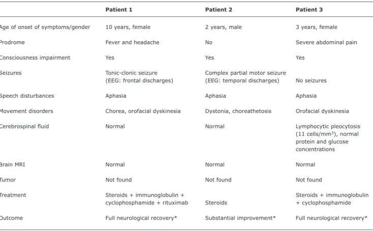
Table 1 - Patients’ characteristics
Patient 1 Patient 2 Patient 3
Age of onset of symptoms/gender 10 years, female 2 years, male 3 years, female
Prodrome Fever and headache No Severe abdominal pain
Consciousness impairment Yes Yes Yes
Seizures Tonic-clonic seizure Complex partial motor seizure
(EEG: frontal discharges) (EEG: temporal discharges) No seizures
Speech disturbances Aphasia Aphasia Aphasia
Movement disorders Chorea, orofacial dyskinesia Dystonia, choreathetosis Orofacial dyskinesia
Cerebrospinal luid Normal Normal Lymphocytic pleocytosis
(11 cells/mm3), normal protein and glucose concentrations
Brain MRI Normal Normal Normal
Tumor Not found Not found Not found
Treatment Steroids + immunoglobulin + Steroids + immunoglobulin
cyclophosphamide + rituximab Steroids + cyclophosphamide
Outcome Full neurological recovery* Substantial improvement* Full neurological recovery*
EEG = electroencephalogram; MRI = magnetic resonance imaging.
* According to Florance et al.,4patients were considered to have “full neurological recovery” if they were able to return to all their activities; “substantial improvement” if they returned to their homes with mild deficits and were improving; and “limited improvement” if they were at home, in the hospital, or in a rehabilitation center with minimal change in the neurological status 3 months after neurological symptom presentation.
Patient 3
A 3-year-old girl with unremarkable previous medical history presented severe abdominal pain followed by mood oscillation, inconsolable crying, agitation, and insomnia alternating with drowsiness 3 days before being admitted into the hospital. The patient did not present seizure,
motor deicits or meningism. She underwent a spinal tap and the CSF showed the following results: 11 cells/
mm3 and normal protein and glucose levels. Fungal and bacterial cultures were negative. Brain MRI was normal. EEG showed slow background activity without epileptiform discharges. After the diagnosis of encephalitis, the patient was empirically treated with acyclovir for 21 days. Polymerase chain reaction for herpes simplex was negative. The patient developed bradycardia, aphasia, and orofacial dyskinesia. Behavior abnormalities became worse and there was impairment in consciousness level. Complementary investigation for rheumatologic disorders and infective diseases were negative. After the exclusion of major causes of encephalitis, NMDAr antibodies were
identiied in serum and CSF, and neoplasm screening did
not detect any tumor. The patient was treated with cycles
of IV methylprednisolone (30 mg/kg/day) for 5 days associated with ciclophosphamide and followed by oral prednisone (2 mg/kg/day). After 5 months of treatment, she has presented normal neurological examination.
Discussion
This paper characterizes the irst description of patients
with a serologically-proven diagnosis of anti-NMDAr encephalitis in Brazil. This entity represents an important differential diagnosis of encephalitis not only in adults, but also among the pediatric population. We observed a wide variety of symptoms among the three cases that we have described, but they share some common traits, and all of them presented normal investigation for the other common causes of encephalitis in childhood.
Based on data published in literature in the last 3 years, it is possible to estimate that anti-NMDAr encephalitis seems to be more common than any other paraneoplastic encephalitis. In the United Kingdom, for instance, a prospective study found a prevalence of 4% of anti-NMDAr encephalitis among all patients with encephalitis.6
278 Jornal de Pediatria - Vol. 88, N° 3, 2012
Correspondence: Felippe Borlot
D. Adma Jafet, 74, Cj. 121
CEP 01308-050 - São Paulo, SP - Brazil Fax: +55 (11) 3259.8574
E-mail: [email protected] References
1. Dalmau J, Tüzün E, Wu HY, Masjuan J, Rossi JE, Voloschin A, et
al. Paraneoplastic anti-N-methyl-D-aspartate receptor encephalitis
associated with ovarian teratoma. Ann Neurol. 2007;61:25-36.
2. Dalmau J, Gleichman AJ, Hughes EG, Rossi JE, Peng X, Lai M, et al. Anti-NMDA-receptor encephalitis: case series and analysis of the effects of antibodies. Lancet Neurol. 2008;7:1091-8. 3. Prüss H, Dalmau J, Harms L, Höltje M, Ahnert-Hilger G, Borowski
K, et al. Retrospective analysis of NMDA receptor antibodies in encephalitis of unknown origin. Neurology. 2010;75:1735-9. 4. Florance NR, Davis RL, Lam C, Szperka C, Zhou L, Ahmad S, et
al. Anti-N-methyl-D-aspartate receptor (NMDAR) encephalitis in
children and adolescents. Ann Neurol. 2009;66:11-8.
5. Wong-Kisiel LC, Ji T, Renaud DL, Kotagal S, Patterson MC, Dalmau J, et al. Response to immunotherapy in a 20-month-old boy with
anti-NMDA receptor encephalitis. Neurology. 2010;74:1550-1.
6. Granerod J, Ambrose HE, Davies NW, Clewley JP, Walsh AL, Morgan D, et al. Causes of encephalitis and differences in their clinical presentations in England: a multicentre, population-based prospective study. Lancet Infect Dis. 2010;10:835-44.
7. Dalmau J, Lancaster E, Martinez-Hernandez E, Rosenfeld MR, Balice-Gordon R. Clinical experience and laboratory investigations in patients with anti-NMDAR encephalitis. Lancet Neurol. 2011;10:63-74.
Florance et al. reported the irst pediatric series of
anti-NMDAr encephalitis cases.4 Thirty-two patients under the age of 18 years were studied. Before this study, only
sporadic cases had been reported. The irst symptoms that these authors identiied were behavioral changes and agitation. Seizures, speech impairment, movement disorders
such as dystonia, orofacial dyskinesia, and chorea are the commonest presentations in young children. Twenty-eight patients presented with mood, behavior, or personality changes. Most patients became increasingly anxious and agitated. Only six patients had early seizures, and a total of 77% presented seizures during the evolution of symptoms. Movement disorders such as orolinguofacial dyskinesia, dystonic posture, increased tone, and choreoathetoid
movements were identiied in 84% of patients. Central
hypoventilation was seen only in seven children.4 Even though autonomic dysfunction and hypoventilation are less frequent in childhood, those potentially fatal complications should be seriously considered as part of clinical presentation
of anti-NMDAr encephalitis. Severe cardiac arrhythmia has
not been described in children.4,7 We observe in the literature of the pediatric population that anti-NMDAr encephalitis has a predominance of the female sex (81%), the age of the onset of symptoms ranges from 23 months to 18 years (median:
14 years), and prodromal symptoms are non-speciic and identiied in less than 50% of the affected children.4
Therefore, the presence of movement disorders following an impairment of consciousness and/or speech disturbances in a child must direct us to the possibility of anti-NMDAr encephalitis.
The importance of acknowledging this clinical setting becomes even more relevant considering that the initial complementary tests may be normal. In adults, MRI is normal in 50% of patients and other 50% may present
mild T2 or luid-attenuated inversion-recovery (FLAIR) signal
hyperintensity in hippocampus, cortex, frontobasal and insular regions, in basal ganglia and in the brainsteam.1 In children, transient FLAIR hyperintensity in one or more
areas (medial temporal lobe, periventricular, cerebellar) may be present in less than one-third of cases.4 Thus, it makes the measurement of antibody titers in serum
and/or CSF by ELISA essential for the diagnosis of
anti-NMDAr encephalitis.
Contrary to the original idea of an obligatory association between this condition and ovarian tumors, a recent review of 400 cases demonstrated that the younger the patient, the less likely a tumor will be detected.4,7 The presence of ovarian teratomas becomes more relevant only after the age of 13 years.4 Nevertheless, screening using computed tomography scan and pelvic ultrasound are still recommended in all patients. Testicular germ-cell tumor, neuroblastoma, and Hodgkin’s lymphoma are other associated tumors. If no tumor is found, the approach of this encephalitis must be only directed to immunotherapy.7
The irst-line immunotherapy treatments are either
IV high-dose corticosteroids, immunoglobulin, or plasma
exchange. In the cases without a proper response to
irst-line therapy, cyclophosphamide or rituximab may be added.7 Recovery time is slow, usually taking months, but a favorable outcome, with full recovery or mild sequelae, is the rule for 75% of patients.2,5 Overall mortality rate is around 4% and is usually associated with secondary comorbidities acquired in the intensive care unit.7
Clinical signs of encephalitis, including seizures, behavior abnormalities, speech disturbances, and movement disorders are the hallmarks of anti-NMDAr encephalitis in childhood. Tumors are not usually detected by this age. The diagnosis of anti-NMDAr encephalitis must be addressed only after the exclusion of infectious and other recognizable causes of encephalitis. Emergency pediatricians, intensive care unit pediatricians and pediatric neurologists should be aware of this treatable autoimmune condition.
Acknowledgements
The authors thank Drs Denise A. De Oliveira, Pollyanna B. Lima, Karin R. Koladicz, and Larissa A. Mehl, who provided clinical assistance for the patients reported,
and Dr Caio Quaio for the technical assistance. Special
acknowledgement to Dr Josep Dalmau, who provided
conirmatory tests in cases 1 and 3.