Bull Parr A/n Health Or&w 14(2). 1980
SEROLOGIC INVESTIGATION OF HERPES SIMPLEX VIRUS: USEFULNESS OF THE DOUBLE IMMUNODIFFUSION (DID) TECHNIQUE IN EPIDEMIOLOGIC SURVEYS’
J&e’ Manuel lkhevarrk, 2 Maria Dolores Bermfidez de Castro,3 Enrique N5jera,4 and Rzifael Nfijeras
Testing of sera from 511 Spanish patients indicates that the double immunodiffusion technique is as sensitive as the neu- tralization test for detecting antibody to herpes simplex virus (HSV). The former method would also seem to have potential for distinguishing between antibodies to HSV types 1 and 2-a
significant distinction because of the possible connection between HSV-2 and cancer of the human cervix.
Introduction
The double immunodiffusion (DID)
technique has been used in recent years to
study the antigenic composition of herpes
simplex viruses (1-4). Nevertheless, little
attention has been paid to the potential use- fulness of this procedure for seroepidemi-
ologic work or diagnosis of recent infec-
tions. Since the DID technique is simple
and yields quick results compared to the
neutralization (N) and complement fixa-
tion (CF) tests, it would be of interest to
know how similar its results are to those of
these other techniques-in order to gauge
its potential usefulness for seroepidemiol-
ogy and diagnosis. The work reported here
had two purposes: to examine the distribu-
tion of neutralizing, precipitating, and
1 Also appearing in Spanish in the R&tin de la Ofictna
Sanitaria Panamericana 88(2), 127.136, 1980. ZAssistant, Respiratory Virus Service; National Cen- ter for Sanitary Microbiology, Virology and Immunol- ogy; Madrid, Spain.
3Chief, Cell Cultures Section: National Center for Sanitary Microbiology, Virology, and Immunology.
%hief, Epidemiology Service: National Center for Sanitary Microbiology, Virology, and Immunology.
SChief, Respiratory Virus Service: National Center for Sanitary Microbiology, Virology, and Immunol- WY.
complement-fixing antibodies to the herpes
simplex viruses in the Spanish population,
and to compare the results obtained by the
DID technique with those obtained by the
N and CF methods.
Materials and Methods
Viruses and Antigens Employed
The antigen used in all the tests was
herpes simplex virus type 1 (HSV-I), strain
HFEM, obtained from an Australian sub-
ject (Hilda F.) in 1937. P. Wildy, who
cloned this strain three times consecutively
in chick embryo chorioallantoic membranes
in 1953, graciously supplied us with this
material.
To reproduce the virus-so as to obtain
antigen lots for DID, N, and CF testing-
the strain was inoculated onto confluent
monolayers of BHK-21* cells grown in
Winchester flasks. After allowing 2 hours
for adsorption (at 37”(Z), enough medium
199 (maintenance medium for tissue cul-
ture) was added to bring the total volume to
200 ml. Later, when a generalized cyto-
pathic effect was observed (normally in 24
*Baby hamster kidney.
Echevuvria et al. l SEROLOGIC INVESTIGATION OF HERPES SIMPLEX VIRUS 117
to 48 hours) the cells were detached from the glass of the flask with a rubber scraper, cen-
trifuged at 700 revolutions per minute
(rpm) for 20 minutes, resuspended in 2 ml
of medium 199, and sonicated for 20 seconds at 60 watts in a B-12 Sonifier (Branson Sonic
Power Co., Danbury, Connecticut). The
extracts of infected cells thus obtained were centrifuged again at 700 rpm for 30 minutes
to eliminate membrane fragments, and the
resulting clear supernatent fluid was used as antigen in the DID and N tests. Antigen for
the CF test was prepared from these cell
extracts by treating them with 5 per cent
chloroform, incubating them for 20 hours
at 4’%, and centrifuging them at 700 rpm
for 15 minutes. Between preparation and
use, all the antigens were stored undiluted
I at -70%.
Cell Cultures
VERO cells, used as indicators of neu-
tralization in the N tests, were grown on
Eagle’s Minimum Essential Medium(MEM)
in Earl’s saline containing 5 per cent calf
serum (Bio-Cult Laboratories, Scotland),
20,000 units/ml of penicillin, 10 mg/ml of
streptomycin, and 0.1 mg/ml of NaOH.
Until used in the N tests, these cultures were
maintained by successive passages-in
which the cells were dispersed by treatment with a 9.25 per cent trypsin solution (Difco,
1:250) and with 0.02 per cent EDTA in
PBS,* until complete dispersal of the mono- layer was achieved.
The BHK-21 cell monolayers used for
production of antigen were grown on the
same medium as described above, except
that bovine fetal serum (Flow Laboratories,
Irvine, Scotland) was substituted for calf
serum.
*Edetate-ethylenediaminotetra-acetate; phosphate buffered saline.
Neutralization (N) Tests
These tests employed the Takatsy micro-
technique (1954) as modified by Sever in
1962 (5), following the guidance provided
by Kalter in 1973 (6). The tests were per-
formed with presterilized cell culture
microtiter plates (see photo) of the “Cook
Microtiter” type (C.A. Greiner, West Ger-
many). The test sera were titered without
prior inactivation.
Complement Fixation (CF) Tests
These tests utilized the 1974 Grist tech- nique (7), a modification of the procedure
described by Bradstreet and Taylor in 1962
(8), and employed U-plates (model IS-MAC-
96) manufactured by the Linbro Scientific
Co. in Hamden, Connecticut.
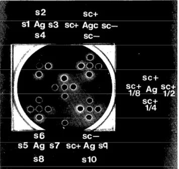
Double Immunodiffsion (DID) Tests
Glass 9 x 8 cm plates and plastic 7 x 2 cm
slides (Div. Travenol Laboratories, Cali-
fornia) were used in these tests. The gel
employed was made up with a 1 per cent
suspension of Ionagar (Difco) in distilled
water’containing 0.9 per cent NaCl and
0.1 per cent sodium azide. The thickness of the gel on the plates was 2 mm.
The sera were put in wells 4 mm in dia-
meter that were regularly arranged, in
groups of four or six, around a central well
the same size-where the antigen was
placed. The distance between the edge of
the central well and the nearest edge of
each peripheral well was 2 mm.
The wells were inoculated with .03 ml of
serum or antigen, and the antigen was
allowed to diffuse a little, after which .025 ml of a 2 per cent sodium lauryl sarcosinate
solution in distilled water was added to
each antigen well. The plates were then
left in a humid chamber at room tempera-
ture for 48 hours, after which they were
washed by immersion in 0.85 per cent
118 PAHO BULLETIN . vol. 14, no. 2, 1980
Neutralization testing of sera with HSV-1 antigen on microtiter plates.
stained by immersion for 5 minutes in a
solution of 1 per cent amido Schwartz stain
(prepared with 6 per cent glacial acetic
acid and 1.36 per cent sodium acetate) in
distilled water. Excess stain was removed by washing the plates in a solution of 5 per cent glacial acetic acid and distilled water for 24 hours. As shown in the photograph,
control samples of antigen (Age), positive
sera (SC+), and negative sera (SC-) were in-
cluded in each test. The control antigen
was prepared with BHK-21 cells from un-
infected cultures, using the same method
employed to prepare the extracts of infected cells that were used as antigen.
Test Sera
A total of 511 sera from healthy individ-
uals or patients with nonherpetic condi-
tions were used in the survey. The sera,
chosen from the clinical samples received
by our respiratory virus service during
1974-1976, came from various provinces of
Spain, They were stored at -20°C from the time of receipt until use and were divided into 12 groups according to their respective subjects’ age.
Results
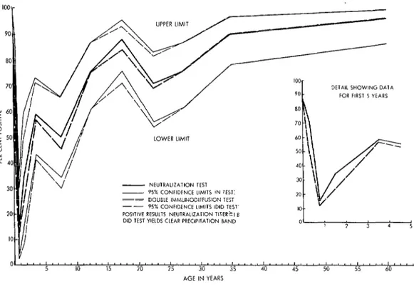
The results of the N and DID tests are
shown in Table 1 and Figure 1. As the fig- ure indicates, after showing a rapid decline during the first 7 to 8 months of life, the
percentage of positive sera6 rose sharply
‘Sera yielding a clean precipitation band in the DID test, aneutralization titer21:8 in the N test, or a titer ~1:8 in the CF test were considered positive by each of
Echevarria et al. l SEROLOGIC INVESTIGATION OF HERPES SIMPLEX VIRUS 119
Double immunodiffusion testing of sera with HSV-1 antigen on glass 9 x 8 cm plates.
SC + = positive control serum SC- = negative control serum Ag = antigen
Age = control antigen
Table 1. Percentages of sera yielding positive N and DID test results, by age group.
Age group
No. of
El-a
tested
Positive sera (N test)
No. %
Positive sera (DID test)
NO. %
95 per cent 95 per cent confidence limits confidence limits
for N test for DID test
results results
O-1 month 49 42 85.7 40 81.6
1-6 months 31 22 71.0 15 48.4
6-12 months 26 4 15.4 3 11.5
l-2 years 20 7 35.0 5 25.0
2-5 years 46 27 58.7 26 56.5
5-10 years 42 21 50.0 19 45.2
lo-15 years 49 37 75.5 37 75.5
15-20 years 50 44 88.0 42 84.0
20-25 years 48 34 70.8 33 68.8
25-30 years 50 38 76.0 38 76.0
30-40 years 50 45 90.0 45 90.0
40-80 years 50 48 96.0 48 96.0
O-80 years 511 369 72.2 351 68.7
72.8-94.1 70.0-91.2
52.0-85.8 31.1-67.0
4.3-34.9 2.4-30.1
15.3-59.2 8.6-49.1
43.2-73.0 41.1-71.1 34.2-65.8 29.8-65.3
61.1-86.6 61.1-86.6
75.7-95.5 70.9-92.8
55.9-83.0 53.7-81.3
120 PAHO BULLETIN l vol. 14, no. 2, 1980
UPPER LIMIT
DETAIL SHOWING DATA FOR FIRST 5 YEARS
LOWER LIMIT
- NEUTRAUZATION TEST
- 95% CONFIDENCE LIMITS IN TEST1
-- DOUBLE IMMUNODFFUSION TEST
-- 95% CONFIDENCE LlMlT5 (DID TEST
POSITlYE RESULTS NEUTRALIZATION TITER?1 8 DID TEST YlEtDS CLEAR PRECIPITATION BAND
Ol.~~““““.“~““~..“~....~“.~~....~....~.~..~...~~....~~...,
5 IO 15 20 25 30 35 40 45 50 55 60
AGE IN YEARS
Figure 1. Percentages of sera from different age groups yielding positive N and DID test results. The thin lines show 95 per cent confidence limits for these data.
until the subjects reached 2-5 years of age. Then, from 5-10 years onward it continued rising, but more slowly, until the subjects
were 15-20 years of age. Finally, beyond
the 20-25 year group there was a very slow rise that ended, in the oldest age group, with 96 per cent of the subjects yielding seropositive results.
Of the sera giving positive DID results, 200 (57 per cent) showed a single precipita- tion band and 151 (43 per cent) showed two bands. Of the sera yielding a double band,
only 23 (15 per cent) were obtained from
subjects between 6 months and 15 years of
age. These results are shown in Table 2
and Figure 2.
The CF test results are shown in Table 3
and Figure 3. The percentage of positive
sera was found to decline rapidly at first, falling to 8 per cent in the 6-12 month age group. After that it rose rapidly (exceeding 40 per cent in the 2-5 year age group), re-
mained at about the same level in the 5-16
and lo-15 year age groups, dropped slowly
to 24 per cent in the 20-25 year group, and then rose slowly again, reaching a plateau
around 51 per cent among subjects above
30 years of age. This latter percentage is
roughly comparable to the 62 per cent
positivity found in the sera from subjects less than 1 month old.
Discussion
The curves obtained with the N and
DID techniques are clearly similar and,
indeed, virtually identical-a fact con-
firmed by estimating the 95 per cent con-
fidence limits of these results. None of the differences in the results of the two tests- with respect to each of the age groups in-
volved-were found to be statistically sig-
Echevarria et al. l SEROLOGIC INVESTIGATION OF HERPES SIMPLEX VIRUS 121
Table i. Percentages of sera yielding two precipitation bands upon DID testing, as compared to sem yielding positive DID test results, by age group.
Age group
No. of sera tested
Sera yielding positive DID test results
NO. %
95 per cent confidence limits
for DID test results
Sera showing 2 precipitation
bands
No. %
95 per cent confidence limits
for sera showing 2 bands
O-l month 1-6 months 6-12 months l-2 years 2-5 years 5-10 years IO-15 years 15-20 years 20-25 years 25-30 years 30-40 years 40-80 years O-80 years
49 40 81.6 70.0-91.2
31 15 48.4 30.1-67.0
26 3 11.5 2.4-30.1
20 5 25.0 8.6-49.1
46 26 56.5 41.1-71.1
42 19 45.2 29.8-65.3
49 37 75.5 61.1-86.6
50 42 84.0 70.9-92.8
48 33 68.8 53.7-81.3
50 38 76.0 61.8-86.9
50 45 90.0 78.2-96.7
50 48 96.0 86.3-99.5
511 351 68.7
16 32.6
4 12.9
1 3.8
1 5.0
5 10.9
9 21.4
7 14.3
21 42.0
18 37.5
20 40.0
29 58.0
20 40.0
151 29.5
19.9-47.5 3.6-29.8 0.1-19.6 0.1-24.9 2.4-20.8 10.3-36.8 5.9-27.2 28.2-56.8 24.0-52.6 26.4-54.8 43.2-71.8 26.4-54.8
0 ,
5 IO 15 20 25 30 35 40 45 50 55 60
AGE IN YEARS
Figux 2. Percentages of sera fmm different age groups yielding positive results and two precipitation bands on DID testing. The thin lines show 95 per cent
122 PAHO BULLETIN l vol. 14, HO. 2, I980
Table 3. Percentages of sera yielding positive CF test results (titers 2 1:8), by age group.
No. of sera tested
Positive sera (titers t 1:s)
No. %
95 per cent confidence limits
of CF test red ts
O-1 month 48 30 62.5 47.3-76.0
1-6 months 31 6 19.3 7.4-37.5
6-12 months 24 2 8.3 1.0-27.0
1-2 years 19 3 15.8 3.4-39.5
2-5 years 46 19 41.3 27.0-56.7
5-10 years 42 18 42.9 27.7-59.0
IO-15 years 48 20 41.7 27.6-56.8
15-20 years 48 18 37.5 24.0-52.6
20-25 years 46 11 24.0 12.6-38.8
25-30 years 47 15 31.9 19.0-47.1
30-40 years 43 22 51.2 35.5-66.7
40-80 years 45 23 51.1 35.8-66.3
O-80 years 487: 187 38.4
*Twenty-four sera (out of 511 samples) were not tested by CF (4.6 per cent of the total) because of lack of sera in these cases.
AGE IN YEARS
Echevarria et al. . SEROLOGIC INVESTIGATION OF HERPES SIMPLEX VIRUS 123
curred in sera from subjects 1-6 months of
age-at a time when antibodies acquired
from the mother were disappearing and
when the highest percentage of low neu-
tralizing titers (less than or equal to 1:s) were detected.
In view of the fact that the DID test was capable of detecting the sera yielding neu- tralization titers 1 1:8, these results clearly indicate very similar levels of sensitivity. In addition, the DID test employs far sim-
pler procedures than the N test, and un-
like the latter requires no sterile materials or maintenance of cell cultures. Therefore,
it is felt that the DID technique could
prove useful in seroepidemiologic studies of
herpes simplex virus and also in laboratory
diagnosis of recent herpes simplex infec-
tions.
As Figure 2 indicates, there is some evi- dent difference between the overall percent-
ages of sera yielding positive DID results
and the percentages yielding two precipita-
tion bands. The chart also shows that in
terms of subject age groups there may have been some time-lag involved in development
of seropositivity with two precipitation
bands as compared with one. Because of the
small numbers of sera tested in each age
group, the confidence limits of the two
curves were too broad to confirm existence of this time-lag. If the apparent lag is real, however, it could be accounted for in either of the following ways:
l The second band could arise from a
component of the same virus that is less anti- genie or more difficult to detect than that responsible for the first band.
l The second band could be specific for a
particular type of herpes simplex virus.
Since the curve in Figure 3 showing the per-
centage of double-band sera in each age
8-roup appears to lag somewhat behind that
showing the overall percentage of positive
sera in each group, it appears plausible that the second band could be specific for type 2 herpesvirus (HSV-Z), i.e., that it could have
arisen from a HSV-2 component cross-react-
ing with our HSV-1 antigen because this
antigen is not type-specific.
One particular precipitation band was
observed in all the sera that yielded positive DID results. In 1974 Skinner et al. (I) de-
iscribed one precipitation band, called
“Band II,” as being common to all human
sera reacting positively when tested by DID against preparations of either type of herpes
simplex virus. To find out whether our
common band corresponded to this one, 50
of the positive sera were tested in parallel
with “Band II” antiserum graciously sup-
plied by Dr. Skinner. In all cases the bands
produced by the antiserum and by the test
sera were found to be the same. Hence it
could be concluded that this common band
represents “anti-Band II” antibodies.
The CF test results clearly reinforce those of the N and DID tests. All the results suggest
that herpes simplex primoinfection occurs
most commonly between the first and fifth
years of life, since all three tests showed a
rapid rise during these years in levels of
seropositivity. Among older age groups the
level of CF positivity declined slightly, but the level rose again among subjects over 25
years of age and then remained stable at
about 50 per cent for subjects over age 30. This behavior of the CF curve is highly con- sistent with the fact that the N and DID
curves never clearly leveled off but con-
tinued to rise, even though the rise was rela- tively slight after 5-10 years of age.
Either of two hypotheses could account
for the second rise of the CF curve:
1) It may be that herpes simplex primo- infection is most common in the first five years of life, after which it falls off, and that its effects are reinforced among older
age groups by recurrences, which in some
cases prompt the reappearance of comple-
ment-fixing antibodies.
2) Backward (and downward) projection
of the second rise in the CF curve intersects the horizontal axis at a place corresponding to lo-12 years of age. This could indicate
124 PAHO BULLETIN l vol. 14, no. 2, 1980
begins to be detected, so that in effect the curve obtained is the sum of two curves- each indicating antibodies to one of the two types of virus. That is, during the first 10 or 12 years of life we would see a curve re-
sulting exclusively from circulation of
HSV-1, and after that we would see a curve
resulting from circulation of both HSV-1
and HSV-2. This hypothesis appears con-
sistent with the available data on surveys
carried out in healthy populations using
the kinetic neutralization test (9, 10). The
results of these surveys indicate that specific
HSV-2 neutralizing antibodies begin to
appear around 13 years of age and reach
positivity levels of 20 per cent in groups of subjects above 20-25 years of age.
Differentiation of HSV-1 and HSV-2
antibodies in human sera is a subject that
has aroused great worldwide interest be-
cause of the possible connection between
HSV-2 and cancer of the human cervix.
Considerable emphasis is therefore being
placed on the need for a quick and simple
technique capable of distinguishing the two
types of HSV.
The results presented here clearly show that the DID technique is fully applicable
to detection of antibodies against HSV in
human sera at a level of sensitivity compa-
rable to that attained by the neutralization test. Moreover, at this point it appears that a
DID test improved in terms of sensitivity could help to identify some of the polypep-
tides described as carriers of total or partial
type-specificity-such as VP19e (11) or VP7
and VP8. This would make it possible to use
the DID technique for detection of type-
specific antibodies, thereby taking advan-
tage of that technique’s simplicity and
speed.
ACKNOWLEDGMENTS
We are grateful to F. de Ory Manchbn, P. laboration on the serologic tests, and to A.
Anda Ferngndez, M. P. Marcos Moreno; Llacer Gil de Ramales for assistance with
and M. D. Garcia Rodriguez for their col- statistical analysis of the results.
The work reported here was designed to test the potential usefulness of the double immuno- diffusion technique for seroepidemiologic and diagnostic work on herpes simplex virus (HSV).
A total of 511 sera from subjects attending Spain’s Respiratory Virus Service in 1974-1976 were tested for HSV antibodies by double immu- nodiffusion (DID), neutralization (N), and com- plement fixation (CF). The DID test results closely paralleled those of the N test, both of these detecting considerably more positive sera than the less sensitive CF test.
In addition, some of the positive sera tested by DID yielded two precipitation bands. While the
second band could have arisen merely from a viral component that was less antigenic or harder to detect, it could also have been produced by antibody to a particular type of HSV. Specifical- ly, it seems possible that the second band could have been caused by antibody to a HSV type 2 component cross-reacting with the HSV type 1 test antigen employed.
Echevarria et al. l SEROLOGIC INVESTIGATION OF HERPES SIMPLEX VIRUS 125
REFERENCES
(I) Skinner, R.B.G., J. Taylor, and D. J. Edwards. Precipitating antibodies to herpes sim- plex virus in human sera: Prevalence of antibody to common antigen (Band II). Intervirology 4:
320-324, 1974.
(2) Watson, D. H., W.I.H. Shedden, A. Elliot, T. Tetsuka, P. Wildy, D. Bourgaux-Ramoisy, and E. Gold. Virus specific antigens in mamma- lian cells infected with herpes simplex virus. Zm- munology 11:399-408, 1966.
(3) Watson, D. H., and P. Wildy. The prep- arations of “monoprecipitin” antisera to herpes virus specific antigens. J Gen Vi701 4:163-168, 1969.
(4) Watson, D. H. The separation of herpes virus-specific antigens by polyacrylamide gel e1ectrophoresis.J Gen Viral 4:151-161, 1969.
(5) Sever, J. L. Application of a microtechnique to viral serological investigations. J Zmmunol 88: 320-329, 1962.
(6) Kalter, S. S. Microculture procedures. In Kruse, P. F., Jr., and M. K. Patterson, Jr. (eds.),
Tissue Culture Methods and Applications. New
York, Academic Press, 1973.
(7) Grist, N. R., C. A. Ross, and E. J. Bell.
Diagnostic Methods in Clinical Virology (2nd ed.). Oxford, Blackwell Scientific Publications, 1974.
(8) Bradstreet, C. M., and C. E. Taylor. Technique of complement-fixation test applica- ble to the diagnosis of virus diseases. Month Bull Min Health (London) 21:96-104, 1962.
(9) Nahmias, A. J., W. E. Josey, Z. M. Naib, C. F. Lute, and A. Duffey. Antibodies to Herpes- tirus hominis types 1 and 2 in humans: I Patients with genital herpetic infections. Am J Epidemiol
91:539-546, 1970.
(10) Rawls, W. E., W.A.F. Tompkins, and J. L. Melnick. The association of herpesvirus type 2 and carcinoma of the uterine cervix. Am J Epidemiol 89:547-554, 1969.
(II) Honess, R. W., and D. H. Watson. Herpes simplex virus-specific polypeptides studied by polyacrylamide gel electrophoresis of immune precipitates.j Gen Viral 22:171-185, 1974.
BIOLOGICAL CONTROL OF DISEASE VECTORS*
The World Health Organization is encouraging and coordinating interna- tional research on the biological control of vectors as part of a special pro- gram of research and training in tropical diseases. The program is the out- come of a collective effort by many countries and international agencies to make better use of existing control methods, to train personnel, and to develop research on these diseases.
Specialized scientific working groups deal with the development of new control methods for each group of tropical diseases, in particular leprosy, malaria, filariasis, schistosomiasis, trypanosomiasis, and leishmaniasis, or are responsible for activities, such as the biological control of vectors that cover all or most of these diseases.
The objectives of the Working Group on the Biological Control of Vectors are to identify, evaluate, and develop biological control agents for the safe and effective control of invertebrate vectors and intermediate hosts of human diseases, with special emphasis on bacteria, fungi, protozoa, and nematodes.
The Steering Committee of the Working Group has drawn up a plan of ac- tion for 1980 and 1981 in the light of the latest developments in biological control and the expected progress in research in this field.