Key words:
Cryptorchidism; anatomy and histology; Epididymis; abnormalities
Int Braz J Urol. 2013; 39: 240-7
__________________ Submitted for publication: October 03, 2012
__________________ Accepted after revision: January 10, 2013
Objectives: Report the incidence and structure of testicular appendices (TAs) in patients with cryptorchidism, comparing their incidence with epididymal anomalies (EA) and pa-tency of the vaginal process (PVP) and analyzes the structure of TAs.
Material and Methods: We studied 72 testes of patients with cryptorchidism (average of 6 years), and 8 testes from patients with hydroceles (average of 9 years). We analyzed the relations among the testis, epididymis and PVP and prevalence and histology of the TAs. The appendices of 10 patients with cryptorchidism and 8 with hydrocele were dissected and embedded in paraffin and stained with Masson trichrome; Weigert and Picro-Sirius Red with polarization and immunohistochemistry analysis of the collagen type III fibers to observe collagen. The stereological analysis was done with the software Image Pro and Image J, using a grid to determine volumetric densities (Vv). Means were statistically compared using the ANOVA and unpaired T test (p < 0.05).
Results: Of the 72 testes with cryptorchidism, 20 (27.77%) presented EA, 41 (56.9%) had PVP and 44 (61.1%) had TAs. Of the 44 testes with cryptorchidism and appendices, 30 (68.18%) presented PVP and 11 (25%) presented EA. There was no alteration of the epithelium in the appendices of patients in both groups. Stereological analysis documen-ted the prevalence of ESFs (mean of 1.48%), prevalence of veins (mean of 10.11%) and decrease (p = 0.14) of SMCs in the TAs of patients with cryptorchidism (mean = 4.93%). Collagen III prevailed in the TAs of patients with cryptorchidism.
Conclusion: The testicular appendices presented significant structural alteration in the patients with cryptorchidism, indicating that TAs present a structural remodeling.
INTRODUCTION
Testicular and epididymal appendices have been considered congenital anomalies (1). The appendix testis or hydatid of Morgagni is believed to be the embryologic remnant of the cranial end of the Mullerian or paramesonephric duct (2). It is present in more than 90% of males, it varies in size from 1-10 mm in diameter and it is usually pedunculated, which predisposes to torsion. It is
the most frequently twisted of the four testicular appendages (3-5). The other are the appendix of the epididymis, which is a remnant of the wolffian duct, the paradidymis and the vas aberrant. These vestigial structures have a similar histology, com-posed of gelatinous and vascular connective tissue covered with a columnar epithelium (6).
The functions of testicular appendices are controversial, but they may control the amount of serous fluid within the space of the vaginal
Structural analysis of testicular appendices in patients
with cryptorchidism
_______________________________________________
Guilherme D. Tostes, Suelen F. Costa, João P. de Carvalho, Waldemar S. Costa, Francisco J.B.
Sampaio, Luciano A. Favorito
Urogenital Research Unit, Rio de Janeiro State University, Rio de Janeiro, Brazil
ABSTRACT
ARTICLE
INFO
process (7). Another hypothesis suggests that the surface epithelium, subepithelial capillaries and lymphatic vessels of testicular appendices form a functional unit (8). Some studies have analyzed the embryology (9) and structure of the testicular and epididymal appendices (3,10).
Studies of the incidence of these structures in patients with cryptorchidism and comparison with individuals without testicular position ano-malies are rare in the literature (11). There are no published studies analyzing the structure of testi-cular appendices in patients with cryptorchidism.
This work reports the incidence and struc-ture of testicular appendices (TAs) in patients with cryptorchidism, comparing their incidence with epididymal anomalies and patency of the vaginal process and analyzes the structure of testicular appendices to assess whether their architecture is altered when cryptorchidism is present.
MATERIALS AND METHODS
The present work received institutional re-view committee and parent approval and has been carried out in accordance to the ethical standards of the responsible institutional committee on hu-man experimentation.
We analyzed 72 testes from 55 patients with cryptorchidism (17 with bilateral cryptorchi-dism) and compared them with a control group of 8 testes from patients with hydroceles. The patients with cryptorchidism had ages from 1 to 13 years (average of 6 years), while those in the control group ranged from 1 to 14 years old (ave-rage of 9 years). In the two groups, we analyzed the relations among the testis, epididymis and pa-tency of the vaginal process and the incidence and histology of the testicular appendices.
To analyze the relations between the testis and epididymis during surgery, we used a previous classification (12,13). By this classification, there are six types of relations between the testis and epididymis: Type I - epididymis attached to the testis at the head and tail; Type II - epididymis totally attached to the testis; Type III - epididymis attached to the testis only at the head; Type IV - epididymis attached to the testis only at the tail; Type V - no visible connection between the testis
and epididymis; and Type VI - epididymal atresia. Type I and II relations are considered normal whi-le the other types are considered to be anatomical anomalies (12). To analyze the vaginal process we considered two situations: (a) complete oblitera-tion of the vaginal process between the internal inguinal ring and the superior pole of the testis; and (b) complete patency of the vaginal process.
In relation to the testicular appendices, we analyzed the following situations in the two groups during the surgeries: I) absence of testi-cular and epididymal appendices; II) presence of testicular appendix only; III) presence of epidi-dymal appendix only; IV) presence of testicu-lar and epididymal appendices; V) presence of two epididymal appendices and one testicular appendix; and VI) presence of paradidymis or vas aberrans of Haller.
The appendices of 10 patients with cryp-torchidism and 8 patients with hydrocele were separated from the other structures and fixed in 10% buffered formalin, and routinely processed for paraffin embedding. Then 5 µm thick sections were obtained at 200 µm- intervals. Smooth mus-cle cells, connective tissue and elastic system fi-bers were studied by histochemical and immuno-labeling methods.
fibers (EF), Monoclonal Elastin Antibody ab 9519 (Abcam Laboratories, Cambridge, MA, United Sta-tes) was used.
Means were statistically compared using the unpaired t-test and linear regression was per-formed when applicable. Statistical relevance was accepted if p < 0.05.
RESULTS
Of the 72 testes with cryptorchidism, 7 were abdominal (9.7%), 54 (75%) were located in the inguinal canal and 11 (15.3%) were supra-scro-tal. Of the testis with cryptorchidism, 20 (27.77%) presented epididymal anomalies, 41 (56.9%) had patent vaginal process and 44 (61.1%) had testi-cular appendices. The prevalence of the testitesti-cular appendices in the testis with cryptorchidism and their relation with the testicular position are re-ported in Table-1. There was no statistically signi-ficant difference in relation to the location of the appendices (p = 0.0652).
Of the 7 abdominal testes, all had patent vaginal process and 4 (57%) had testicular appen-dices. Of the 54 testes located in the canal, 31 (57%) had appendices, and of these, 21 (38.8%) presented patent vaginal process and 19 (35.8%) had epididymal anomalies. Of the 11 cases of supra-scrotal testes, 9 (82%) had appendices, 5 (55.5%) had patency of the vaginal process and 1 (11.1%) presented epididymal anomaly. Finally, of the 8 cases in the control group, 7 had appendices
(87.5%), 1 (14.28%) had epididymal anomaly and 1 (14.28%) had patent vaginal process.
Of the 44 testes with cryptorchidism and appendices, 30 (68.18%) presented patent vaginal process and 11 (25%) showed epididymal ano-malies. Of the 28 testes with cryptorchidism and without appendices, 11 (39.28%) presented patent vaginal process and 9 (32.14%) showed epididymal anomalies. The relation between the type of testicu-lar appendix found and the patency of the vaginal process and the presence of epididymal anoma-lies is shown in Table-2. There was no statistically significant difference in relation to the patency of vaginal process between the patients with cryptor-chidism and appendices with the control group (p = 0.053) and with the patients with cryptorchidism without appendices (p = 0.252). Also there was no statistically significant difference in relation to the epididimal anomalies between the patients with cryptorchidism and appendices with the control group (p = 0.305) and with the patients with cryp-torchidism without appendices (p = 0.6830). Of the 55 patients, 17 had bilateral cryptorchidism, for a total of 34 testes. Of these, 1 was abdominal, 24 were located in the canal and 9 were supra-scrotal. Of the 34 testes of this group, 25 (73.5%) had testi-cular appendices, with 13 being right testes and 12 left ones, with no statistical difference.
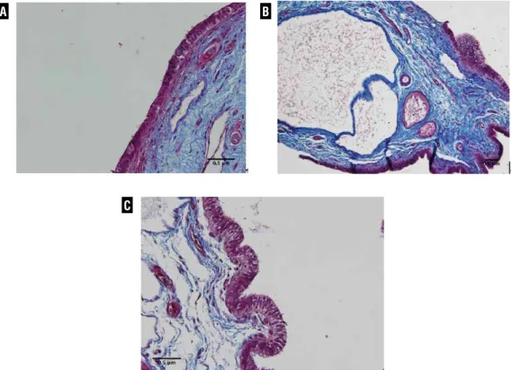
Sections stained with Masson’s trichrome from the testicular appendix demonstrated a vascu-lar stroma lined with pseudocolumnar epithelium. The stroma consisted of loose connective tissue
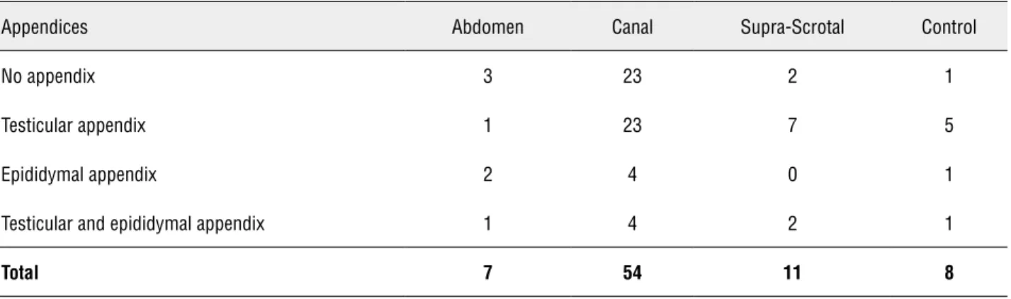
Table 1 – Incidence of the testicular and epididymal appendices studied with respect to position of the testes with cryptor-chidism and the testes in the control group. There was no statistically significant difference in relation to the location of the appendices (p = 0.0652).
Appendices Abdomen Canal Supra-Scrotal Control
No appendix 3 23 2 1
Testicular appendix 1 23 7 5
Epididymal appendix 2 4 0 1
Testicular and epididymal appendix 1 4 2 1
containing blood vessels, fibroblasts and varying numbers of acini (gland-like structures), which were lined with a non-ciliated columnar epithe-lium (Figure-1A). The epididymal appendices were vesicular structures. The cavity of the vesicle was
lined with a pseudostratified columnar epithelium (Figure-1B). There was no apparent alteration be-tween the epithelium in the appendices of patients with cryptorchidism and in the control group, as seen in Figures 1A and 1C.
Table 2 – Relation between the type of appendix found and the patency of the vaginal process and presence of epididymal anomaly. There was no statistically significant difference in relation to the patency of vaginal process (p = 0.252) and epidi-dymal anomalies (p = 0.6830) between the patients with cryptorchidism and appendices with the patients with cryptorchidism without appendices.
Appendices Patent vaginal process Epididymal anomaly
Testicular (31) 20 5
Epididymal (6) 5 4
Testicular and Epididymal (7) 5 2
Total (44-100%) 30 (68.18%) 11(25%)
Figure 1 - Appendix epithelium. A) Patient with cryptorchidism with 3-years-old, showing the vascular stroma lined with pseudos-tratified epithelium of the testicular appendix. Masson’s trichrome x200. B) Patient of the control group with 13-year-old, showing epididymal appendix with cavitation covered by pseudostratified epithelium. Masson’s trichrome x 200. C) Patient of the control group with 13-years-old, showing pseudostratified epithelium of the entire testicular appendix. Masson’s trichrome x200.
A
C
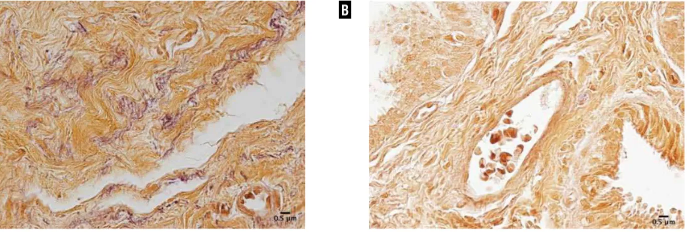
Stereological analysis documented a pre-valence of elastic fibers in the testicular appen-dices of the patients with cryptorchidism (mean of 1.48%) in comparison to the appendices of the patients with a hydrocele testis (mean of 0.29%), a result that was not statistically different (p = 0.22). Figure-2 shows the testicular appendices in patients with cryptorchidism and in the control group stained by Weigert’s resorcin-fucsin with oxidation. The testes of the patients with cryptor-chidism contain a larger quantity of elastic fibers. Stereological analysis also showed a grea-ter incidence of vessels in the testicular
appendi-ces of the patients with cryptorchidism (mean of 10.11%, standard deviation of 6.88) in relation to the control group (mean of 4.77%, standard devia-tion of 1.9). This difference was statistically sig-nificant (p < 0.001). Figure-3 shows the testicular appendices in patients with cryptorchidism and those of the control group, stained by Masson’s trichrome. A greater number of vessels can be ob-served in the testes of patients with cryptorchi-dism.
Stereology analysis showed a decrease wi-thout significance (p = 0.14) of smooth muscle cells in the appendices of patients with
cryptor-Figure 2 - Elastic system fibers of the testicular appendix. A) Testicular appendix of 13-year-old patient with cryptorchidism. Weigert x400. B) Testicular appendix of 13-year-old patient of the control group. Weigert x400. Note the marked increase of elastic fibers (brown) in the patient with cryptorchidism when compared to the control.
Figure 3 - Testicular appendix vessels. A) Testicular appendix of 2 years-old patient with cryptorchidism. Masson’s trichrome X200. B) Testicular appendix of 1 year-old patient in the control group. Masson’s trichrome X200. Note the marked increase of vessels in the appendix of the cryptorchidic patient.
A
A
B
chidism (mean = 4.93%) in comparison with the control group (mean = 9.032%). Figure-4 contains photomicrographs comparing the smooth muscle arrangement in the cryptorchidism and the con-trol group appendices.
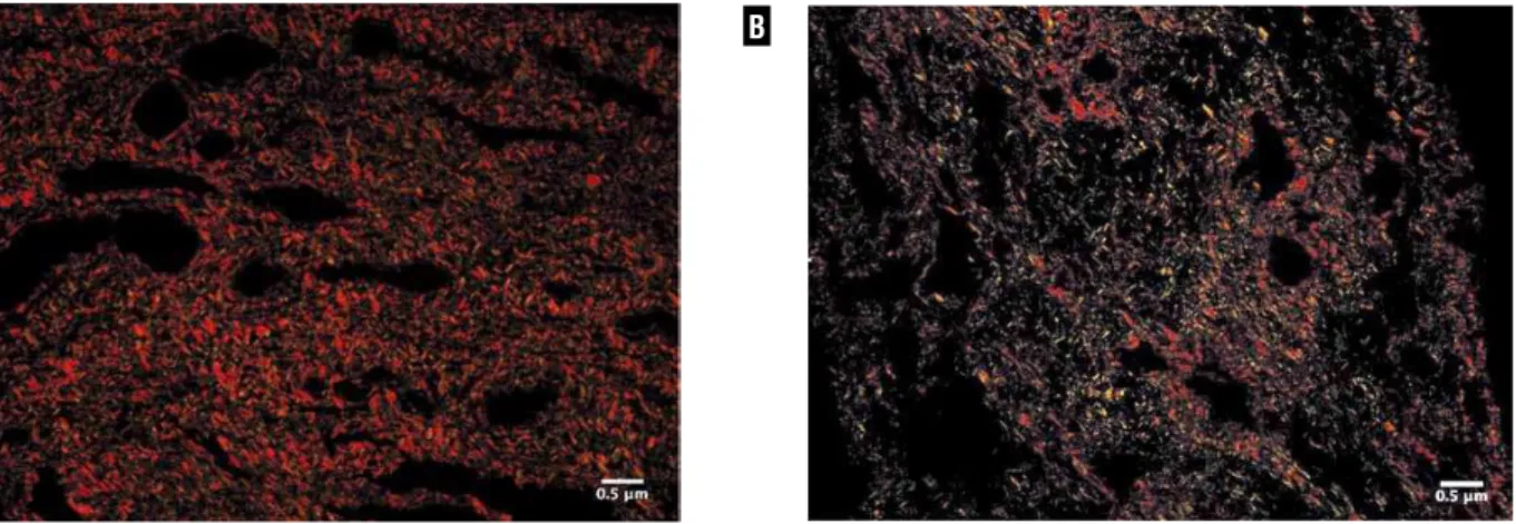
In the qualitative analysis, type III colla-gen was observed in both groups, although type III collagen was prevalent in the testicular appen-dices of patients with cryptorchidism and type I was prevalent in the patients of the control group. Figure-5 shows examples of the two groups
stai-ned with Picro Sirius Red with polarization. The photomicrographs presented a large difference in colors between the groups. This difference can su-ggest changes in the collagen fiber organization.
DISCUSSION
Knowledge of the presence, form and loca-tion of testicular and epididymal appendices is im-portant in clinical practice because of the possibility of torsion of these structures, their association with
Figure 4 – Distribution of smooth muscle cells of the testicular appendix. A) Testicular appendix of 3-year-old patient with cryp-torchidism. Imunohistochemical staining for alfha-actin X200. B) Testicular appendix of 11-year-old patient of the control group. Imunohistochemical staining for alfha-actin X200. Note the marked increase of SMCs in the appendix of the control patient when compared to the cryptorchidic patient.
Figure 5 – Qualitative collagen distribution in testicular appendices. A) Testicular appendix of 3-year-old patient with cryptorchidism showing prevalence of yellowish color. Picrosirius-polarization method, X400. B) Testicular appendix of 11-years- old patient of the control group showing prevalence of greenish color. Picrosirius- polarization method, X400.
A
A
B
anatomical anomalies and the higher incidence of tumors (3,15). Cryptorchidism is one of the most common congenital anomalies among males, with a rate between 2 and 5% of full-term births, a rate that can reach 30% in premature babies (16-18). There are few studies in the literature on the in-cidence and analysis of the structure of testicular appendices in patients with cryptorchidism and their relation with epididymal anomalies and pa-tency of the vaginal process (11).
Previous studies have shown that testicular appendices occurs significantly less often in pa-tients with cryptorchidism. This can indicate a pos-sible role of testicular appendices in the testicular migration process (19). In the present study, 61.1% of the testes with cryptorchidism presented testicu-lar appendices, a much higher rate than the 24% reported by Jozsa (19). We did not find a significant difference in the number of appendices in the tes-tis with cryptorchidism in relation to those of the control group, and also did not find a significant difference in the incidence of appendices in relation to the testicular position in the patients with cryp-torchidism.
Cryptorchidism can be associated with va-rious anatomical anomalies, but epididymal anoma-lies and patency of the vaginal process are among the most frequent (20-22). Epididymal anomalies are associated with cryptorchidism, with prevalence that ranges from 36 to 79% (20,21). In this study we found 27.7% of the testes with cryptorchidism also had epididymal anomalies and 56.9% had patency of the vaginal process. Of the 44 testes with appen-dices, 68.18% had patency of the vaginal process and 25% presented epididymal anomalies. There was no significant difference between the incidence of epididymal anomalies or patency of the vaginal process in the patients with cryptorchidism with and without appendices.
Previous studies have shown that the tes-ticular appendices of patients with cryptorchidism show significant alterations in the estrogen and an-drogen receptors (19,23). Patients with hydroceles also present alteration in the expression of andro-gen receptors and epithelial destruction in the testi-cular appendices (19). These alterations are argued to be associated with increased hydrostatic pressure in the scrotum of patients with hydroceles (19,23).
Reports of structural and ultrastructural changes in the testicular appendices of patients with cryptorchidism are rare (10,19). In this study we did not observe any alterations in the epithe-lium of the testicular and epididymal appendices between the patients with cryptorchidism and tho-se in the control group, where the majority of ca-ses were made up of patients with hydroceles. We found a large quantity of veins and connective tissue in both the testicular and epididymal appen-dices. These findings are in agreement with those of Sahni et al. (10) and can confirm the theory that the epithelial layer of the appendices along with the subepithelial capillaries and lymphatic vessels form a functional unit (8,19).
In our samples, we observed the presence of elastic fibers in the appendices, more so in those of patients with cryptorchidism. The increased synthe-sis of elastic fibers may be associated with excessive distension of an organ (24). We also observed a de-crease in the quantity of smooth muscle fibers and predominance of type III collagen in the patients with cryptorchidism. We can speculate that the smooth muscle fiber reduction is the primary event in fibrotic tissue formation. This event is probably correlated to a hydrodistension process. It has been variously shown that a shift towards greenish color in the Picrosirius polarization method is associated with less organized and/or degraded collagen (25). This color change can also occur in earlier phases of the remodeling and repair of connective tissues, when the synthesis of type III collagen is enhanced. Thus, based on these findings, the results of the Pi-crosirius-polarization method suggest that collagen matrix at the testicular appendices in patients with cryptorchidism is disrupted or degraded, rather than fibrotic, which is consistent with higher hydrostatic pressure. These findings lead us to suggest that in testes without complete migration, testicular appen-dices may result from the higher intra- abdominal pressure to which these testes are exposed.
pa-tients with cryptorchidism: although the epithelium was not changed, the testicular appendices of the patients with cryptorchidism had a larger quanti-ty of elastic fibers and smaller quantiquanti-ty of smooth muscle cells and predominance of type III collagen, remodeling in patients with cryptorchidism.
ACKNOWLEDGEMENT
By grants from the National Council for Scientific and Technological Development (CNPQ - Brazil) and the Rio de Janeiro State Research Foun-dation (FAPERJ), RJ, Brazil
CONFLICT OF INTEREST
None declared.
REFERENCES
1. Johansen TE: Anatomy of the testis and epididymis in cryptorchidism. Andrologia. 1987; 19: 565-9.
2. Nöske HD, Kraus SW, Altinkilic BM, Weidner W: Historical milestones regarding torsion of the scrotal organs. J Urol. 1998; 159: 13-6.
3. Rolnick D, Kawanoue S, Szanto P, Bush IM: Anatomical in-cidence of testicular appendages. J Urol. 1968; 100: 755-6. 4. Vermeulen CW, Hagerty CS: Torsion of the appendix testis
(hydatid of Morgagni) report of two cases with a study of the microscopic anatomy. J Urol. 1945; 54: 459-65. 5. Krukowski ZH, Auld CD: Torsion of the appendix epididymis
in a maldescended testis. Br J Urol. 1983; 55: 244-5. 6. Grosfeld JL, O’neil JA, Coran AG, Fonkalsrud EW:
Unde-scended Testis, Torsion and Varicocele; In: Pediatric Sur-gery 6th (ed.), Chapter 75. Mosby. 2006; pp. 1193-214. 7. Józsa T, Csízy I, Kutasy B, Cserni T, Flaskó T: Decreased
incidence of appendix testis in cryptorchidism with intra-operative survey. Urol Int. 2008; 80: 317-20.
8. Posinovec J: Is the unstalked hydatid a functioning organ?. Verh Anat Ges. 1969; 63: 751-9.
9. Jacob M, Barteczko K: Contribution to the origin and devel-opment of the appendices of the testis and epididymis in humans. Anat Embryol (Berl). 2005; 209: 287-302. 10. Sahni D, Jit I, Joshi K, Sanjeev: Incidence and structure of
the appendices of the testis and epididymis. J Anat. 1996; 189: 341-8.
11. Favorito LA, Cavalcante AG, Babinski MA: Study on the inci-dence of testicular and epididymal appendages in patients with cryptorchidism. Int Braz J Urol. 2004; 30: 49-52.
12. Turek PJ, Ewalt DH, Snyder HM 3rd, Duckett JW: Normal epididymal anatomy in boys. J Urol. 1994; 151: 726-7. 13. Favorito LA, Sampaio FJ: Anatomical relationships between
testis and epididymis during the fetal period in humans (10-36 weeks postconception). Eur Urol. 1998; 33: 121-3. 14. Mandarim-de-Lacerda CA, Fernandes-Santos C, Aguila MB:
Image analysis and quantitative morphology. Methods Mol Biol. 2010; 611: 211-25.
15. Sundarasivarao D: The Müllerian vestiges and benign epi-thelial tumours of the epididymis. J Pathol Bacteriol. 1953; 66: 417-32.
16. Kogan S, Hadziselimovic F, Howards SS, Snyder III HM, Huff D: Pediatric andrology. In: Adult and pediatric urology, 3rd (ed.), St Louis. Mosby Year Book. 1996.
17. Gill B, Kogan S: Cryptorchidism. Current concepts. Pediatr Clin North Am. 1997; 44: 1211-27.
18. Cendron M, Huff DS, Keating MA, Snyder HM 3rd, Duckett JW: Anatomical, morphological and volumetric analysis: a review of 759 cases of testicular maldescent. J Urol. 1993; 149: 570-3.
19. Józsa T, Dienes B, Telek A, Hargitai Z, Pór A, Kiss C: Differ-ential expression of androgen and estrogen receptor of ap-pendix testis in patients with descended and undescended testes. Int J Urol. 2008; 15: 171-4.
20. Marshall FF: Anomalies associated with cryptorchidism. Urol Clin North Am. 1982; 9: 339-47.
21. Mollaeian M, Mehrabi V, Elahi B: Significance of epididymal and ductal anomalies associated with undescended testis: study in 652 cases. Urology. 1994; 43: 857-60.
22. Barthold JS, Redman JF: Association of epididymal anom-alies with patent processus vaginalis in hernia, hydrocele and cryptorchidism. J Urol. 1996; 156: 2054-6.
23. Samnakay N, Cohen RJ, Orford J, King PA, Davies RJ: An-drogen and oestrogen receptor status of the human appen-dix testis. Pediatr Surg Int. 2003; 19: 520-4.
24. Cortivo R, Pagano F, Passerini G, Abatangelo G, Castellani I: Elastin and collagen in the normal and obstructed urinary bladder. Br J Urol. 1981; 53: 134-7.
25. Ushiki T: Collagen fibers, reticular fibers and elastic fibers. A comprehensive understanding from a morphological viewpoint. Arch Histol Cytol. 2002; 65: 109-26.
_____________________
Correspondence address:Dr. Luciano Alves Favorito Rua Professor Gabizo 104/201 Tijuca, Rio de Janeiro, RJ, 20271-062, Brazil