Elizabeth de Abreu Lopes
Licenciatura em Química
Spirooxadiazoline oxindoles:
synthesis and evaluation of anticancer
and antimalarial activities
Dissertação para obtenção do Grau de Mestre em Química Bioorgânica
Orientador: Maria M. M. Santos, Ph.D, FFULisboa
Co-orientador: Lídia M. Gonçalves, Ph.D, FFULisboa
Setembro, 2017
Elizabeth de Abreu Lopes
Licenciatura em Química
Spirooxadiazoline oxindoles:
synthesis and evaluation of anticancer
and antimalarial activities
Dissertação para obtenção do Grau de Mestre em Química Bioorgânica
Orientador: Maria M. M. Santos, Ph.D, FFULisboa
Co-orientador: Lídia M. Gonçalves, Ph.D, FFULisboa
Presidente: Prof. Doutora Paula Cristina Sério Branco, FCT-UNL Arguente(s): Doutor Vasco Daniel Bigas Bonifácio, IST-CQFM Vogal(ais): Prof. Doutora Maria M. M. Santos, FFULisboa
Setembro, 2017
Spirooxadiazoline oxindoles: synthesis and evaluation of
anticancer and antimalarial activities
Copyright © Elizabeth de Abreu Lopes, Faculdade de Ciências e Tecnologia da Universidade Nova de Lisboa
A Faculdade de Ciências e Tecnologia e a Universidade Nova de Lisboa têm o direito, perpétuo e sem limites geográficos, de arquivar e publicar esta dissertação através de exemplares impressos reproduzidos em papel ou de forma digital, ou por qualquer outro meio conhecido ou que venha a ser inventado, e de a divulgar através de repositórios científicos e de admitir a sua cópia e distribuição com objetivos educacionais ou de investigação, não comerciais, desde que seja dado crédito ao autor e editor.
Dedico esta tese ao meu pai, uma das pessoas mais importantes da minha vida e de quem as saudades são cada vez maiores. Se não fosse por ti, nunca teria chegado até aqui. Talvez não tenhas vivido e não tenhas feito tudo o que querias fazer, para eu pudesse escrever esta tese hoje e por isso agradeço-te, por todo o sacrifício. Gostava que estivesses aqui para ver até onde cheguei e até onde vou chegar. Obrigada por tudo. Amo-te
Poster communications
IX
Poster communications
Lopes E., Uyar D., Gonçalves L., Santos M. M. M. “Spirooxadiazoline oxindoles as anticancer agents” 9thIMed.ULisboa Postgraduated Students Meeting & 2nd i3du Meeting. Lisboa, Portugal. 13-14/07/2017
Lopes E., Uyar D., Gonçalves L., Santos M. M. M. “Synthesis of novel spirooxadiazoline oxindoles and evaluation as anticancer agents” XXV Encontro Nacional da Sociedade Portuguesa de Química. Lisboa, Portugal. 16-19/07/2017
Lopes E., Uyar D., Gonçalves L., Santos M. M. M. “Spirooxadiazoline oxindoles: potential anticancer agentes” 1º Encontro do Colégio da Química. Lisboa, Portugal. 20-21/07/2017
Lopes E., Uyar D., Gonçalves L., Santos M. M. M. “Spirooxadiazoline oxindoles – a novel family of anticancer agents” 2nd Training School of Cost Action CM1407. Lisboa, Portugal. 18-20/09/2017.
Lopes E., Uyar D., Gonçalves L., Santos M. M. M. “Spirooxadiazoline oxindoles – a novel family of anticancer agents” 4th Meeting of Cost Action CM1407. Lisboa, Portugal. 21-22/09/2017
Table of contents
XI
Table of contents
Poster communications ... IX
Table of contents ... XI
Figure index ... XIII
Table index... XV
Scheme index ... XVII
Agradecimentos ... XIX
Abbreviations ... XXI
Abstract ... XXV
Resumo ... XXVII
Chapter 1. Natural products: indoles and spirooxindoles
1.1. Indole derivatives ... 4
1.1.1. Spirooxindole natural products ... 6
1.2. Previous work in our research group and scope of the thesis ... 7
Chapter 2. Synthesis of spirooxadiazoline oxindoles 2.1. Introduction ... 13
2.2. Synthesis of spiro[indoline-3,2'-[1,3,4]oxadiazoline]-2-ones ... 17
2.2.1. Traditional synthetic route... 17
2.2.2. Microwave assisted route ... 21
2.3. Stability studies in NMR ... 23
Chapter 3. Small molecules acting as p53 reactivators 3.1. Introduction ... 27
3.1.1. p53 protein... 27
3.1.2. p53-MDM2 inhibition ... 29
Table of contents
XII
3.1.4. Spirooxindole core ... 33
3.2. Evaluation of anti-proliferative activity of spirooxadiazoline oxindoles 26 in breast cancer cells ... 36
3.2.1. Assessment of cell viability and SAR study ... 37
3.2.2. Comparison of spirooxadiazoline oxindole family with [1,2,4] and [1,3,4]-oxadiazole scaffolds ... 39
3.3. Final remarks ... 40
Chapter 4. Small molecules for targeting P. falciparum 4.1. Introduction ... 43
4.1.1. Life cycle of malaria parasite ... 44
4.1.2. Strategies for antimalarial drug development ... 45
4.2. Screening against Plasmodium falciparum strains and SAR study ... 50
4.3. Final remarks ... 52
Chapter 5. Final considerations ... 55
Chapter 6. Experimental Section 6.1. Chemistry: solvents and instruments ... 59
6.1.1. Procedure for the synthesis of 6-chloroindolin-2,3-dione (27a)... 59
6.1.2. General procedure for the synthesis of hydrazones 30a-g ... 60
6.1.3. General procedure for the synthesis of hydrazonyl chlorides 29a-g ... 60
6.1.4. General procedure for the synthesis of spirooxadiazoline oxindoles 26a-u .... 60
6.1.5. General procedure for the microwave-assisted synthesis of spirooxadiazoline oxindoles 26 ... 73
6.2. Biology ... 74
6.2.1. Anti-proliferative assays ... 74
6.2.2. P. falciparum assays ... 74
Figure index
XIII
Figure index
Fig. 1.1 – Chemical structures of natural products with different biological targets (1-5). .... 3
Fig. 1.2 – Natural products and drugs with indole-derivative scaffolds 6-11. ... 5
Fig. 1.3 – Indole derivatives with antiplasmodial activity. ... 5
Fig. 1.4 – Spirooxindole natural products 15-19. ... 6
Fig. 1.5 – Drugs inspired in spirooxindole natural products. ... 6
Fig. 1.6 – Optimization of spirooxindole scaffold by our research group. ... 8
Fig. 1.7 – Design of the new spirooxadiazoline oxindoles family. ... 9
Fig. 2.1 – Difference in the spiro carbon for both regioisomers. In regioisomer 26, the spiro carbon is linked to a nitrogen (orange) and in regioisomer 42, the spiro carbon is linked to another carbon (brown). ... 18
Fig. 2.2 – 13C NMR of 26c. (yellow – spiro carbon, reddish brown – C=O, pink – C=N, green – equivalent carbons). ... 20
Fig. 2.3 – IR spectrum of 26i. Red, green and purple peaks are common to all family. ... 21
Fig. 2.4 – Structure of 26w... 23
Fig. 2.5 – Spectra of 26w in DMSO-d6 every seven days. ... 23
Fig. 3.1 – p53 regulates cellular response to stress. Several stress factors activate p53 and lead to a stabilization and accumulation of it in cell nuclei. Some genes are activated and trigger several responses. [Adapted from [43, 46]] ... 28
Fig. 3.2 – MDM2 (surface)-p53 complex (PDB ID: IYCR). The main interactions are made by Leu26, Phe19 and Trp23 from a small amphipathic p53 derived α-helix of p53 and the MDM2 pocket. [Adapted from [44]]... 30
Fig. 3.3 – p53-MDM2 small molecule inhibitors that are in preclinical and clinical trials (compounds 20 and 45-47).[39] ... 32
Fig. 3.4 – A) MDMX (surface)-p53 complex (PDB ID: 3DAB). The main interactions are made with Leu26, Phe19 and Trp23 from a small amphipathic p53 derived α-helix of p53 and the MDMX pocket. B) Superimposition of the two complexes. MDM2 is in white, MDMX is in yellow and p53 are in pink and blue, respectively. C) Superimposition of the two complexes. The residues that are non-identical are identified. [Adapted from [44]] ... 33
Fig. 3.5 – Spirooxindoles optimization to SAR405838 (or MI-77291) (20). [adapted from [44]] ... 34
Fig. 3.6 – Co-crystal structure of MDM2 (surface) and SAR405838 (18) (green). [Adapted from [44, 59]] ... 35
Figure index
XIV
Fig. 3.8 – Most potent analogues from both spirooxadiazoline oxindole families 25 and 26. ... 40
Fig. 4.1 – Countries endemic for malaria in 2000 and 2016. Since 2000, 17 countries are no longer endemic, mostly because of malaria control interventions[67] ... 43 Fig. 4.2 – Increasing of investments. Governments of endemic countries provided 32% of total malaria funding in 2015, followed by international funds. USA and UK are in the ranking of the largest malaria funders, 35% and 16%, respectively.[67] ... 44 Fig. 4.3 – Life cycle of malaria parasite[adapted from [66]] ... 45 Fig. 4.4 – Several strategies for new antimalarial drugs.[73] ... 46 Fig. 4.5 – Structural representation of chloroquine (57), atovaquone (58) and mefloquine (59).[73] ... 46 Fig. 4.6 – Optimization of spiroindolinones to NITD609 (21). [Adapted from [70]] ... 49 Fig. 4.7 – Compounds with potency against Plasmodium falciparum. ... 51 Fig. 5.1 – Further optimizations that can be performed to increase potency against cancer and malaria. ... 56
Table index
XV
Table index
Table 2.1- Synthesis of hydrazones 30. ... 15 Table 2.2 – Synthesis of hydrazonyl chlorides. ... 16 Table 2.3 - Library of spirooxadiazoline oxindoles 26a-u and respective yields. ... 19 Table 2.4 – Vantages and disadvantages of traditional and microwave assisted reactions. .. 21 Table 2.5 – Spirooxadiazoline oxindoles synthesized by microwave assisted reaction ... 22 Table 3.1 – Compounds that act in wt and mut p53.[48] ... 29 Table 3.2 – In vitro anti-proliferative activity at 50 µM. ... 37 Table 3.3 – Comparison of cell viability of spirooxadiazoline oxindoles 26 in colon and breast cancer cell lines. ... 38 Table 3.4 – In vitro anti-proliferative activity of spirooxadiazoline oxindoles with [1,2,4]-oxadiazole core. ... 39
Table 4.1 – In vitro activity of spirooxadiazoline oxindole derivatives 26c-v against Plasmodium falciparum. ... 50
Scheme index
XVII
Scheme index
Scheme 2.1 – Retrosynthetic route to obtain spirooxadiazoline oxindoles derivatives,
highlighting the commercial available compounds. ... 14
Scheme 2.2 – Formation of the Corey-Kim reagent. ... 15
Scheme 2.3 – Mechanism proposed by Patel et al. for halogenation of hydrazones.[29] ... 16
Scheme 2.4 – Synthesis of 6-chloroisatin (27a). ... 17
Scheme 2.5 – 1,3-dipolar cycloaddition of isatin derivatives 27 as dipolarophiles and nitrile imines 28 as dipoles. ... 18
Scheme 2.6 –Synthesis of spirooxadiazoline oxindole 26 by microwave assisted reaction. . 22
Agradecimentos
XIX
Agradecimentos
Em primeiro lugar, quero agradecer à minha orientadora, Dra. Maria M. M. Santos, por me ter aceite no seu grupo e por todo o apoio, críticas e exigência que me tornaram melhor química do que alguma vez fui. Agradeço à minha co-orientadora, Dra. Lídia Gonçalves, por toda a ajuda nos ensaios biológicos e ao grupo de investigação do Dr. Philip Rosenthal por ter realizado os ensaios biológicos de P. falciparum. Os meus agradecimentos estendem-se ainda à Dra. Noélia Duarte e Dra. Paula Nobre pelos ensaios de LC-MS.
Agradeço também aos meus colegas da “MS Team”, Dário Silva, Valentina Barcherini, Margarida Espadinha e Alessandro Liteanu, por todo o apoio, bom ambiente e ajuda durante este ano. Um agradecimento especial para a Margarida por todas as discussões e ajuda no laboratório quando a inspiração desaparecia.
Neste seguimento, agradeço também ao Jorge Grilo pela preciosa ajuda em todas as vezes que precisava de iluminação racional.
Agradeço ainda à Ana Rita Pratas e à Lara Fidalgo por todos os jantares e copos durante este ano e todas as corridas matinais para começar o dia, mesmo quando a preguiça era demasiada.
Obrigada a todos os colegas de laboratório por tornarem este ano muito mais agradável, com todas as brincadeiras, companheirismo e cumplicidade que não se encontram em mais lado nenhum, inclusive ao Colega (ele sabe quem é) por todas as imitações que, no fundo, faziam-me rir.
Fora do círculo da faculdade, agradeço à Carolina Queiroz e à Mariana Reis, as minhas colegas de casa, pela amizade e companheirismo que pode ser muito difícil encontrar quando se mora fora de casa.
Obrigada também ao meu grupo de amigos em Viana (Ricky, Freixo, Baptista, Motas, Vieira e Cuco) por todos os bons momentos e por toda a cumplicidade demonstrada desde o 12º ano. E ainda, à Jéssica Fernandes pelo simples fato de ser minha amiga e estar ao meu lado nos momentos mais difíceis.
Por último, mas não menos importante, queria agradecer à minha família, em especial à minha mãe por todo o esforço financeiro que tem feito para sustentar-nos sozinha, assim como por todo o apoio nestes últimos dois anos que têm sido tão difíceis. Ao meu irmão por ser simplesmente meu irmão e aos meus avós paternos por toda a ajuda que vêm dando desde o meu primeiro ano de faculdade.
Abbreviations
XXI
Abbreviations
3D7 Plasmodium falciparum chloroquine-sensitive strain
µM Micromolar
Å Ångström
ACN Acetonitrile
AML Acute myeloid leukaemia
Asn Aspartame
Asp Aspartate
ATP Adenosine triphosphate
br Broadened Cmpd Compound cyB Cytochrome B Cys Cysteine d Doublet dd Doublet of doublets DHFR Dihydrofolate reductase DHPS Dihydropteroate synthase
DMSO Dimethyl sulfoxide
DNA Deoxyribonucleic acid
Dox Doxorubicin
dt Doublet of triplets
EC50 Concentration of 50% of maximal effectiveness
Equiv Equivalents
ERBB2 Erb-b2 receptor tyrosine kinase 2 ESI Electrospray ionization
Et Ethyl
EtOAc Ethyl acetate
FDA Food and Drug Administration
Fig. Figure
G1 phase Growth 1/Gap 1 phase
GI50 Concentration for 50% of maximal inhibition of cell proliferation
Gln Glutamine
HCT116 p53(+/+) Human colorectal cancer cell line with wild-type p53
His Histidine
Abbreviations
XXII
Hz Hertz
IC50 Concentration for 50% of maximal inhibition
Ile Isoleucine
IR Infra-red
J Coupling constant
K1 Plasmodium falciparum multidrug-resistant strain
kDa kiloDalton
LC Liquid chromatography
Leu Leucine
m Multiplet
MCF-7 Human breast adenocarcinoma cell line MDA-MB-231 Human breast adenocarcinoma cell line
MDM2 Minute Double Murine 2
MDMX Minute Double Murine X
Met Methionine
MHz Megahertz
mM Millimolar
MP Melting point
mRNA Messenger ribonucleic acid
MS Mass spectrometry
MTT 3-(4,5-dimethylthiazol-2-yl)-2,5-diphenyltetrazolium bromide
Mut p53 Mutated p53
MW Microwave
NCS N-chlorosuccinimide
NMR Nuclear Magnetic Resonance
NP Natural products ºC Celsius degrees P. Plasmodium Ph Phenyl Phe Phenylalanine PMM Pentamethylmelamine
Ppm Parts per million
Pro Proline
p-TLC Preparative thin layer chromatography
q Quartet
Abbreviations
XXIII
RNA Ribonucleic acid
s Singlet
SAR Structure-activity relationship
SD Standard deviation
Ser Serine
SW620 Human colorectal cancer cell line with mutated p53
t triplet TEA Triethylamine Thr Threonine TLC Thin-layer chromatography Trp Tryptophan Tyr Tyrosine UK United Kingdom
USA United States of America UV-Vis Ultraviolet-visible
Val Valine
W2 Plasmodium falciparum chloroquine-resistant strain WHO World Health Organization
Abstract
XXV
Abstract
Cancer is one of the modern world’s most common and deadly non-infectious disease. According to WHO Cancer Report of 2015, it is one of leading causes of morbidity and mortality worldwide with 8.8 million deaths in 2015 and it is expected to rise about 70% over the next 20 years. The non-selectivity and acute toxicity of many anticancer agents has prompted the search for new alternatives with improved tumour selectivity, efficiency and safety.
Spirooxindole alkaloids are a family of natural products that have a spiro ring fusion at position 3 of the oxindole core. Several natural products that possess this heterocyclic core, such as alstonisine, horsfiline, strychnofoline and spirotryprostatin A and are described as having interesting bioactivities. These scaffolds have been described with different biological activities, including in vitro anticancer activities in several cancer cell lines.
In this thesis, we report the development of a novel library of spirooxadiazoline oxindoles derivatives with an [1,3,4]-oxadiazole ring and evaluation of its anticancer and antimalarial activities. With this goal, twenty spirooxadiazoline oxindole compounds were synthesized by reaction of isatin derivatives and nitrile imines (formed in situ from the corresponding hydrazonyl chlorides), in moderate to high yields (42-90%).
The anti-proliferation activity of this library was tested in two breast cancer cell lines, MCF-7 and MDA-MB-231, at a concentration of 50 µM. In MCF-7, the p53 retains wild type conformation and in MDA-MB-231, the p53 protein has the mutation R280K. Five compounds showed to inhibit around 40-50% the cancer cells growth, at 50 µM concentration. Two of them showed to act in both tested cell lines, other two compounds showed to be more selective for MDA-MB-231 cell line and one showed to be selective for MCF-7 cell line. These five compounds show the importance of some substituents such as the meta-substituted phenyl ring in position 3’, the substitution in position 5 and the alkyl group in position 5’.
Comparing the anti-proliferation activity of this library with its regioisomer, the [1,2,4]-oxadiazole scaffold shows higher inhibition of cancer cells growth than the [1,3,4]-[1,2,4]-oxadiazole scaffold, for both cancer lines. This can be explained by the different position of the substituents in space that fill the binding site.
It has been reported that some compounds, for example artemisinin, have both anticancer and antimalarial activity. For this reason, this family was also tested in Plasmodium falciparum strains, W2 and 3D7. Six compounds showed to be very potent for a chloroquine-resistant strain (W2), with IC50 values between 1.4 and 9.0 µM. In addition, two compounds showed to be potent for both chloroquine resistant and sensitive strains, W2 and 3D7, respectively.
Overall, in this work it is reported for the first time the synthesis of a new family of spirooxadiazoline oxindoles. It is also the first time reported 1,3-dipolar cycloadditions by
Abstract
XXVI
reacting the hydrazonyl chlorides with isatin derivatives, reducing the number of reaction steps. The anti-proliferative activity is also reported for the first time in two breast cancer cell lines and, more importantly, the discovery that these small molecules represent useful compounds for the development of novel antimalarials.
Resumo
XXVII
Resumo
O cancro é a doença não infeciosa mais comum e mortal (a nível mundial). De acordo com o relatório de 2015 da Organização Mundial de Saúde, é uma das principais causas de mortalidade em todo o mundo, com cerca de 8.8 milhões de mortes em 2015, sendo expectável que aumente 70% nos próximos 20 anos. A falta de seletividade e toxicidade de vários agentes anticancerígenos tem levado à pesquisa de novos agentes que tenham seletividade para células cancerígenas sem comprometer a sua eficácia e toxicidade.
Os alcalóides spirooxindóis são uma família de produtos naturais que contêm um carbono spiro na posição 3 do oxindole. Vários produtos naturais têm esta estrutura heterocíclica, tais como a alstonisina, a horsfilina, a strichnofolina e a spirotriprostatina A. Os spirooxindóis têm sido descritos como tendo diferentes atividades biológicas, incluindo atividade anticancerígena sobre diversas linhas celulares.
Durante o trabalho experimental desta tese, sintetizou-se uma biblioteca de vinte novos derivados de spirooxadiazolina oxindóis. Estes compostos foram posteriormente submetidos a ensaios de anti-proliferação em linhas celulares de cancro da mama, MCF-7 e MDA-MB-231, e a ensaios contra P. falciparum. Esta família de spirooxadiazolina oxindóis, com um núcleo de [1,3,4]-oxadiazolina, foi sintetizada a partir de derivados de isatina e nitrilo iminas (formadas in situ a partir dos cloretos de hidrazonilo correspondentes), com rendimentos moderados a elevados (42-90%).
A atividade anti-proliferativa desta biblioteca foi avaliada em duas linhas celulares de cancro da mama, MCF-7 e MDA-MB-231, numa concentração de 50 µM. Na linha MCF-7, a p53 mantém a conformação nativa e na linha MDA-MB-231, a p53 está mutada. Cinco compostos mostraram 40-50% de inibição do crescimento de células cancerígenas, a uma concentração de 50 µM. Dois compostos mostraram atuar nas duas linhas celulares testadas, outros dois compostos mostraram seletividade para a linha celular MDA-MB-231 e um composto mostrou seletividade para a linha celular MCF-7. Estes compostos têm substituintes semelhantes, tais como, o anel aromático da posição 3’ substituído na posição meta, a substituição do anel indólico na posição 5 e o grupo alquilo na posição 5’, o que leva a crer que são importantes para a interação com o alvo biológico.
Comparando as atividades anti-proliferativas desta família com spirooxadiazolina oxindole com um núcleo de [1,2,4]-oxadiazolina (regioisómero), este último tem um maior taxa de inibição de crescimento de células cancerígenas. A maior diferença entre os regioisómeros é a posição dos substituintes no espaço que preenchem o sítio ativo.
Têm sido descritos alguns compostos, como por exemplo a artemisina, que têm atividade anticancerígena e antimalárica. Por esta razão, as spirooxadiazolina oxindóis foram também
Resumo
XXVIII
testadas em estirpes de Plasmodium falciparum, W2 e 3D7. Seis compostos mostraram ser muito potentes para a estirpe resistente à cloroquina (W2), com valores de IC50 entre 1.4 e 9.0 µM. Adicionalmente, dois compostos mostraram ser potentes tanto para a estirpe resistente como para a estirpe sensível à cloroquina, W2 e 3D7, respetivamente, alargando o espetro de atividade da família.
Em resumo, nesta tese é descrita pela primeira vez a síntese de uma nova família de spirooxadiazolina oxindóis, a sua atividade anti-proliferativa em duas linhas celulares de cancro da mama e, mais importante, a descoberta que estes compostos têm potencial para o desenvolvimento de novos agentes antimaláricos.
Palavras-chave: spirooxadiazolina oxindóis; cicloadição 1,3-dipolar; cancro da mama; p53; malária
NATURAL PRODUCTS: INDOLES
AND SPIROOXINDOLES
Chapter
Chapter 1. Natural products: indoles and spirooxindoles
3
Chapter 1. Natural products: indoles and spirooxindoles
Nature has been a valuable source of compounds to develop drugs.[1] The extracts of the plants represent a large family of chemical entities with several biological activities such as anticancer, antimalarial, antiviral and antipyretic 1-5. They are originated mostly from bacteria, fungi, plants and marine organisms.[2]
According to WHO, in 1985, approximately 65% of the world population had plant-derived traditional medicine for their primary health care. Since the discovery of penicillin, in 1928, more than 23000 natural products have been characterized. Most of them are from bacterial source, mainly from the family Actinomycetaceae. After the discovery of streptomycin by the Selman Waksman group at Rutgers University, a lot of efforts in pharmaceutical companies and academic laboratories have been made for natural products (NPs) discovery. [3, 4]
NPs have exceptional characteristics such as chemical diversity and significant number of stereospecific carbon centres. This last feature attracts synthetic chemists interested in developing routes for total synthesis.
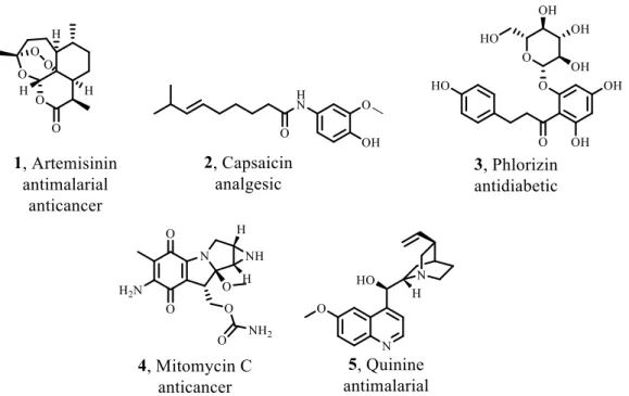
Until 2013, 1453 new chemical entities have been approved by FDA, which 40% are natural products or their derivatives. This number has risen 50% in the last 30 years, from which 74% are for anticancer applications. However, the anti-infective therapy is the area of highest use of NPs. For example, artemisinin (1) is a NP, widely used as an antimalarial drug and it is produced by the yeast Saccharomyces cerevisiae. Quinine (5) is another antimalarial drug, first isolated from the bark of Cinchona species and reported in 1820. The bark was used by indigenous groups from Amazon for the treatment of fevers (Fig. 1.1).[3, 5]
Chapter 1. Natural products: indoles and spirooxindoles
4
Usually, the isolated NPs are not the drug to be used for treatment of a disease. However, they can be used as lead compounds for the development of analogues. Furthermore, NP are selective to bind biological targets, becoming privileged structures as templates for the synthesis of biologically active NP-based molecules.[4] Indoles and spirooxindoles are scaffolds often found in NP. Dragmacidin E, jasplakinolide, elacomine and alstonisine are examples of these scaffolds.[6, 7]
1.1.
Indole derivatives
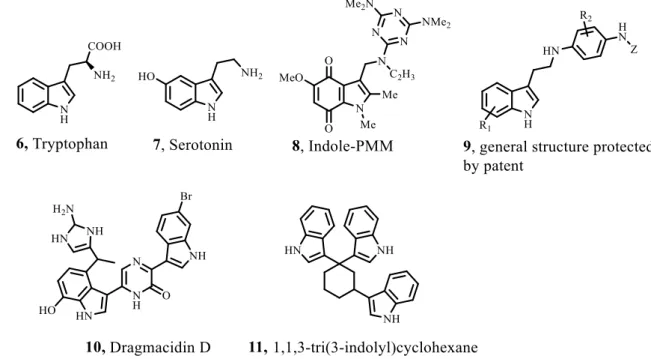
Indole based derivatives occur usually in natural products from plants, animals and marine sources. Tryptophan (6), for example, is an essential amino acid that is included in several biological processes. Serotonin (7) is a neurotransmitter and is derived from tryptophan. The indole core is known as a “privileged scaffold” in medicinal chemistry, because of its features and biological processes that are involved. Indole derivatives mimic the structure of peptides and are able to bind reversibly to enzymes.[2] Indoles have been reported to induce apoptosis in breast, squamous cell carcinoma, cholangiocarcinoma, colon, cervical, ovarian, pancreatic and prostate cancer cells.[8]
Pentamethylmelamine (PMM) is attached to an indole-2,3-dione moiety 8. It entered in clinical trials in the 1970s for the treatment of ovarian carcinoma, but it was abandoned because of the lack of solubility in water. An indole derivative family 9 was patented in 2011 and it was reported for the treatment of several cancers (Fig. 1.2).[8] Dagmacidin D (10) has two indole groups bonded to a piperazine ring. It shows in vitro cytotoxicity with IC50 values of 15 µg/mL against P-388 cell lines (leukemia) and 1-10 µg/mL against A-549 (human lung), HCT-8 (human colon) and MDA-MB (human breast) cancer cell lines. The 1,1,3-tri(3-indolyl)cyclohexane (11) inhibits cancer cell growth in lung cancer cells, having positive pharmacologic properties. However, it triggers DNA damage and leads to the production of reactive oxygen species.[9]
Chapter 1. Natural products: indoles and spirooxindoles
5
Fig. 1.2 – Natural products and drugs with indole-derivative scaffolds 6-11.
The indole scaffold is also known for its antiparasitic activity. For example, usambarensine (12) and usambarine (13) are indole alkaloids, which are isolated from Entamoeba histolytica. These natural products have antiplasmodial activity with IC50 values of 0.023 and 0.13 µM. A derivative from usambarine, dihydrousambarensine (14), was tested against multidrug-resistant P. falciparum strain, K1, showing to be 5 times more potent than chloroquine, the most used drug against this strain (Fig. 1.3). However, this last one showed to be inactive in vivo in mice affected with P. berghei chloroquine-sensitive strain. This means that dihydrousambarensine is selective to chloroquine-resistant strain.[10]
Fig. 1.3 – Indole derivatives with antiplasmodial activity.
Overall, among all natural products, indole alkaloids family is a very privileged scaffold, having very interesting anticancer and antimalarial activities.[10]
Chapter 1. Natural products: indoles and spirooxindoles
6
1.1.1. Spirooxindole natural products
Based on tryptamine, spirooxindole alkaloids 15-19 belong to a family that was first isolated from plants of the Paocynaceae and Rubiaceae. The main feature of this family is the spiro carbon in the position 3 of the oxindole core which gives several degrees of substitution around both rings linked to the spiro carbon. This carbon possesses a tetrahedral geometry, so the two rings can be almost perpendicular to each other, which gives unique conformational characteristics (Fig. 1.4).[11]
Fig. 1.4 – Spirooxindole natural products 15-19.
Several new drugs with different biological targets have been synthesized taking spirooxindole natural products as lead compounds (Fig. 1.5).
Chapter 1. Natural products: indoles and spirooxindoles
7
1.2.
Previous work in our research group and scope of the thesis
In our research group, we have been studying new indole-based scaffolds to reactivate p53 function. Different analogues of pyrrolidinyl-spirooxindole natural product, containing other 5-membered rings were developed.
The first family developed was the spiroisoxazoline oxindoles 23. Eighteen new spiroisoxazoline oxindole were synthesized by reaction of hydroximoyl chlorides and 3-methyleneindoline-2,3-diones. This synthesis requires several steps, including the preparation of the dipolarophile. This family was tested in colon cancer cell lines, HCT116 and SW620. From them, one compound showed to have a GI50 value of 26.56 µM in HCT116 p53(+/+). They showed that any substituent in the aromatic rings increased the potency and the chlorine in the 6th position of the indole is important for the interaction with MDM2, corroborating with the co-crystal structure that was previously published. Furthermore, this atom occupies an unoccupied small pocket by indole and mimics the side chain of Trp23 of p53.[12]
Further optimization of spiroisoxazoline oxindoles led to the spiropyrazoline oxindole scaffold 24 by changing the isoxazoline oxygen to a N-phenyl group, resulting in an extra substituent. This family showed a high anti-proliferative activity in breast cancer cell lines, MCF-7 and MDA-MB-231, with GI50 values around 7.0 µM for MCF-7, showing greater selectivity for this cell line. The use of bromine atom in this scaffold showed to increase the potency as anticancer agent. Spiropyrazoline oxindole 24b was also tested in human colon cancer cell line HCT116 to determine the DNA cell content. The experiment showed that most of cells were accumulated in G1 phase after 24h. Spiropyrazoline oxindoles showed to induce apoptosis.[13-15]
Taking in considerations that position 5 and 6 of the oxindole ring increase anticancer potency, new optimizations led to the spirooxadiazoline oxindoles 25. Sixteen new spirooxadiazoline oxindoles with an [1,2,4]-oxadiazole ring were synthesized by reacting 3-imino-indolin-2-ones with nitrile oxides. In this family, the different positions of the halogens used as substituents were tested, including the 5th and the 6th positions of the oxindole and the orto, meta and para positions of the aromatic rings. This family was tested in human colon cancer cell lines, HCT116 and SW620. The most active compounds have both aromatic rings substituted in meta and para positions, with 8.5-fold and 4.3-fold increase in potency when compared to the non-halogens substituents. Spirooxadiazoline oxindole 25b showed to inhibit p53-MDM2 interaction in the same extent as nutlin-3 at 10 and 20 µM. This same derivative also showed to be responsible for the accumulation of cancer cells in the cell cycle G1 phase.[16]
The family of spirooxadiazoline oxindoles 26 is a regioisomer of the family of spirooxadiazoline oxindoles 25. The biggest difference between both families is the position of the heteroatoms in the oxadiazole ring, resulting in spatial orientation of the substituents. Damla Uyar, in her Master thesis, synthetized a small library of twelve spirooxadiazoline oxindoles 26
Chapter 1. Natural products: indoles and spirooxindoles
8
and tested its anti-proliferative activity in human colon cancer cell lines. Spirooxadiazoline oxindoles 26b showed to have more selectivity for SW620 cancer cell line, which has p53 with R273H mutation (Fig. 1.6).[17, 18]
Fig. 1.6 – Optimization of spirooxindole scaffold by our research group.
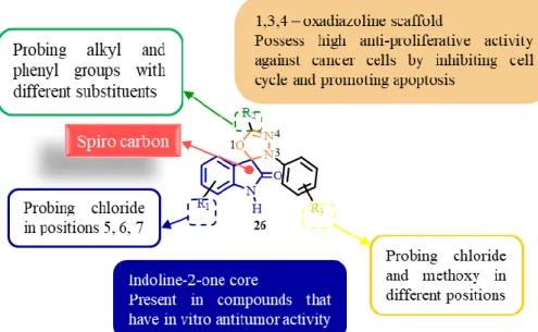
In this thesis, the main goal was the development of a new library of spirooxadiazoline oxindoles with a [1,3,4]-oxadiazole ring, instead of an [1,2,4]-oxadiazole core to be tested in human breast adenocarcinoma cell lines, MCF-7 and MDA-MB-231, and P. falciparum chloroquine-resistant and sensitive strains (Fig. 1.7).
Chapter 1. Natural products: indoles and spirooxindoles
9
SYNTHESIS OF
SPIROOXADIAZOLINE OXINDOLES
Chapter
Chapter 2. Synthesis of spirooxadiazoline oxindoles
13
Chapter 2. Synthesis of spirooxadiazoline oxindoles
2.1.
Introduction
The anticancer activity of the previous chemical families synthetized by our group has increased the interest in developing new spirooxindoles with five-membered ring.[12, 13, 16] As previously reported, the spirooxadiazoline oxindole family with an [1,2,4]-oxadiazole core has shown promising results in colon cancer cell lines, HCT116 and SW620 with wt p53 and mut p53, respectively.[16] Besides anticancer activity, oxadiazole and oxindole derivatives were described to have biological activity against therapeutically targets involved in other diseases.[19-22] For these reasons, joining these two scaffolds can lead to compounds with very interesting biological activities.
Based on all the results obtained by our research group, it was synthesized a library of twenty new spirooxadiazolines oxindoles with [1,3,4]-oxadiazole core.[12, 13, 15, 16, 23] The compounds were synthesized by 1,3-dipolar cycloaddition of isatin derivatives and nitrile imines (formed in situ from the corresponding hydrazonyl chlorides) derivatives, according to the Scheme 2.1.
The biggest advantage of these synthetic route is the commercial availability of the starting materials. The hydrazones 30, that are chlorinated to afford the hydrazonyl chlorides 29, are obtained by the reaction of aldehyde 31 and hydrazine 32 derivatives, which are commercially available. Several isatin derivatives 27 are also available, which can reduce several synthetic steps for the preparation of the dipolarophiles. In this library of spirooxadiazoline oxindoles 26, only electron donating groups were tested. Besides, the substituents were tested in several positions: 5th, 6th and 7th positions of the oxindole ring, meta and para-substituted phenyl ring and alkyl group as R2 and meta and para substitution as R3.
The anti-proliferative assays in human breast adenocarcinoma cell lines and a screening in P. falciparum chloroquine-resistant and sensitive strains were later performed.
Chapter 2. Synthesis of spirooxadiazoline oxindoles
14
Scheme 2.1 – Retrosynthetic route to obtain spirooxadiazoline oxindoles derivatives, highlighting the commercial available compounds.
Hydrazones were synthetized by condensing aldehyde 31 and hydrazine 32 derivatives in EtOH 20% for approximately 3 h (Table 2.1). This reaction involves the nucleophilic attack of the lone pair of electrons from the terminal nitrogen atom of the hydrazine to the carbonyl carbon of aldehyde with elimination of water. The precipitation of the product and its easy filtration are advantages in this procedure, giving very high yields (55-95%).[24]
Chapter 2. Synthesis of spirooxadiazoline oxindoles
15
Table 2.1- Synthesis of hydrazones 30.
Compd R2 R3 Yield/% 30a H m-Cl 95 30b p-Cl H 95 30c H o-Cl 56 30d p-OMe H 96 30e m-Cl m-Cl 98 30f m-Cl H 55 30g H H 92
The hydrazones 30a-g were characterized by 1H NMR. In all spectra, a deshielded singlet appears around 9.0 ppm corresponding to the hydrogen of the hydrazone group. In addition, the aromatic protons of the aromatic phenyl rings appear between 8.0 and 6.5 ppm, together with the proton of carbon 1 (assigned in Scheme 2.1). All NMR spectra were in accordance with the ones reported in the literature.[23, 25-28]
The second step of the synthetic route was the chlorination of the hydrazones 30 (Scheme. 2.1).
Hydrazonyl chlorides have been used for the synthesis of 5-membered rings (e. g. spirotriazoline oxindoles and spiropyrazoline oxindoles).[12-14] In order to substitute the hydrogen by a chlorine atom, N-chlorosuccinimide (33) and dimethyl sulfide (34) form the Corey-Kim reagent 37 (Scheme 2.2). This complex is also used for the oxidation of alcohols to aldehydes and ketones. It is a good alternative to the Swern oxidation, since it can be used in temperatures above -25ºC.
Scheme 2.2 – Formation of the Corey-Kim reagent.
Chapter 2. Synthesis of spirooxadiazoline oxindoles
16
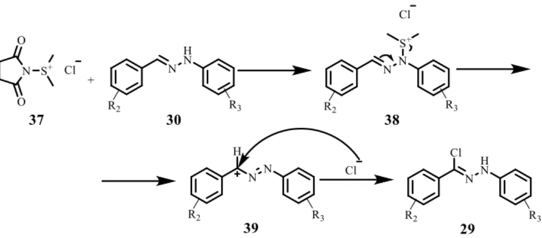
Scheme 2.3 – Mechanism proposed by Patel et al. for halogenation of hydrazones.[29]
This mechanism involves the formation of a benzylic cation 39 which is stabilized by the adjacent nitrogen atom. Then, the cation undergoes nucleophilic attack by the counteranion Cl- to give a phenylazobenzyl chloride, which tautomerizes to compound 29.[29]
This reaction occurs in very low temperature (-78ºC), using dichloromethane as solvent with moderate to excellent yields (49-92%) (Table 2.2).
Table 2.2 – Synthesis of hydrazonyl chlorides.
Compd R2 R3 Yield/% 29a H m-Cl 92 29b p-Cl H 72 29c H o-Cl 85 29d p-OMe H 49 29e m-Cl m-Cl 53 29f m-Cl H 70 29g H H 92
The 1H NMR spectra of the hydrazonyl chlorides 29a-g are very similar to the ones obtained for the corresponding hydrazones 30a-g. However, since the hydrogen was replaced by a chloride, all protons are slightly less shielded. The biggest difference is the singlet of the proton of the hydrazone that appears around 10.0 ppm. All spectra are in accordance with the ones reported in the literature.[23, 29, 30]
An additional step was made for the synthesis of the 6-chloroindoline-2,3-dione (27a) from the 6-chlorooxindole. This compound was synthetized because of the high availability of the corresponding oxindole. This is a two-step reaction. First occurs the bromation of the position 3 of the indole derivative and then the oxidation of this position (Scheme 2.4).
Chapter 2. Synthesis of spirooxadiazoline oxindoles
17
Scheme 2.4 – Synthesis of 6-chloroisatin (27a).
6-chloroisatin (27a) was obtained with a yield of 46% and the chemical structure was confirmed by 1H NMR and it is in accordance with reported literature.[31]
2.2.
Synthesis of spiro[indoline-3,2'-[1,3,4]oxadiazoline]-2-ones
2.2.1. Traditional synthetic route
The spirooxadiazoline oxindoles library was formed by reacting isatin and nitrile imines derivatives (formed in situ by the dichlorination of the corresponding hydrazonyl chloride) (Scheme 2.5 and Table 2.3). To the date, the 1,3-dipolar cycloadditions of hydrazonyl chlorides with 3-imino-indolinones and 3-methylene indolinones are the most common. In this thesis, it is reported for the first the reaction of hydrazonyl chlorides with isatin derivatives. The biggest advantage of the use of isatin derivatives is their commercial availability, so the number of reaction steps is reduced.
Chapter 2. Synthesis of spirooxadiazoline oxindoles
18
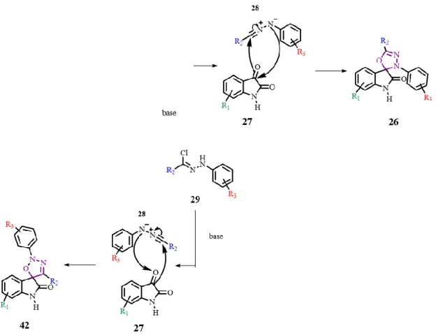
Scheme 2.5 – 1,3-dipolar cycloaddition of isatin derivatives 27 as dipolarophiles and nitrile imines 28 as dipoles.
In this reaction, two regioisomers can be formed. They can be distinguished by NMR. For the regioisomer 26, the spiro carbon and the C=N bond have their signals around 96.0 and 150.0 ppm in the 13C NMR, respectively[17]. For the regioisomer 42, since it has a different chemical environment, the signal of the spiro carbon should appear much more shielded, around 88.0 ppm.[32] The spiro carbon of the regioisomer 26 is bonded to two heteroatoms (oxygen and nitrogen) and two carbons, while the spiro carbon of the regioisomer 42 is bonded to an oxygen and three carbons. (Fig. 2.2)
Fig. 2.1 – Difference in the spiro carbon for both regioisomers. In regioisomer 26, the spiro carbon is linked to a nitrogen (orange) and in regioisomer 42, the spiro carbon is linked to another carbon (brown).
Chapter 2. Synthesis of spirooxadiazoline oxindoles
19
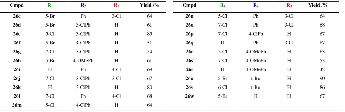
Table 2.3 - Library of spirooxadiazoline oxindoles 26a-u and respective yields.
Cmpd R1 R2 R3 Yield /% Cmpd R1 R2 R3 Yield /% 26c 5-Br Ph 3-Cl 64 26n 5-Cl Ph 3-Cl 64 26d 5-Br 3-ClPh H 61 26o 7-Cl Ph 3-Cl 68 26e 5-Cl 3-ClPh H 85 26p 7-Cl 4-ClPh H 67 26f 5-Br 4-ClPh H 51 26q H Ph 3-Cl 87 26g 7-Cl 3-ClPh H 54 26r 5-Cl 4-OMePh H 63 26h 5-Br 4-OMePh H 61 26s 7-Cl 4-OMePh H 53 26i H Ph 4-Cl 68 26t H 4-OMePh H 42 26j 7-Cl 3-ClPh 3-Cl 67 26u 5-Br t-Bu H 90 26k H 3-ClPh H 80 26v 6-Cl t-Bu H 86 26l 7-Cl Ph 4-Cl 68 26w 5-Br H H 67 26m 5-Cl 4-ClPh H 64
Chapter 2. Synthesis of spirooxadiazoline oxindoles
20
The spirooxadiazoline oxindoles 26a-x were obtained with moderate to excellent yields (42 to 90%). In most of the reactions, the starting materials were not consumed completely. This can be explained by the possibility of the nitrile imine to dimerize.[17]
Compounds 26a-v were characterized by NMR, infra-red and mass spectrometry.
The 1H NMR spectra showed an increase of aromatic protons relatively to the hydrazonyl chloride spectra. The proton of the NH from the oxindole moiety appears around 10.0 ppm.
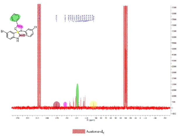
For all reactions, only regioisomer 26 was formed (the spiro carbon signal appeared between 95.5 and 97.5 ppm) as observed by 13C NMR of 26c as example (Fig. 2.2).
Fig. 2.2 – 13C NMR of 26c. (yellow – spiro carbon, reddish brown – C=O, pink – C=N, green – equivalent
carbons).
Another two signals are characteristic: the carbonyl carbon, and the carbon linked to the nitrogen by a double bond at around 170.0 and 152.0, respectively.[17]
IR spectra were obtained. Three main peaks were identified: the carbonyl stretch (C=O) around 1740 cm-1, amine stretch (NH) around 3275 cm-1 and C=N stretch around 1600 cm-1. Other peaks were identified as aromatic bonds. In the case of compounds 26u and 26v, an extra peak was identified at 2970 cm-1 corresponding to the alkyl C-H bond (Fig. 2.3).
Chapter 2. Synthesis of spirooxadiazoline oxindoles
21
Fig. 2.3 – IR spectrum of 26i. Red, green and purple peaks are common to all family.
2.2.2. Microwave-assisted route
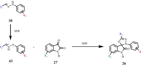
Microwave assisted synthesis is known to be a green and faster way to perform several reactions (Table 2.4), including the formation of 5-membered heterocycles.[33] For hydrazones, it has been reported some reactions for the formation of 5-membered rings from electron deficient dipolarophiles to form bipyrozolyl under microwave irradiation and solvent-free conditions.[34, 35] In this reaction, azomethine imines 43 are formed from the hydrazones 30 (instead of nitrile imines) and posterior 1,3-dipolar cycloaddition by reacting the azomethine imine 43 with the dipolarophile 27 (Scheme 2.6).
During the course of this thesis, microwave-assisted reactions to form spirooxadiazoline oxindole 26 derivatives were tried, in an attempt to synthetize for the first time spirooxindoles by MW, using also for the first time an isatin as dipolarophile.
Table 2.4 – Vantages and disadvantages of traditional and microwave assisted reactions.
Traditional Microwave
• Room temperature reaction • Big scale reactions
• No pressure • Long time reaction • Addition of TEA
• Use of dichloromethane as solvent • Synthetic route with 3 steps
• High temperature reaction • Small scale reactions • Use of pressure • Short time reaction • No need of base • Solvent free
Chapter 2. Synthesis of spirooxadiazoline oxindoles
22
Scheme 2.6 –Synthesis of spirooxadiazoline oxindole 26 by microwave assisted reaction.
The reactions of the 5-bromoisatin with the hydrazones 30a-g were tested under several different conditions: time (30 min to 2 h); temperature (90ºC to 180ºC); the use of solvent (dioxane) and free solvent reaction. Only two products 26f and 26f were synthetized by microwave assisted reaction (Table 2.5).
Table 2.5 – Spirooxadiazoline oxindoles synthesized by microwave assisted reaction
Cmpd R1 R2 R3 Traditional Yield /% MW Yield /% 26d 5-Br 4-ClPh H 61 71 26h 5-Br 4-OMe H 51 79
It was already reported the 5-membered cycle formation from olefins, maintaining the same bond order. According to the mechanism described, the double bond is used for the formation of the ring as a normal 1,3-dipolar cycloaddition. However there is an autooxidation of the 5-membered ring with the oxygen that exists in the microwave vessel to form again the double bond.[36] In the case of this reaction, it is possible that before the rearrangement, the 5-membered ring 44 is too unstable and degrade in the system (Scheme 2.7). The stability of the hydrazonyl chlorides to high temperatures can also contribute to the degradation of the product, since the hydrazonyl chlorides used in this thesis have never been tested in microwave assisted reactions.[33-36]
Chapter 2. Synthesis of spirooxadiazoline oxindoles
23 Other conditions can be tested in the attempt to obtain the nitrile imines such as addition of NCS and triethylamine.[33]
2.3.
Stability studies in NMR
The stability of the spirooxadiazoline oxindoles 26 were studied before the evaluation of biological activity to assure that compounds 26 do not degrade in solution during the biological assays. For this reason, in a NMR tube, spirooxadiazoline oxindole 26w (Fig. 2.4) was dissolved in DMSO-d6 (solvent used in biological assays) and kept at room temperature. The stability was performed using NMR spectroscopy, obtaining a new 1H NMR of the solution every week (Fig. 2.5).
Fig. 2.4 – Structure of 26w.
Fig. 2.5 – Spectra of 26w in DMSO-d6 every seven days.
Comparing the five spectra, it is possible to conclude that compound 26w is very stable in DMSO-d6. Only after the 5th week, it was observed very small extra peaks in the spectrum.
Chapter 2. Synthesis of spirooxadiazoline oxindoles
24
Despite, these small changes, the compound does not suffer degradation in DMSO, so it can be safely used in biological assays.
SMALL MOLECULES ACTING AS
p53 REACTIVATORS
Chapter
Chapter 3. Small molecules acting as p53 reactivators
27
Chapter 3. Small molecules acting as p53 reactivators
3.1.
Introduction
3.1.1. p53 protein
The p53, a short-lived protein, known as the guardian of the human genome, was discovered in 1979 as a cellular 53 kDa nuclear phosphoprotein bond to the large transforming antigen of the SV40 DNA virus.[37-39] The p53 gene (TP53) was first cloned in 1983.[40] Six years later, p53 was shown to block the transformation of rat embryo fibroblasts. In 1990, TP53 was identified in several Li-Fraumeni syndrome, a rare condition which entire families are affected by several types of cancers. In 1993, p53 was considered the molecule of the year.[40]
It belongs to a small family of related proteins which includes p63 and p73. While these two proteins have important roles in the normal development, p53 has a special role to prevent tumour development.[41] The wild type (wt) p53 protein binds to specific nucleotide sequences termed p53-responsive elements that, when together, stimulate a p53-dependent expression.[42] This tumour suppressor is regulated by degradation via the ubiquitin-proteasome pathway and monitors the integrity of the cells, and is one of the responsible to control cell cycle, DNA repair and synthesis, cell differentiation, genomic plasticity, senescence, angiogenesis and programmed cell death, including apoptosis and cell cycle arrest (Fig. 3.1).[37, 38, 43, 44]
High concentrations of p53 protein are found during the meiotic process of spermatogenesis. At that moment, the genome recombines and is responsible for genetic diversity. DNA damage in somatic cells caused by radiation or topoisomerase inhibitors leads to stabilization of wt p53 protein. p53 arrests cells in G1 phase of cell cycle, which is the moment when the DNA is repaired, avoiding mutations.[37]
The p53 pathway uses several mechanisms to arrest cell cycle progression, preventing the propagation of DNA damage and repairing it. If the damage is too severe, p53 leads to apoptotic cell death to prevent any malignant proliferation of damaged cells. Therefore p53 plays a protective role in normal cells by limiting the propagation of damaged cells.[43] Since this type of cell death requires a genetic program, mutations in apoptotic pathways produce drug resistant tumours. p53 has a main role in apoptosis induced by anticancer agents in damaged cells.[45] When p53 is mutated (mut p53), its function of tumour suppressor can be affected.
Chapter 3. Small molecules acting as p53 reactivators
28
Fig. 3.1 – p53 regulates cellular response to stress. Several stress factors activate p53 and lead to a stabilization and accumulation of it in cell nuclei. Some genes are activated and trigger several responses.
[Adapted from [43, 46]]
The p53 tumour suppressor gene is mutated or deleted in half of all types of cancer.[38, 40, 43, 47] When mutated, p53 protein has a different conformation, failing to bind DNA and regulate the transcription of p53 responsive genes. This phenomenon is known as loss of function mutation. Wt p53 is the most common in malignancies, such as acute myeloid leukaemia (AML) and melanoma. In these cases, cellular oncogene products of DNA tumour viruses bind to p53 protein and block its ability of transcription.[40, 47] Murine Double Minute 2 (MDM2) and Murine Double Minute X (MDMX) are two examples of these inhibitors.
Chapter 3. Small molecules acting as p53 reactivators
29
Table 3.1 – Compounds that act in wt and mut p53.[48]
Molecule/ compound Mechanism of action Target Stage of development Nutlins RG7112 Inhibits p53-MDM2 interaction MDM2 Phase I clinical trials Benzodiazopinediones Inhibits p53-MDM2 interaction MDM2 Preclinical Spirooxindoles Inhibits p53-MDM2 interaction MDM2 Phase I clinical trials
RITA Inhibits p53 binding Wt and mut
p53 Preclinical Serdemetan Inhibits p53-MDM2 interaction MDM2 Phase I clinical trials SJ-172550 Inhibits p53-MDM2/X interaction MDMX Preclinical RO-2443/RO-5693 Inhibits p53-MDM2/X interaction MDMX Preclinical XI-011 Repression of MDMX promoter MDMX Preclinical
3.1.2. p53-MDM2 inhibition
MDM2 is a phosphoprotein of 54 kDa that has a putative nuclear localization signal and two zinc fingers. The MDM2 gene is located on chromosome 12q13-14 and encodes for 491 amino acid protein.[39] It can be associated with mut and wt p53 and its excess functions as a negative regulator of this protein.[40, 49] It is an E3 ubiquitin ligase that targets p53 degradation by the proteasome pathway.[40, 44]
Inactivation of the p53 tumour suppressor by mutation or overexpression of negative regulators occurs frequently in cancer. As explained before, p53 plays an important role in the regulation of proliferation and apoptosis in DNA damaging chemotherapies, so the reactivation of p53 by disruption of p53-MDM2 protein interaction is an interesting strategy for targeted anticancer therapy.[38, 47]
According to the X-ray structure, the p53-MDM2 edge bears a total of 1498 Å of surface area. The interface has a steric complementarity between the MDM2 cleft and the hydrophobic face of α-helix of p53, which includes Phe19, Leu22, Trp23 and Leu26. These four amino acids make a
Chapter 3. Small molecules acting as p53 reactivators
30
sequential and extensive van der Waals interaction with the MDM2 pocket. They are complemented with two intermolecular hydrogen bonds: the interaction of the Phe19 backbone amide of p53 with the Gln72 side-chain of MDM2 and the nitrogen of p53 of the Trp23 indole group with the Leu54 backbone carbonyl moiety of MDM2 (Fig. 3.2).[38, 40]
The core of p53 is a region which can interact with DNA in a sequence specific-manner. Usually the mutations observed in tumours occur in regions where critical residues of DNA contact or their conformation are changed. For optimal binding to DNA, the protein is in a tetrameric state since there are four interactions of four separate p53 molecules via the tetramerization domain. The C-terminal region is responsible for regulatory properties and has mainly basic residues. This region suffers modifications by acetylation, phosphorylation, O-glycosylation and RNA binding. The DNA binding domain is separated from the transcriptional domain by several proline residues which can interact with signal transduction molecules that have SH3 binding domain. On the other side, the acidic N-terminal domain activates the expression of target genes while giving the support for transcribing the new mRNA. This region is also important for stability and activity of the interactions of p53 and other proteins as MDM2, forming specific complexes, which allows targeting of ubiquitin-mediated proteolytic machinery.[49, 50] Modifications by phosphorylation in this region change the properties of this area, including its conformation, and as a consequence, the interaction of p53 with proteins.[50] According to the studies by Lane’s laboratory, the disruption of p53-MDM2 interactions leads to an uncontrolled increase of p53, since MDM2 takes p53 from the nucleus to the cytoplasm, where it should be inaccessible to target DNA for transcription and it should be degraded by ubiquitination through its E3 ubiquitin ligase activity.[44, 51-53] MDM2 can also auto ubiquitinate itself, leading to self-degradation.[39]
Fig. 3.2 – MDM2 (surface)-p53 complex (PDB ID: IYCR). The main interactions are made by Leu26, Phe19 and Trp23 from a small amphipathic p53 derived α-helix of p53 and the MDM2 pocket. [Adapted from [44]]
Chapter 3. Small molecules acting as p53 reactivators
31 p53 is a transcription factor for MDM2 which results in an autoregulatory feedback loop, so p53 protein regulates the transcription of MDM2 gene and, in turn, MDM2 protein regulates the activity of p53 protein.[39, 40, 42-44]
The gene transcription by p53 is influenced by modifications such as phosphorylation or interactions with other proteins. MDM2 binds to p53 at its N-terminal domain and inhibits its transcriptional activity. Its amplification on chromosome 12q12-13 and excess of the gene product inactivates p53, so its tumour suppressor function is also shut down. Sequence-DNA binding and transcriptional activity of the wt p53 protein are also reduced in the presence of oncoproteins produced by some DNA viruses.[37, 43]
3.1.2.1. Small molecules inhibitors of p53-MDM2 interaction
MDM2 overexpression is oncogenic and is associated with the late stage disease, resistance to chemotherapy and radiotherapy and poor prognosis.[39] Furthermore, genetic studies have shown that loss of p53 activity induces tumour formation, while its activity restoration leads to the tumour regression.[40] p53 and MDM2 are both valuable targets for developing anticancer agents. One of the most popular strategies for reactivation of p53 activity is the use of non-peptidic small molecule inhibitors targeting the van der Waals protein-protein interactions site between MDM2 and p53 by mimicking the three critical amino acids residues from p53 pocket.[43, 54] The use of small molecules inhibitors for targeting p53-MDM2 and p53-MDMX interactions in a dual inhibition is another attractive strategy.[39, 40] Other approaches are blocking MDM2 expression, inhibiting the E3 ubiquitin ligase activity of MDM2 and targeting the protein-protein complex of proteins that interact with MDM2.[39]
Several study groups have shown the use of antisense oligonucleotides to inhibit MDM2 expression. They also showed that both wt p53 and mut p53 respond to MDM2 inhibition. These same groups have investigated the use of natural products such as curcumin, genistein and ginsenosides to down-regulate MDM2 expression.[39, 55-57]
Recently, small molecules were also reported to target E3 ligase activity of MDM2. They inhibit the ubiquitination of p53 in vitro, allowing the activation of p53 signalling, inducing cell cycle arrest and/or apoptosis. The first groups of these kind of inhibitors were the arylsulfonamide, bisaryluarea and acylimidazolone, first described in 2002. They allosteric inhibit MDM2 by blocking rearrangements of MDM2 that can be necessary for p53 ubiquitination.[39]
The most important families of inhibitors of p53-MDM2 interaction are cis-imidazolines 45 (or also known as nutlins), spirooxindoles 20, piperidinones 48, 1,4-diazepines and isoindolinones (Fig. 3.3).
Chapter 3. Small molecules acting as p53 reactivators
32
Fig. 3.3 – p53-MDM2 small molecule inhibitors that are in preclinical and clinical trials (compounds 20 and 45-47).[39]
3.1.3. p53-MDMX inhibition
MDMX is an additional MDM2 family member of which the 6-chlorotryptophan residue projects into the Trp23 binding site and optimizes the steric complementarity of p53-MDMX interface. It was discovered in 1996 as a close homolog protein of MDM2 that potentiates the ubiquitin ligase activity of MDM2 and blocks its p53 transcriptional activity without its degradation.[44] Like MDM2, it is overexpressed in several tumours. Due to its characteristics, it is important for targeting p53-MDMX interaction.
Despite being similar, MDM2 and MDMX have some conformational differences as well as some different amino acids which lead to a poorly inhibition by p53-MDM2 small molecules inhibitors.[38, 40] The critical residues for binding p53 are identical of MDM2, however the residues around Leu26 pocket are different, leading to different shape and size. These residues include Met53 (Leu54 in MDM2), Leu98 (Ile99 in MDM2), Leu102 (Ile103 in MDM2) and Pro95 (His96 in MDM2), resulting in a smaller binding groove in MDMX. Around the Trp23 pocket, the Phe86 residue of MDM2 is replaced by Leu85 residue in MDMX (Fig. 3.4).[44] Both have an acidic N-terminal p53 binding domain, C-terminal ring domain and a zinc finger domain.[44] The ring-finger domain located at the C-terminal end of both proteins is a well-conserved region. The integrity of this region is required for heterodimerization. The C4 zinc finger is also conserved in both proteins. MDMX was shown to be a cytoplasmic protein and it is dependent on other
Chapter 3. Small molecules acting as p53 reactivators
33 proteins, such as MDM2, for nuclear localization.[38-40] Disrupting the MDM2/MDMX heterocomplex inhibits the activity of p53 and cannot be reversed. Because of their critical and non-redundant role in p53 inhibition, targeting both MDM2 and MDMX can fully activate wt p53 in tumour cells.[44]
Fig. 3.4 – A) MDMX (surface)-p53 complex (PDB ID: 3DAB). The main interactions are made with Leu26, Phe19 and Trp23 from a small amphipathic p53 derived α-helix of p53 and the MDMX pocket. B) Superimposition of the two complexes. MDM2 is in white, MDMX is in yellow and p53 are in pink and blue, respectively. C) Superimposition of the two complexes. The residues that are non-identical are identified.
[Adapted from [44]]
Because of the differences of the binding site of MDM2 and MDMX, inhibitors of p53-MDM2 interactions have low affinity for MDMX. For this reason, the search of MDMX antagonists is mandatory for a more robust p53 activation.
SJ-172550 was the first molecule to be identified to kill cells by reversibly bind to MDMX. Two compounds of a series of indolyl hydantoyn compounds, RO-2443 and RO-5693, showed also to bind to the MDMX pocket, being then described as potent inhibitors of MDMX. Another compound, XI-011 was reported to activate p53 in breast cancer cells, by inhibiting its interaction with MDMX. This inhibition is through the transcriptional repression of MDMX promoter. All these compounds are still in a preclinical stage.[48]
3.1.4. Spirooxindole core
In 2005, the Wang group at the University of Michigan reported the structure-based design of spirooxindole as a new class of potent small-molecule inhibitors of p53-MDM2 interaction. The oxindole group was found to mimic the Trp23 moiety (6) and spirooxindole-containing natural products 49 were identified and docked to MDM2 protein (Fig. 3.5).
MI-5 (51) was designed to interact effectively with MDM2. The oxindole moiety inserts into the Trp23 pocket, the phenyl group occupies the Phe19 pocket and the isopropyl group occupies the Leu26 pocket. The substitution of isopropyl group to t-butyl group and the introduction of a chlorine atom in position meta in the phenyl group lead to MI-17 (52) which is 100-fold more potent than MI-5.
Chapter 3. Small molecules acting as p53 reactivators
34
Chapter 3. Small molecules acting as p53 reactivators
35 Further optimizations were made leading to MI-63 (53). Despite having good water solubility, it has a very modest oral bioavailability. To overpass this problem, more modifications were made leading to MI-219 (54). This compound was proved to induce activation of p53 by blocking p53-MDM2 interaction in cells with wt p53. It was shown to inhibit cell growth in cancer cells and has more than 10-fold selectivity in tumour cells with mutated or deleted p53. Changing the stereochemistry, MI-888 (56) has a much higher affinity to MDM2, selectivity in inhibition of tumour cell growth in cancer cells with wt p53, oral bioavailability and it is capable of achieving total and durable tumour regression.[44]
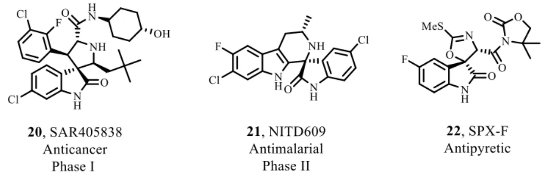
Currently, SAR405838 (20), an analogue of MI-888, is in Phase I of clinical trials. It was developed by Sanofi-Aventis as a derivative of MI-219. It works as a MDM2 antagonist, binding to the p53 pocket of MDM2 molecule with specificity and selectivity, resulting in p53 reactivation at low nanomolar concentrations and induces cell-cycle arrest and apoptosis.[54, 58] Spirooxindole molecules were inspired by the presence of indoles in natural products. The starting point that led to SAR405838 was to mimic the three most important amino acids and their van der Waals and hydrogen bond interactions. The X-ray structure of MDM2 complexed with an spirooxindole analogue showed that the oxindole moiety inserts deeply into the Trp23 pocket of MDM2. The 2-fluoro-3-clorophenyl ring binds to the Leu26 binding site. Its rotative plane allows the halogens to reach the bottom of the MDM2 pocket. The Phe19 cleft is filled by the neopentyl side chain which requires an induced fit. For that, the backbone of His73 must retract outward from the pocket and allows the side chain of Tyr67 to rotate (Fig. 3.6).[38]