Universidade de Lisboa
Faculdade de Medicina de Lisboa
Papel do peptídeo natriurético cerebral de tipo B na
classificação do tipo de AVC isquémico
Ana Catarina Gaspar Fonseca
Doutoramento Medicina Clinica
Neurologia
Universidade de Lisboa
Faculdade de Medicina de Lisboa
Papel do peptídeo natriurético cerebral de tipo B na
classificação do tipo de AVC isquémico
Ana Catarina Gaspar Fonseca
Dissertação orientada por:
Professor Doutor José Manuel Ferro
Professora Doutora Dulce Brito
Medicina Clinica
Neurologia
Todas as afirmações efetuadas no presente documento são da exclusiva responsabilidade do seu autor, não cabendo qualquer responsabilidade à Faculdade de Medicina de Lisboa pelos conteúdos nele apresentados.
A impressão desta dissertação foi aprovada pelo Conselho
Cientifico da Faculdade de Medicina de Lisboa em reunião de 16
de Julho de 2013
7
Acknowledgments
During the time I worked on this thesis there were several people that I had the opportunity to come across. During this time I learned new technical knowledge, but I realized that one of the most important and interesting points of scientific work is the possibility to discover different people, exchange ideas and discuss views. Some of these people impressed me by their accessibility, generosity, intelligence, for their advices and for the respect that they have for those that surround them. Therefore I would like to thank to:
- Professor José Ferro, for his support, time, expertise, advice and for setting an outstanding example of clinical practice and research.
- Professora Dulce Brito for the concern , help and expertise.
- Drª. Teresa Pinho e Melo and Professor Patricia Canhão for their collaboration, suggestions and support.
- Dr. António Samapaio Matias for his time and collaboration. - Drª Luisa Albuquerque Drª Ruth Geraldes for their help and support.
- Harvard Medical School Portugal Program for their interest in my project and for giving me the opportunity to live the day-to-day life at the Harvard School of Public Health
- Professor Louis Caplan for his patience and help. Having the opportunity to learn with him is a great honor.
- Professor Ajay Singh for his support, for trying to explaining me, after all, what means to be a clinical researcher and for teaching me some of the “tricks”
- To the patients and their families
- My friends for understanding and for being there whenever I need them
9
Index
INDEX ... 9 ABSTRACT ... 13 RESUMO ... 15 INTRODUCTION ... 17 1.1STROKE ... 19 1.2ATRIAL FIBRILLATION ... 211.3ATRIAL FIBRILLATION DIAGNOSIS ... 25
1.4 BIOMARKERS ... 28
1.5BRAIN NATRIURETIC PEPTIDE AND N-TERMINAL PROBRAIN NATRIURETIC PEPTIDE ... 32
1.6 NT-PROBNP AND BNP IN ISCHEMIC STROKE ... 35
REFERENCES ... 49
HYPOTHESIS ... 71
AIMS ... 73
1. TIME COURSE OF NT-PROBNP LEVELS AFTER ACUTE ISCHEMIC STROKE ... 75
INTRODUCTION ... 77 METHODS ... 78 RESULTS ... 82 DISCUSSION ... 88 CONCLUSION ... 90 REFERENCES ... 91
2. VALIDATION OF NT-PROBNP CUT-OFF VALUES FOR THE DIAGNOSIS OF CARDIOEMBOLIC STROKE RELATED TO ATRIAL FIBRILLATION ... 95
INTRODUCTION ... 97 METHODS ... 98 RESULTS ... 101 DISCUSSION ... 107 CONCLUSION ... 109 REFERENCES ... 109
3. NT-PROBNP ACCURACY FOR DETECTING ATRIAL FIBRILLATION IN CRYPTOGENIC STROKE ... 113
INTRODUCTION ... 115 METHODS ... 115 RESULTS ... 118 DISCUSSION ... 122 CONCLUSION ... 125 REFERENCES ... 125
4. POTENTIAL OF BRAIN NATRIURETIC PEPTIDE TO IDENTIFY TIA AND STROKE DUE TO OCCULT ATRIAL FIBRILLATION: POPULATION-BASED STUDY ... 129
10 INTRODUCTION ... 131 METHODS ... 131 TYPE OF STUDY ... 131 SAMPLE ... 131 STUDY DESIGN ... 132 RESULTS ... 133 DISCUSSION ... 136 CONCLUSION ... 137 REFERENCES ... 137 DISCUSSION ... 139 CONCLUSIONS... 147 ATTACHMENTS ... 151
11 This study was funded by grants from:
• “Fundação para a Ciência e Tecnologia” (FCT), in the Framework of the Cooperation Agreement between Portugal and Harvard Medical School – 2010 (HMSP-ICJ/0003/2010)
• Anual research grant “D. Manuel de Mello” of the “Fundação Amélia da Silva de Mello”
• “Bolsa para a Formação Avançada em Investigação Clínica de Internos-Doutorandos da Fundação para a Ciência e Tecnologia” (SFRH/BD/60058/2009)
13
Abstract
Even after an extensive etiological investigation in about one third of ischemic strokes it is not possible to establish a cause. A fraction of these strokes could have been due to an episode of paroxysmal atrial fibrillation which could not be identified. Studies show that the current available methods to diagnose paroxysmal atrial fibrillation have a low sensitivity. N-terminal of the brain natriuretic peptide (NT-proBNP) could be useful as a biomarker of cardioembolic stroke.
NT-proBNP serum levels were initially evaluated in 66 ischemic atroke patients. Patients with cardioembolic stroke had higher levels of NT-proBNP than patients with non-cardioembolic stroke. NT-proBNP had a very good accuracy to diagnose cardioembolic stroke related to atrial fibrillation (AUC-0.92) Two cut-off points of serum NT-proBNP levels with a high sensitivity (265.5 pg/mL) and specificity (912 pg/mL) for the diagnosis of paroxysmal atrial fibrillation were established. Although NT-proBNP levels started to decrease 72 hours after stroke onset, the diagnostic accuracy of NT-proBNP to diagnose cardioembolic stroke was similar in the first 72 hours after ischemic stroke. In a validation study performed in 184 patients the cut-off points of 265.5 pg / mL and 912.0 pg / mL of NT-proBNP had a sensitivity, specificity, positive predictive value and negative predictive value in the first study versus validation study of respectively (94.4% vs 100%, 72.9% vs 70.5%, 56.6% vs 59.1%, 97.2% vs 100%) versus (55.5% vs 81.8%, 97.9% vs 87.5, 90.9% vs 73.8%, 83.9 vs 91.9%) for the diagnosis of ischemic stroke of cardioembolic etiology associated to atrial fibrillation. A prospective study including 80 patients was done to evaluate the cut-off points in patients with cryptogenic stroke. The Area Under the Curve of the Receiver Operating Curve of NT-proBNP for the diagnosis of paroxysmal atrial fibrillation in patients with cryptogenic stroke was good - 0.83, 95% CI (0.73-0.92), the cut-off point of 265.5 pg/mL had a sensitivity of 88.2% 95% CI (65.7-96.7%). The cut-off point of 912 pg/mL had a specificity of 88.9% 95% CI (78.8-94.5%).
14
NT-proBNP may be useful as a biomarker for paroxysmal atrial fibrillation in ischemic stroke and therefore helpful to reclassify strokes initially categorized as cryptogenic.
15
Resumo
Mesmo após uma extensa investigação etiologica, em aproximadamente um terço dos acidentes vasculares cerebrais (AVC) isquemicos não é possibel estabelecer uma causa. Uma fracção destes AVCs pode ter sido causada por episodios de fibrilhação auricular paroxistica que não foram identificado. Os estudos mostram que os métodos actuais de diagnostico de fibrilhação auricular paroxistica têm uma baixa sensibilidade. O N-terminal do peptideo natriuretico cerebral (NT-proBNP) pode ser util como biomarcador de AVC cardioembolico.
Os valores séricos de NT-proBNP forem inicialmente determinados em 66 doentes com AVC isquémico. Os doentes com um AVC de etiologica cardioembolica tinham valores de NT-proBNP superiores aos de doentes com uma etiologia não-cardioembolica. O NT-NT-proBNP teve uma boa precisão para diagnosticar AVC cardioembolico relacionado com fibrilhação auricular (AUC-0.92). Dois pontos de corte de valores séricos de NT-proBNP com uma elevada sensibilidade (265.5 pg/mL) e especificidade (912 pg/mL) para o diagnostico de fibrilhação auricular paroxitica foram estabelecidos. Apesar de os valores séricos de NT-proBNP começarem a diminuir 72 horas após o inicio do acidente vascular cerebral, a precisão diagnostica do NT-proBNP para o diagnostico de AVC cardioembolico foi semelhante nas primeiras 72 horas após inicio do AVC. Num estudo de validação realizado em 184 doentes, os pontos de corte de 265.5 pg / mL e 912.0 pg / mL de NT-proBNP tiveram uma sensibilidade, especificidade, valor preditivo positive e valor preditivo no primeiro estudo versus estudo de validação de respectivamente (94.4% vs 100%, 72.9% vs 70.5%, 56.6% vs 59.1%, 97.2% vs 100%) versus (55.5% vs 81.8%, 97.9% vs 87.5, 90.9% vs 73.8%, 83.9 vs 91.9%) para o diagnostico de AVC isquémico de etiologia cardioembolic associado a fibrilhação auricular. Um estudo prospectivo que incluiu 80 doentes foi realizado para avaliar os pontos de corte em doentes com AVC criptogénico.
A área sob a curva ROC da precisão diagnostica dos valores séricos de NT-proBNP para o diagnostico de fibrilhação auricular paroxistica nos doentes com AVC criptogenico foi boa - -
16
0.83, 95% IC (0.73-0.92). O ponto de corte de 265.5 pg/mL teve uma sensibilidade de 88.2% 95% IC (65.7-96.7%). O ponto de corte de 912 pg/mL teve uma especificidade de 88.9% 95% IC (78.8-94.5%).
O NT-proBNP pode ser util como um biomarcador de fibrilhação auricular paroxística nos doentes com AVC isquémico. Poderá também ajudar a reclassificar AVCs inicialmente categorizados como criptogénico.
19
1.1 Stroke
Stroke is a leading cause of disability and mortality. It is the third cause of death worldwide, the first cause of acquired disability, the second cause of dementia and is associated to significant direct and indirect costs [1]. In Portugal, it has an estimated incidence of 187 per 100000 inhabitants and it is the first cause of death and dependency in patients older than 65 years. A cross-sectional study of a representative sample of the Portuguese population including subjects aged 40 reported a prevalence of atrial fibrillation of 2.5% (2.2-2.8%: 95% CI) [2]. With growing ageing of the world population and increasing incidence of vascular risk factors, the prevalence of stroke-related burden is expected to increase over the next two decades. Although there has been in the last decade an increased use of treatment during ischemic acute stroke, such as thrombolysis (intravenous or intraarterial) and implementation of stroke unit care, these are poorly effective and are available to only a small portion of the population. Resources should be potentiated to improve primary and secondary prevention of stroke. The majority of strokes worldwide (87%) are ischemic. After the diagnosis of stroke, a thorough investigation should be undertaken in order to identify a specific etiology. The identification of a specific etiology has important clinical implications in terms of prognosis, recurrence risk and influences the short term management and the prescription of secondary prevention interventions.
Ischemic stroke is often classified regarding etiology into cardioembolic, large vessel, small vessel, rare causes and cryptogenic causes. It is estimate from large studies that the cause of ischemic stroke is cardioembolic in 17-46% of cases [3].
A cardioembolic etiology is attributable when the embolus that causes the occlusion of a cerebral vessel has a cardiac origin or when there is the passage of embolic material from the venous system through the heart (paradoxical embolism).
Currently the most used ischemic stroke etiology classification is the “Trial of Org 10172 in Acute Stroke Treatment” (TOAST) criteria that first appeared in 1993 [4]. Using this
20
classification, in order to be classified as having a cardioembolic stroke etiology, patients have to have an arterial occlusions presumably due to an embolus arising in the heart. Cardiac sources are divided into high-risk and medium-risk groups based on the evidence of their relative propensities for embolism. At least one cardiac source for an embolus must be identified for a possible or probable diagnosis of cardioembolic stroke. Evidence of a previous transient ischemic attack or stroke in more than one vascular territory or systemic embolism supports a clinical diagnosis of cardiogenic stroke.
In the A-S-C-O (Phenotypic) classification [5], which was defined in 2009, patients are evaluated for 4 predefined phenotypes, atherosclerosis (A), small vessel disease (S), cardiac disease (C) and other causes (O). The first step is to ‘grade’ every patient in each of the 4 main
ischemic groups. Three grades of likelihood are considered: • Grade 1: definitely a potential cause of the index stroke; • Grade 2: causality uncertain;
• Grade 3: unlikely a direct cause of the index stroke (but disease is present). Three levels are added for the diagnostic instruments used:
• Level A: direct demonstration by gold-standard diagnostic tests or criteria; • Level B: indirect evidence or less sensitive or specific tests or criteria; • Level C: weak evidence in the absence of specific tests or criteria.
The cardiac pathologies that may cause brain embolism may be systematized in seven main groups [6]:
a) Arrhythmias, especially atrial fibrillation (AF), atrial flutter and sick sinus disease;
b) Valve heart diseases including mitral stenosis, prosthetic heart valves, infectious endocarditis and marantic endocarditis;
21
c) Changes in ventricular myocardium leading to ventricular dilatation, these may can be caused by cardiomyopathies, ischemic heart disease or myocarditis;
d) Intracardiac masses, such as thrombus or tumoral masses, namely atrial myxomas;
e) Intracardiac shunts, specially “patent foramen ovale” and interatrial septal defects, leading to a paradoxical embolism mechanism;
f) Atrial lesions such as left atria enlargement;
g) Aortic lesions, namely aortic atherosclerotic plaques;
The most frequent cardiac source of embolism is the left atrium and the left appendage.
The most common underlying disorder responsible for cardioembolism in ischemic stroke in developed countries is non-valvular atrial fibrillation [3,7].
1.2 Atrial fibrillation
Atrial fibrillation is a supraventricular arrhythmia characterized by an unorganized activation of the electrical atrial activity leading to an inefficient atrial contraction [8].
There are two mechanisms that have been proposed to underlie the initiation and perpetuation of atrial fibrillation: first, rapid ectopic activity may trigger and maintain atrial fibrillation. Second, sustained atrial fibrillation may depend on single or multiple electrical re-entrant circuits resulting from shortening of effective refractory periods and from localized deceleration of intra-atrial conduction. In addition to initiation by electrical trigger beats, re-entry requires a susceptible substrate such as fibrosis. Both mechanisms are not mutually exclusive [9]. There
22
are also genetic mutations as well as several loci associated with atrial fibrillation that suggest increased individual susceptibility to this arrhythmia [10]. These were identified through analysis of rare monogenetic hereditary atrial fibrillation conditions or using genome-wide association studies. The resulting decrease in atrial contraction leads to blood stasis and to an increased probability of thrombus formation. In locations with stasis, a low laminar blood flux and other factors activate the classical coagulation cascade, leading to thrombus formation. In patients with atrial fibrillation without valve disease, stasis takes place mainly in the atrial appendages. More than 90% of thrombi detected in transesophageal echocardiograms in patients with non-valvular atrial fibrillation were located in the left atrium [11]. It has also been suggested that in patients with atrial fibrillation there is an increase in platelet activation and in thrombin activation that may contribute to a hypercoagulability state [12]. Various inflammatory markers (C-reactive protein, tumor necrosis factor-α, interleukin-2, interleukin-6, and interleukin-8) have been associated to atrial fibrillation. Proposed mechanisms linking inflammation and the prothrombotic atrial fibrillation state include endothelial activation/damage, production of tissue factor from monocytes, increased platelet activation, and increased expression of fibrinogen [13].
Atrial fibrillation has a prevalence of 0.4 to 1% in the population of developed countries in general. The prevalence of atrial fibrillation increases with age, and is higher in men than in women [7,14]. The percentage of strokes attributable to atrial fibrillation increases from 1.5% at 50 to 59 years of age to 23.5% at 80 to 89 years of age [15]. About 70% of patients with atrial fibrillation have an age comprised between 65 and 85 years old [16]. Atrial fibrillation is an established risk factor for ischemic stroke and therefore one of the most important findings in the context of the cardiac investigation of patients with ischemic stroke.
Patients with atrial fibrillation have an associated five-fold increased risk of stroke than patients without this arrhythmia [17]. Stroke related to atrial fibrillation is generally severe with an estimated mortality of 50% in the first year after stroke, has a high recurrence rate and results in significant costs, mobility and mortality [8]. No strategy to pursue a normal sinus rhythm after
23
atrial fibrillation, including cardioversion, antiarrhythmic drugs or ablation, has been shown to reduce the risk of stroke. In the study ATHENA (A Placebo-Controlled, Double-Blind, Parallel Arm Trial to Assess the Efficacy of Dronedarone 400 mg bid for the Prevention of Cardiovascular Hospitalization or Death from Any Cause in Patients with Atrial Fibrillation/Atrial Flutter), the maintenance of a sinus rhythm following atrial fibrillation by Dronedarone resulted in a decrease in hospitalization by cardiovascular causes in which stroke or transient ischemic attack (TIA) were included [18].
Regarding its temporal pattern, atrial fibrillation can be classified as paroxysmal, persistent or permanent. It is classified as paroxysmal whenever it terminates spontaneously or by an induced mechanism in less than seven days after its onset. Paroxysmal atrial fibrillation may present as a brief single episode of arrhythmia or as clusters of abnormal rhythm of variable duration. It usually lasts less than 48 hours. Whenever atrial fibrillation lasts more than seven days but is ended by a spontaneous or induced way it is classified as persistent. Whenever atrial fibrillation is maintained, regardless of attempted cardioversion or not, it is designated as permanent [8]. Paroxysmal atrial fibrillation comprises between 25% to 62% of atrial fibrillation cases. The natural history is for paroxysmal episodes to increase until they become persistent. Paroxysmal atrial fibrillation has the same risk as persistent or permanent atrial fibrillation to cause an ischemic stroke [19]. It was generally considered that only episodes of paroxysmal atrial fibrillation with more than 30 seconds of duration were of prognostic importance. However, a recent study showed that even high atrial rates of short duration comprising a few seconds are associated with higher risk for acute and chronic brain infarcts [20,21].
Previously, the main cause of atrial fibrillation was rheumatic fever. Currently, due to a decrease in the incidence of rheumatic fever, atrial fibrillation is mainly related to ischemic heart disease, hypertensive heart disease, congestive heart failure and diabetes mellitus. When atrial fibrillation is related to one of these causes it is designated as rheumatic or non-valvular atrial fibrillation [8].
24
Although it is frequently related to structural heart disease, about 45% of patients with paroxysmal atrial fibrillation and 25% with persistent atrial fibrillation have no echocardiographic detectable heart disease [8]. In elderly patients the development of atrial fibrillation is mostly related to cardiac disorders, whereas younger patients may develop atrial fibrillation in the absence of underlying heart disease (“lone AF”) [9]. Lone AF usually becomes manifest earlier in life and may have a stronger genetic predisposition than common atrial fibrillation [22].
There are echocardiograph changes that indicate a particular high risk of embolism in patients with atrial fibrillation such as: increased left atria size, decreased flux velocities in the left atrial appendage, mitral valve annulus calcification and left ventricular dysfunction [23]. About 13% of patients with non-rheumatic atrial fibrillation have detectable thrombus in a transesophageal echocardiogram [24]. It is unknown if these patients have a definitely higher risk of ischemic stroke than patients with no detectable thrombus [25].
Some clinical classification schemes have been proposed for predicting stroke in patients with non-valvular atrial fibrillation, one of the most frequently used in clinical practice is the CHADS2 score (Congestive Heart Failure, Hypertension, Age, Diabetes Mellitus and Stroke). This score was proposed in 2006 assigns 1 point for the presence of each of the included risk factors for stroke except for a history of stroke or transient ischemic attack that is assigned with a 2 points score. Patients with a CHADS2 score of 0 or 1 have a 1% annual risk of stroke. CHADS2 scores of 2 have a an annual risk of stroke of 2.5% and patients with a score equal or higher than 3 have an annual risk of stroke of more than 5%. [26]. Since 2006, stronger evidence has accumulated that additional risk factors should be considered in assessing thromboembolic risk and would be of value in identifying those patients at truly low risk. The additional risk factors have been expressed in the CHA2DS2-VASc (Congestive heart failure, Hypertension, Age≥75 years, Diabetes mellitus, previous Stroke/transient ischemic attack, Vascular disease, Age 65-74 years, Sex category. In this score, age≥75 years and previous stroke carry doubled risk weight [27]. The diagnosis of a cardioembolic etiology
25
related to atrial fibrillation is particularly important for secondary stroke prevention. Although the majority of patients after an ischemic stroke receive antiplatelets as a secondary prevention treatment, including patients with a stroke of undetermined etiology, in the case of ischemic stroke related to atrial fibrillation, Vitamin-K antagonists are clearly superior to aspirin in the secondary prevention of stroke as shown in the European Atrial Fibrillation (EAFT) trial [28, 29]. The new anticoagulant drugs such as Rivaroxaban [30], Dabigatran [31] or Apixaban [32, 33] also reduce the risk of ischemic stroke in patients with atrial fibrillation. Currently, oral anticoagulation with vitamin K antagonists (INR 2.0-3.0) after ischemic stroke in patients with atrial fibrillation is recommend by the European Stroke Organization (ESO) with an evidence level of Class I, Level A [34].
As ischemic stroke in patents with atrial fibrillation is associated with greater disability and mortality than in those without atrial fibrillation, establishing the presence of underlying atrial fibrillation is of clinical importance.
1.3 Atrial fibrillation diagnosis
The gold standard for the diagnosis of atrial fibrillation is the visual inspection of the electrocardiogram. Guidelines from the European Society of Cardiology (ESC) define atrial fibrillation as a cardiac arrhythmia with the following characteristics: the surface ECG shows absolutely irregular RR intervals; there are no distinct P waves on the surface ECG; and the atrial cycle length (ie, the interval between two atrial activations), when visible, is usually variable and less than 200 ms (>300 beats per min) [35]. Due to the importance of the diagnosis of atrial fibrillation in the context of ischemic stroke all patients should do at least one 12 lead ECG in the etiological workup of stroke, as recommended by both the American Heart Association and the European Stroke Organization [34, 36]. The diagnosis of persistent or permanent atrial fibrillation is straightforward. However, the diagnosis of paroxysmal atrial fibrillation can be quite challenging as it is often asymptomatic and can be present for only a
26
few fractions of seconds making it frequently clinically underdetected. In one study, less than one-third (32%) of the palpitation symptoms corresponded to atrial fibrillation, with a greater percentage (39%) being in sinus rhythm [37]. This difficulty in diagnosis paroxysmal atrial fibrillation prevents an early detection and timely beginning of therapeutics [38]. In patients with an undetermined stroke etiology and when a cardioembolic stroke etiology is suspected, it is also recommended to do a 24 hours ECG recording (Holter) [34, 36].
To date, several studies have explored the use of prolonged noninvasive and invasive cardiac monitoring devices to identify atrial fibrillation. However, studies show that the current available methods to detect paroxysmal atrial fibrillation have a low sensitivity. It remains elusive which is the best method and the duration of monitoring to detect paroxysmal atrial fibrillation. It is known that a 24 hours ECG recording has a higher sensitivity for the diagnosis of paroxysmal atrial fibrillation than a routine 12 lead ECG. In a systematic review [39] the detection rate of paroxysmal atrial fibrillation by a 24-72 hours ECG recording was of 4.6%. Due to the low detection rate of this examination, there is some controversy regarding its clinical utility and its routine use in the etiological investigation of ischemic stroke [40]. Serial ECG assessments within the first 72 hours of an acute stroke significantly improve detection of atrial fibrillation [41]. Automated analysis of continuous stroke unit ECG monitoring improves paroxysmal atrial fibrillation detection in patients with stroke on stroke units compared with 24-hour Holter ECG [42].
Longer recordings such as with a 7-day ambulatory ECG monitoring using an event-loop recording (ELR) device detected paroxysmal atrial fibrillation in more 6 to 8% of patients after a non-diagnostic 24 hours ECG monitoring [43]. The use of 7 days event-loop recorders at 0, 3 and 6 months after stroke onset detected paroxysmal atrial fibrillation in 14% of patients with an initial negative 24 hours ECG monitoring [39]. Nevertheless in order to register events using this device it is necessary to have the patient collaboration. The patient activates the registry of events after feeling palpitations. Periods of asymptomatic atrial fibrillation are therefore not registered. A Mobile Cardiac Outpatient Telemetry during a period of 21 days in patients with
27
cryptogenic transient ischemic attack or stroke resulted in a paroxysmal atrial fibrillation detection rate of 23% [44]. One study suggested that in patients with acute ischemic stroke, frequent atrial premature beats (> or = 70/24 hours) could be a marker for individuals who are at higher risk to develop or have paroxysmal atrial fibrillation. For such patients, it was proposed a diagnostic workup with repeated prolonged ECG monitoring to diagnose paroxysmal atrial fibrillation [45]. However the use of long term monitoring was reported to have compliance problems to be cumbersome by both patients and nursing staff [46].
An implantable loop recorder study found paroxysmal atrial fibrillation is 25% of patients with an initial cryptogenic stroke [47]. However, a major limitation of this method is the need of a surgical procedure required for the device implantation.
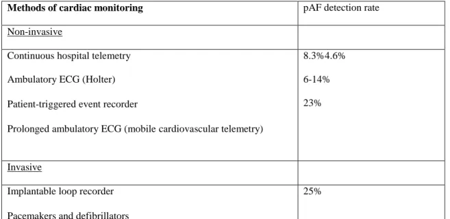
Table 1 summarizes the main methods of cardiac monitoring.
Methods of cardiac monitoring pAF detection rate Non-invasive
Continuous hospital telemetry Ambulatory ECG (Holter) Patient-triggered event recorder
Prolonged ambulatory ECG (mobilecardiovascular telemetry)
8.3%4.6% 6-14% 23%
Invasive
Implantable loop recorder Pacemakers and defibrillators
25%
Table 1 – Detection rates of paroxysmal atrial fibrillation (pAF) of different methods of cardiac monitoring
28
There is currently undergoing a clinical trial “Cryptogenic Stroke and underlying Atrial Fibrillation” (CRYSTAL AF), whose goal is to evaluate the incidence of atrial fibrillation and time to atrial fibrillation detection in patients with cryptogenic stroke using an insertable cardiac monitor during a 1-year period [48].
Currently there is no evidence of prognostic utility in ischemic stroke of electrophysiological studies that measure the atrial refractory period and the times of conduction to define an index of atrial vulnerability to fibrillation (latent atrial vulnerability). In the epidemiological study “Ischemic stroke and atrial vulnerability” (ISAV), the presence of atrial vulnerability in patients with cryptogenic ischemic stroke did not correlate with the occurrence of events such as recurrent stroke or atrial arrhythmia [49].
As patients with paroxysmal atrial fibrillation have a high risk of recurrent stroke and anticoagulation is significantly superior to antiplatelets for the secondary prevention of cardioembolic stroke, alternative ways to detect a possible cardioembolic etiology in cryptogenic stroke must be considered.
Currently, about one third of ischemic strokes are classified as of undetermined etiology. It is possible that a fraction of these strokes named as cryptogenic may be due to an undetected episode of atrial fibrillation [50, 51].
The importance of a correct identification of stroke etiology leads me to posit that biomarkers studied acutely in patients with brain ischemia could identify some cardioembolic sources of embolism.
1.4 Biomarkers
The first reference to the term biomarker appeared in PubMed in 1977 [52]. It was first defined in 1989 in the Medical Subject Heading (MeSH) as a “ Measurable and quantifiable biological
29
parameters (e.g., specific enzyme concentration, specific hormone concentration, specific gene phenotype distribution in a population, presence of biological substances) which serve as indices for health and physiology related assessments, such as disease risk, psychiatric disorders, environmental exposure and its effects, disease diagnosis, metabolic processes, substance abuse, pregnancy, cell line development, epidemiologic studies, etc.” [53]. In 2008, the definition was reviewed and biomarker became defined as “a molecular, biological, or physical characteristic that indicates a specific physiologic state. It is used in clinical practice to identify risk for disease, diagnose disease and its severity, guide intervention strategies, and monitor patient responses to therapy” [54].
Biomarkers have been progressively recognized as important diagnostic tools. Ideally, a biomarker should be highly sensitive, specific, accessible, accurate, reproducible by an analytical method, cost-effective and have a result that can be easily interpreted by a physician [55].
Examples of blood biomarkers used in clinical practice include cardiac troponins in myocardial infarction, human choriogonadotropin to diagnose pregnancy, and creatinine that is used to monitor renal function.
In the context of stroke, biomarkers have been studied for [56]: - Diagnosis of ischemic stroke;
- Diagnosis of ischemic stroke versus hemorrhagic stroke;
- Identification of ischemic stroke phases (as markers of definitive ischemic lesion or of potentially recoverable tissue corresponding to penumbra);
- Determination of stroke etiology;
- Determination of clinical prognosis;
30
Biomarkers under investigation in stroke are or specific to the central nervous system or markers of systemic inflammation, fibrinolysis or hemostasis. Possible biomarkers from the blood, cerebrospinal fluid and brain tissue have been investigated [57]. Preferably a blood or serum derived biomarker should be used, as it is more accessible, less invasive and could be monitored in time through serial measurements.
There are two great obstacles to the use of biomarkers in cerebrovascular pathology:
a) The presence of the blood-brain barrier that difficult and delays the release of proteins of neuronal or glial origin into the bloodstream after stroke;
b) Many of the potential serum biomarkers of cerebral ischemia and inflammation have a low specificity and may also be increased in situations that can be confounded with stroke in their presentation such as acute myocardial infarction or central nervous system inflammation [58]
Presently around 58 possible stroke related biomarkers have been studied. Four of these proteins have been studied as possible blood based biomarkers of cardioembolic stroke. There are also studies investigating if there are gene expressions signatures in blood that could be suggestive of cardioembolic stroke [59,60]. Using whole-genome microarrays, a 40-gene profile that distinguished cardioembolic stroke from large-vessel stroke, and a separate 37-gene profile that distinguished cardioembolic stroke due to atrial fibrillation from nonatrial fibrillation causes was identified. These genes play roles in inflammation [59]. Nevertheless none of these biomarkers can be recommend to be used in clinical practice.
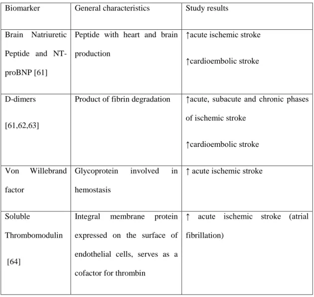
Table 2 displays the characteristics of the main serum biomarkers that have been evaluated in the context of ischemic stroke related to atrial fibrillation.
31
Biomarker General characteristics Study results
Brain Natriuretic Peptide and NT-proBNP [61]
Peptide with heart and brain production
↑acute ischemic stroke
↑cardioembolic stroke
D-dimers
[61,62,63]
Product of fibrin degradation ↑acute, subacute and chronic phases of ischemic stroke ↑cardioembolic stroke Von Willebrand factor Glycoprotein involved in hemostasis
↑ acute ischemic stroke
Soluble
Thrombomodulin
[64]
Integral membrane protein expressed on the surface of endothelial cells, serves as a cofactor for thrombin
↑ acute ischemic stroke (atrial fibrillation)
Table 2 – Resume of the characteristics of the main serum biomarkers that have been studied in the context of ischemic stroke related to atrial fibrillation, ↑ - increased
One of the substances that have been studied as a possible biomarker of cardioembolic stroke is the N-terminal of the brain natriuretic peptide (NT-proBNP).
32
1.5 Brain natriuretic peptide and N-terminal proBrain natriuretic peptide
NT-pro BNP is part of a group of natriuretic peptides, phylogenetically conserved along time that includes peptides such as atrial natriuretic peptide (ANP), natriuretic peptide type C, urodilatin and the Dendroaspis natriuretic peptide [65].
NT-proBNP is coded by a gene composed by three exons and two introns that is located in chromosome 1p36.2. It is initially produced as a prepropeptide of 143 amino acids. This prepropeptide is proteolytic cleaved into a non-active N-terminal fragment composed by 108 amino acids designated proBNP. ProBNP after secretion is divided in two fractions by two proteolytic enzymes. These two fractions are BNP that is biological active (aa 77-108) and the N-terminal-proBNP (proBNP) (aa 1-76) without biological activity [66]. BNP and NT-proBNP concentrations can be determined in blood samples [67]. The half-life of these two peptides differs. NT-proBNP has a half-life superior to BNP. NT-proBNP has a half-life of 120 minutes and BNP a half-life of 22 minutes [68]. It is due to this difference in half-life that most essays measure NT-proBNP instead of BNP.
Although NT-proBNP was initially isolated in porcine brain in 1988 [69], explaining its designation as brain natriuretic peptide, subsequent experiences showed that it also had a cardiac production [70]. Currently it is considered that the heart is its main production site. In the brain, NT-proBNP is mainly produced in the hypothalamus. The cerebral cortex, thalamus, pons have also been pointed as possible production sites [71]. NT-proBNP in normal conditions does not cross the bold-brain barrier [72].
In the heart, BNP can be produced by both atria and ventricles [72]. After being produced NT-proBNP and BNP are mainly secret in “bursts”. Their storage in granules is minimal [73].
Concerning tissue expression, BNP seems to be more present in the atria rather than in the ventricles. However, due to the larger ventricular mass, 70% of all BNP is produced by the
33
ventricle in normal conditions [74]. The observation of ventricular BNP gene expression in ventricular disease may have contributed to the common notion that BNP is mainly a ventricular hormone.
The main stimulus for the synthesis and excretion of BNP is myocites stretching, mainly in the context of volume [75] or cardiac pressure overload [76]. After myocites stretching there is a rapid activation, within hours, of NT-ptoBNP gene expression [77]. Heart fibroblasts can also produce NT-proBNP. Heart hypertrophy, fibrosis and hypoxia can also stimulate NT-proBNP production [78]. Certain hormones (catecholamines, angiotensin II and endothelin-1) may stimulate the production of NT-proBNP through paracrine or endocrine mechanisms [79]. Biological actions of BNP are mediated by the receptor of the natriuretic peptides of type A (NPR-A). This receptor is a guanylyl cyclase receptor. It is a transmembrane receptor with 120 kD. It is composed by an extracellular region that connects to BNP and an intracellular kinase and guanylyl cyclase domain with enzymatic activity. This receptor is mainly located in the cellular membrane of the endothelium of small vessels [80]. BNP connection to the receptor leads to the conversion of guanosine triphosphate (GTP) into cyclic guanosine monophosphate (cGMP) [81]. A kinase protein dependent on cGMP (PKG or cGK) is the principal intracellular mediator of cGMP signals through a catalytic transference of the phosphate of present in TPA to a serine or threonine residue present in the target protein [82]. Signal transduction activated by these receptors is terminated by a GMP phosphodiesterase that modulates the intracellular concentration of cGMP and the duration and magnitude of the response [83].
BNP acts by an endocrine mechanism. It has an important role in the cardiovascular homeostatic regulation and in volume control. BNP biological effects include natriuresis, diuresis, vasodilatation, inhibition of the renin-angiotensin-aldosterone axis and of the sympathetic nervous system [84]. In the heart BNP has a lusitropic and antifibrotic effect [85].
34
Know mechanisms of BNP inactivation include connection to the type C receptor of clearance of the natriuretic peptides and proteolysis by the peptidase NEP 24.1. NT-proBNP is cleared from circulation through the kidney by direct filtration and passive excretion [86].
In healthy individuals, BNP and NT-proBNP plasmatic concentrations are similar. They can be detected in venous blood samples in picomolar concentrations. However, in congestive heart failure patients, NT-proBNP concentration is two to ten times higher than BNP concentration. The explanation for this difference is unknown. Serum NT-proBNP and BNP levels in healthy individuals increase with age and are higher in women than in men [87]. The reason for this age-related increase in BNP or NT-proBNP is presumed to be related to the parallel age increase in subclinical structural heart disease, including heart muscle disease, diastolic abnormalities, valve disease and arrhythmia [88]. An age-related decrease in renal function is also considered to be partially responsible. Ninety percent of young adults have NT-proBNP levels below 70 pg/mL [89]. NT-proBNP plasmatic levels correlate with left ventricular mass. A NT-proBNP cut-off superior to 300 pg/mL has been suggested for the diagnosis of congestive heart failure [90]. NT-proBNP is currently used as a marker of left ventricular dysfunction and of prognosis in patients with congestive heart failure and in acute ischemic heart disease [91]. High NT-proBNP levels have been registered in patients with renal failure, anaemia, acute pulmonary embolism, pulmonary hypertension, sepsis, hyperthyroidism, mitral and aortic regurgitation and dysrhythmias such as atrial fibrillation [92, 93].
Patient with lone paroxysmal atrial fibrillation seem to have increased NT-proBNP levels when compared to patients in sinus rhythm, in the absence of a structural heart disease [94]. Increased plasmatic levels of NT-proBNP have been found in patients with permanent or paroxysmal atrial fibrillation with preserved left ventricular systolic function [95]. One study showed that in 81% of patients NT-proBNP levels increased within four hours of the onset of atrial fibrillation [96]. It has also been documented, after electric cardioversion of atrial fibrillation to sinus rhythm, a return to baseline of NT-proBNP values [97]. NT-proBNP has been shown to be a remarkable predictor of incident atrial fibrillation in the general population, independently of
35
any other previously described risk factor [98]. Recent studies that suggest that NT-proBNP is a predictor of atrial fibrillation following cardiac surgery [99-101] and successful cardioversion [102].
There is currently no established explanation for the increase of BNP in the context of atrial fibrillation. In the isolated atria tissue of rat, alpha1-adrenergic stimulation with phenylephrine induces genetic expression of BNP [103]. It is unknown if alpha1-adrenergic stimulation results in an increase in the synthesis of BNP in patients with atrial fibrillation. A study that analysed human right atrial tissue found that persistent atrial fibrillation was associated to a high expression of proBNP mRNA. However, patients with paroxysmal atrial fibrillation did not have changes in proBNP gene expression [104].
1.6 NT-proBNP and BNP in ischemic stroke
Although NT-proBNP presence was first described in the brain tissue, information regarding NT-proBNP in ischemic stroke is small.
Some studies showed an acute increase in NT-proBNP and BNP serum levels in acute ischemic stroke (Table 3). These studies initially aimed to determine if NT-proBNP or BNP could be used as prognostic markers of acute ischemic stroke along with other markers of myocardial injury such like troponins. It was suggested that it could be used as a marker of long term bad prognosis. It was then noticed that patients with acute ischemic stroke tended to have higher levels of NT-proBNP and BNP when taking in account the cut-off points used for the diagnosis of congestive heart failure.
Four possible explanations for the increase of NT-proBNP and BNP during acute ischemic stroke where proposed in these studies:
36
- Patients with acute ischemic stroke frequently have several vascular risk factors and may have an associated acute or chronic heart failure. Therefore, the increase in NT-proBNP levels could be due to ventricular dysfunction;
- The increase in NT-proBNP levels could be due to atrial fibrillation. Atrial fibrillation is a major risk factor for ischemic stroke and a known cause of NT-proBNP increase;
- As the brain is a site of BNP and NT-proBNP production, although it only produces a small amount of these peptides it is possible that after an acute ischemic lesion there is a release of BNP and NT-proBNP that is measurable in the plasma.
- BNP has an inhibitory action in the central and peripheral sympathetic nervous system. BNP could be increased in acute ischemic stroke as a response to the increase sympathetic activity that occurs after stroke
- Autonomic changes of acute ischemic stroke are frequently associated to right insular cortical ischemia [105]. Insular cortical ischemia has been associated to an increase in the serum levels of norepinephrine, electrocardiographic changes and changes in the circadian patterns of blood pressure. In these cases, there is an inflammatory response with increase of interleukin 6. Interleukin 6 can directly increase BNP and NT-proBNP levels.
Study Nº of
patients
Results
Etgen, 2005 [106]
174 Increase of NT-proBNP levels in acute ischemic stroke, when using as cut-off points the levels used for the diagnosis of congestive heart failure
37
dysfunction or be due to sympathetic nervous system activation
Makikallio, 2005 [107]
51 Increase of NT-proBNP levels in acute ischemic stroke when comparing with healthy controls.
Increase of NT-proBNP could have a brain origin
Nakagawa, 2005 [108]
88 Increase of NT-proBNP in the acute phase of ischemic stroke, decrease in the subacute phase
NT-proBNP levels correlated with mean blood pressure
Giannakoulas, 2005 [109]
30 Higher increase of NT-proBNP in cardioembolic stroke than in large vessels diseases related ischemic stroke
Increase of NT-proBNP due to brain ischemia or direct myocardial dysfunction
Jensen 2006 [110]
250 NT-proBNP levels in the second day after ischemic stroke associated to 6 months mortality
Itumur 2006 [111]
57 Patients with ischemic stroke with insular cortical involvement and with signs of myocardial ischemia had higher levels of NT-proBNP than other patients with ischemic stroke. However this increase was not statically significant
Yip 2006 [112]
86 Ischemic stroke patients with higher NT-proBNP levels had a higher number of unfavorable clinical events (acute myocardial infarction, congestive heart failure, recurrent stroke or any other cause of death)
38
[113] independently associated to congestive heart failure
Di
Angelantonio 2007 [114]
48 BNP is a marker of left atrial dysfunction even in the absence of atrial fibrillation
Montaner 2008 [61]
707 Several potential biomarkers were studied.
Levels of BNP> 76 pg/mL in patients with acute ischemic stroke indicated a cardioembolic etiology with a sensitivity of 72%, a specificity of 69% and a negative predictive value of 82%.
Naya 2008 [115]
76 NT-proBNP levels and the evaluation of flow in the left atria appendage could help to distinguish cardioembolic stroke related to atrial fibrillation from other etiologies
Gartner 2008 [116]
222 A statistically significant association was found between the plasmatic concentration of BNP and the severity of clinically silent brain infarctions related to atrial fibrillation
Tomita 2008 [117]
79 Plasmatic levels of BNP were statistically significantly higher in patients with acute ischemic stroke with a large vessels etiology. Patients with a cardioembolic etiology were excluded from the study. BNP levels were positively correlated with the NIH stroke scale classification and with the volume of the ischemic lesion
Shibazaki K, 2009 [118]
200 Plasma BNP level is significantly higher in cardioembolic patients than in other stroke subtypes, and thus physicians should strongly consider cardioembolism when the plasma BNP level is over 140.0 pg/mL in patients with acute ischemic stroke
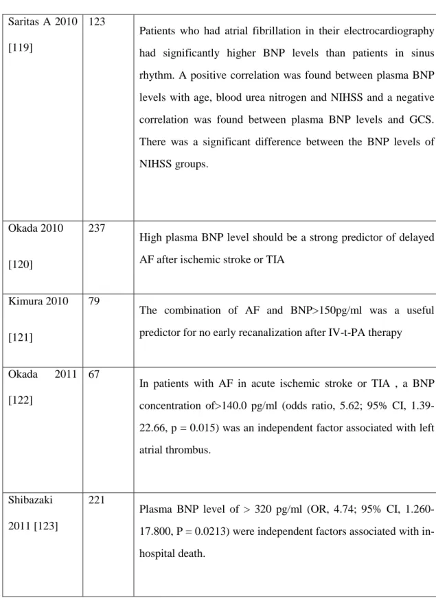
39 Saritas A 2010
[119]
123
Patients who had atrial fibrillation in their electrocardiography had significantly higher BNP levels than patients in sinus rhythm. A positive correlation was found between plasma BNP levels with age, blood urea nitrogen and NIHSS and a negative correlation was found between plasma BNP levels and GCS. There was a significant difference between the BNP levels of NIHSS groups.
Okada 2010
[120]
237
High plasma BNP level should be a strong predictor of delayed AF after ischemic stroke or TIA
Kimura 2010
[121]
79
The combination of AF and BNP>150pg/ml was a useful predictor for no early recanalization after IV-t-PA therapy
Okada 2011 [122]
67
In patients with AF in acute ischemic stroke or TIA , a BNP concentration of>140.0 pg/ml (odds ratio, 5.62; 95% CI, 1.39-22.66, p = 0.015) was an independent factor associated with left atrial thrombus.
Shibazaki 2011 [123]
221
Plasma BNP level of > 320 pg/ml (OR, 4.74; 95% CI, 1.260-17.800, P = 0.0213) were independent factors associated with in-hospital death.
Table 3- Previous studies reporting NT-proBNP or BNP changes in patients with acute ischemic stroke
40
In a previous study [124], we analyzed whether NT-proBNP was elevated in patients with acute ischemic stroke of cardiac cause. We used a sample of consecutive acute stroke patients with ischemic or intracerebral hemorrhage that were admitted from November 2007 to August 2008 to the Stroke Unit of Hospital de Santa Maria, Lisbon, Portugal.
Patients with a history of conditions known to increase NT-proBNP (acute ischemic heart disease, heart failure, heart valve disease, renal failure, cardiomyopathies, pulmonary hypertension, anemia) were excluded. Patients with subarachnoid hemorrhage, cerebral venous thrombosis and transient ischemic attack were also not included in this study.
After patient admission from the emergency department, information on demography, vascular risk factors, or previous atrial fibrillation was collected. A brain CT scan, done after hospital admission, was used to classify strokes as hemorrhagic or ischemic. It was repeated 48–72 h after hospital admission to outline the ischemic area. When the ischemic area was not evident in a CT scan, a 1.5 T magnetic resonance imaging (MRI; including diffusion-weighted imaging) was performed. All patients had transcranial Doppler and carotid and vertebral duplex scanning. Etiological workup included, in all patients with ischemic stroke, complete blood count, erythrocyte sedimentation rate, hepatic and renal function, glucose and lipid levels, protein electrophoresis and coagulation studies. In patients less than 55 years old, workup included autoantibodies, lupus anticoagulant, anticardiolipin antibodies, C and S protein, antitrombin III, fibrinogen levels, HIV 1 and 2, Hepatitis B and C serologies. Transthoracic echocardiogram was performed in all patients. Patients with echocardiographic evidence of heart disease (shortening fraction less than 30%, valve dysfunction, cardiomyopathies, akinetic ventricular wall regions) were excluded.
The following parameters were registered: - left atria autocontrast
- intracardiac thrombus - shortening fraction
41 - akinetic ventricular wall regions.
In patients less than 55 years old a transesophageal echocardiogram was also performed. To determine the presence of atrial fibrillation at least two ECGs were done during hospital stay, and when the stroke cause remained undetermined a 24 h Holter monitoring was also performed. In patients whose stroke cause remained undetermined, lumbar puncture or cerebral angiography was done. Patients with hemorrhagic stroke had blood analysis, ECG and a transthoracic echocardiogram. Ischemic stroke causes were classified according to TOAST [4] classifications in five groups; large-artery atherosclerosis, cardioembolic, small-vessel occlusion, stroke of other determined cause and stroke of undetermined cause. For the present study, patients were subdivided in two groups; cardioembolic cause (corresponding to the cardioembolic classification of TOAST) and noncardioembolic cause (all other four groups of the TOAST classification). Ischemic stroke topography was dichotomized in anterior/carotid territory or posterior/vertebrobasilar territory, accordingly to CT or MRI information. Infarct size was classified according to the ASPECTS scale [125], using CT-scan or MRI information. To evaluate autonomic nervous system activation three measurements of systolic blood pressure (SBP), diastolic blood pressure (DBP) and heart rate (HR) were done at eight-hour intervals after patient admission. In the first 72 h after symptoms onset, four milliliters of blood was drawn from a peripheral vein. Blood samples were immediately centrifuged at 1600 g during 15 min. Serum concentration of NT-proBNP was determined by an electrochemiluminescence assay using the Elecsys 2010 immunoassay analyzer [126].
A descriptive statistical analysis of demographic and vascular risk factors of patients with ischemic stroke, hemorrhagic stroke, cardioembolic and noncardioembolic cause was performed. Data distribution was evaluated using histograms and a one-sample Kolmogorov– Smirnov test. For comparison between groups the w2-test, Fisher exact test, Mann–Whitney test or t-test were used as appropriate. As NT-proBNP values did not follow a normal distribution, a logarithm transformation was done. To compare NT-proBNP values between different timings of blood collection, a one-way analysis of variance was used. t-test was used to compare mean
42
values of NT-proBNP between patients with intracerebral hemorrhage versus ischemic stroke , cardioembolic stroke versus noncardioembolic stroke and cardioembolic stroke related to atrial fibrillation versus noncardioembolic stroke. Receiver operating characteristic (ROC) curves were used to test the ability of NT-proBNP to identify cardioembolic stroke and cardioembolic stroke associated with atrial fibrillation. The area under the curve (AUC) for each ROC curve was determined. Based on the ROC curves, NT-proBNP values with the highest sensitivity and specificity for the diagnosis of cardioembolic stroke and cardioembolic stroke related to atrial fibrillation were determined as well as the corresponding positive and negative predictive values (NPV).
To study the association between NT-proBNP and systolic blood pressure, diastolic blood pressure and heart rate, a simple linear regression was calculated. The corresponding regression coefficients with 95% confidence intervals (CI) were determined. To evaluate differences in NT-proBNP values between arterial territories, t-student test was used. To evaluate the association between ischemic area and NT-proBNP value a Kendall correlation analysis was performed.
From November 2007 to August 2008, 202 stroke patients were admitted to the Stroke Unit. Ninety-two patients were included. The main reasons for exclusion were: heart diseases known to increase NT-proBNP (32.7%), subarachnoid hemorrhage (29.1%) and patient admission 72 h after stroke onset (18.2%). Included patients had a mean age of 58.6 (SD +/- 14.4) years. Women were 64.1% of the patients. Sixty-six (71.7%) patients had an ischemic stroke and 26 (28.3%) an intracranial hemorrhage (Table 4).
Ischemic stroke (n=66) Hemorrhagic stroke (n=26) p
Gender, female (%) 26 (39.4) 7(26.9) 0.26
43
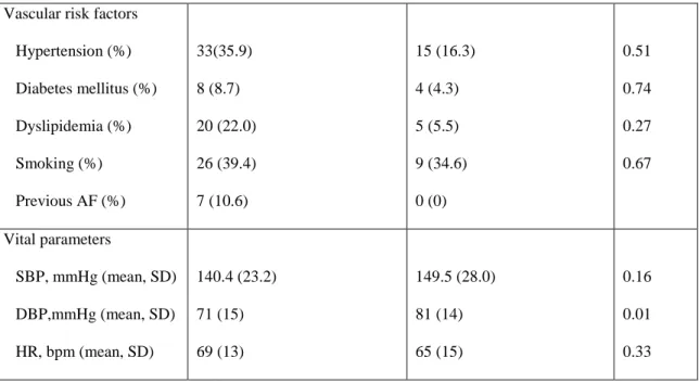
Vascular risk factors Hypertension (%) Diabetes mellitus (%) Dyslipidemia (%) Smoking (%) Previous AF (%) 33(35.9) 8 (8.7) 20 (22.0) 26 (39.4) 7 (10.6) 15 (16.3) 4 (4.3) 5 (5.5) 9 (34.6) 0 (0) 0.51 0.74 0.27 0.67 Vital parameters SBP, mmHg (mean, SD) DBP,mmHg (mean, SD) HR, bpm (mean, SD) 140.4 (23.2) 71 (15) 69 (13) 149.5 (28.0) 81 (14) 65 (15) 0.16 0.01 0.33
Table 4 – Demographic data, vascular risk factors and vital parameters of patients with ischemic and hemorrhagic stroke, SBP – Systolic blood pressure, DBP – diastolic blood pressure, HR - heart rate, SD – standard deviation, AF – atrial fibrillation
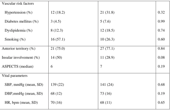
According to the TOAST classification 28 patients had a cardioembolic cause and 38 were noncardioembolic: 12 (18.2%) large arteries; seven (10.6%) small vessels; five (7.6%) other determined; 14 (21.2%) undetermined. Cardioembolic cause included 18 patients with atrial fibrillation (12 paroxysmal, six permanent) and 10 patients with patent foramen ovale. Demographic data, vascular risk factors and vital parameters of patients with cardioembolic ischemic stroke and noncardioembolic ischemic stroke were not significantly different (Table 2). The two subgroups of patients with cardioembolic and noncardioembolic cause were not significantly different concerning arterial territory or insular involvement or infarct size evaluated by the ASPECTS scale (Table 5).
Cardioembolic stroke (n=28) Noncardioembolic stroke (n=38) p
Gender, male (%) 15 (53.6) 25 (65.8) 0.32
44
Vascular risk factors Hypertension (%) Diabetes mellitus (%) Dyslipidemia (%) Smoking (%) 12 (18.2) 3 (4.5) 8 (12.3) 16 (57.1) 21 (31.8) 5 (7.6) 12 (18.5) 10 (26.3) 0.32 0.99 0.74 0.60 Anterior territory (%) Insular involvement (%) ASPECTS (median) 21 (75.0) 14 (50) 6 27 (77.1) 11 (28.9) 7 0.84 0.08 0.19 Vital parameters SBP, mmHg (mean, SD) DBP,mmHg (mean, SD) HR, bpm (mean, SD) 139 (22) 68 (12) 70 (16) 141 (24) 73 (16) 68 (11) 0.68 0.19 0.65
Table 5 – Demographic data, vascular risk factors, stroke characteristics and vital parameters of patients with cardioembolic and noncardioembolic stroke, SBP – Systolic blood pressure, DBP – diastolic blood pressure, HR - heart rate, SD – standard deviation
No intracardiac thrombus or left atria autocontrast were detected during echocardiography. The NT-proBNP values followed a positively skewed distribution and ranged from 8 to 6378 pg/ml with a median of 177.0 pg/ml. After a logarithm transformation, a new variable was obtained, which followed a normal distribution. In 21 patients (22.8%), blood was collected in the first 24 h, in 62 patients (67.4%) in the first 24–48 h and in nine patients (9.8%) in the first 48–72 h. The mean value (95% CI) of NT-proBNP in patients with ischemic stroke was 223.18 (157.42– 316.40) pg/ml and in patients with hemorrhagic stroke was 133.34 (74.13–239.82) pg/ml. However, this difference was not statistically significant (P=0.12). The mean of NT-proBNP values in patients with ischemic stroke in the carotid artery territory was 275.88 (95% CI 179.47–419.89) pg/ml and in patients with stroke in the vertebrobasilar territory was 138.83 (74.44– 259.82) pg/ml. This difference was not statistically significant (P=0.10). No statistically
45
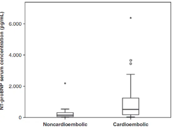
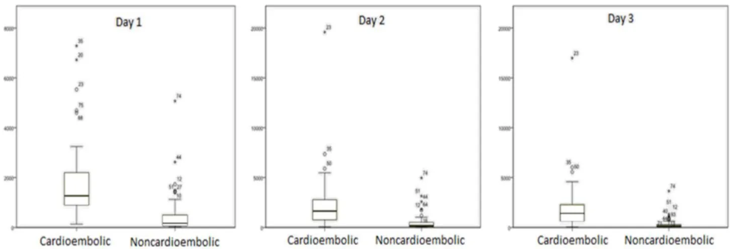
significant association was found between infarct size evaluated by ASPECTS scale and serum values of proBNP (P=0.11). No significant linear relationship was found between NT-proBNP values and systolic blood pressure (P=0.091) or diastolic blood pressure (P=0.26). A significant linear relationship was found between NT-proBNP values and heart rate with a regression coefficient of 0.025 pg/ml/bpm, (P= 0.039). The mean of NT-proBNP values for cardioembolic stroke was significantly higher (P<0.001) (491.6; 95% CI 283.7–852.0 pg/ml) than for noncardioembolic ischemic stroke (124.7; 86.3–180.2 pg/ml) (Figure 1).
Figure 1- Serum concentration of NT-proBNP in patients with noncardioembolic and cardioembolic ischemic stroke. Boxplots present median values and interquartile ranges
The ROC curve of NT-proBNP values for the diagnosis of cardioembolic stroke had an AUC (95% CI) of 0.77 (0.65–0.89). The cut-off point with the highest sensitivity and specificity was set at 265.5 pg/ml (71.4% and 73.7% respectively). This point had a NPV of 77.8%and a
46
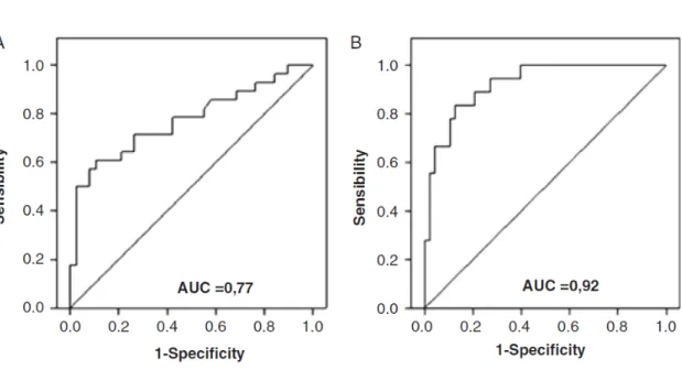
positive predictive value (PPV) of 66.6% (Figure 2). The AUC of NT-proBNP obtained for the diagnosis of cardioembolic stroke related to AF was 0.92 (0.86–0.99). This AUC value was higher than the value obtained for the diagnosis of cardioembolic stroke in general (0.92 versus. 0.77). After analysis of the ROC curve for the diagnosis of cardioembolic stroke related to atrial fibrillation a cut-off point of 265.50 pg/ml was determined (sensitivity of 94.4%, specificity of 72.9%, positive predictive value of 56.6% and a negative predictive value of 97.2%). This cut-off point had an extremely high negative predictive value. However, in the context of clinical decisions, it is more important to confirm the diagnosis of cardioembolism as it leads to a change in clinical decision; therefore another cut-off point with a higher positive predictive value was determined. The cut-off point of 912.0 pg/ml had a sensitivity of 55.5%, specificity of 97.9%, positive predictive value of 90.9%, and a negative predictive value of 83.9% (Figure 2).
Figure 2 –ROC curves illustrating the accuracy of NT-proBNP to identify cardioembolic stroke (A) and cardioembolic stroke associated to atrial fibrillation (B)
This study suggests that the increase of NT-proBNP which occurs during stroke has a cardiac origin and may be due to atrial fibrillation. The mean value of NT-proBNP in patients with
47
cardioembolic ischemic stroke was significantly higher than in patients with noncardioembolic ischemic stroke. No significant association was found between stroke territory or infarct size and proBNP values, or between systolic blood pressure or diastolic blood pressure and NT-proBNP values. The ROC curve for the diagnosis of cardioembolic stroke had an AUC of 0.77 which corresponds to a good ability of NT-BNP to diagnose cardioembolic stroke. The ROC curve for the diagnosis of cardioembolic stroke associated to atrial fibrillation had an AUC of 0.92 which suggests that NT-proBNP has a very good ability to diagnose ischemic stroke associated to atrial fibrillation, being useful to differentiate it from other etiologies. Although previous studies had suggested a cardiac cause they had not excluded the presence of confounding variables, which could have caused the increase of NT-proBNP. Namely Montaner [61] and Shibazaki [118] did not exclude the presence of renal failure, heart failure and ischemic heart disease. In our study, to decrease possible confounding factors, patients with known causes of NT-proBNP increase, such as renal failure and heart failure, ischemic heart disease and valvular heart disease, were excluded, using both clinical and echocardiographic evidence. Also, Montaner [61] and Shibazaki [118] did not analyze if this increase could be due to the large infarct area that patients with cardioembolic stroke tend to have [127]. In their patients, the increase of proBNP could have been due to a large infarct area, as it is established that NT-proBNP is also produced in the brain [69, 128]. In our study, we analyzed this variable and we did not find an association between stroke territory or infarct size and NT-proBNP values. One recent study [122] suggested that the increase of NT-proBNP in stroke could be due to atrial fibrillation. However, the authors did not exclude patients with heart failure and found that the variable ‘congestive heart failure’ was significantly higher in the atrial fibrillation group than in non-atrial fibrillation group. Therefore the increase of the NT-proBNP could not be securely attributed to atrial fibrillation, because heart failure is a cause of NT-proBNP increase [91]. In our study, levels of NT-proBNP were compared between hemorrhagic and ischemic stroke based in the hypothesis that if the increase of NT-proBNP was purely due to a ischemic stroke cause subtype – cardioembolic – and not to other factors such as autonomic activation, there
48
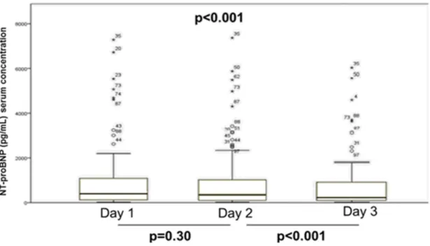
would be higher levels of NT-proBNP in patients with ischemic stroke than in hemorrhagic stroke. In fact, patients with ischemic stroke had higher levels of NT-proBNP (223.18 (157.42– 316.40) pg/ml) than patients with hemorrhagic stroke (33.34 (74.13–239.82) pg/ml). However, this difference was not statistically significant (P=0.12) probably due to the modest number of patients included. When comparing only patients with ischemic stroke, after excluding possible confounding variables, we found that the mean value of NT-proBNP in patients with cardioembolic ischemic stroke was significantly higher than in patients with noncardioembolic ischemic stroke. Due to the exclusion criteria, the number of possible heart embolic sources was restricted to two: atrial fibrillation and patent foramen ovale. Patent foramen ovale can be a cardioembolic source due to paradoxical embolism or due to the induction of changes in the electrical activity of the left atria leading to atrial arrhythmias, such as paroxysmal atrial fibrillation or atrial flutter [129]. It may be therefore able to increase NT-proBNP values. After analysis of the ROC curve for ischemic stroke associated to atrial fibrillation, a cut-off point of 265.5 pg/ml with a high sensitivity (94.4%) and a high negative predictive value (97,2%) was determined. The cut-off point of 912.0 pg/ml had a positive predictive value of 90.9%. If this cut-off point is confirmed in another sample it can lead to a high suspicion of atrial fibrillation in patients with stroke of undetermined cause. A NT-proBNP level above this cut-off may help to select patients for prolonged heart rhythm monitoring to detect paroxysmal atrial fibrillation. In the previously mentioned studies [61, 121, 122], blood was drawn in the first 24 h after stroke onset. In our study, blood was drawn in the first 72 h after stroke onset (although mostly in the first 24–48 h). The option for an enlarged inclusion time was based on the knowledge that a sizable proportion of patients does not go to the hospital in the first 24 h after stroke onset [130]. Available data is unclear regarding the timing when maximum serum concentration of NT-proBNP in ischemic stroke is achieved. Giannakoulas [109] did not find a statistically significant difference between day one and six after stroke onset. Jensen [110] noticed a peak in the second day, with a progressive decrease in NT-proBNP until day five. Iltumur [111] described highest values of NT-proBNP in the day of stroke onset. In our study no significant
49
difference was found between the different timings of drawing blood samples. One limitation of our study is the modest number of patients included; nevertheless it obtained some statistically significant results. Other limitations relate to the use, in the majority of patients, of CT instead of MRI to evaluate the infarct area and location. The study of possible biomarkers of ischemic stroke subtypes may be clinically valuable. Our results suggest that NT-proBNP can be a useful biomarker of certain causes of cardioembolic stroke, namely atrial fibrillation. Our results must be replicated in an independent sample. It is necessary to conduct another study on a different sample, with a larger number of patients to validate the cutoff points established. It is also necessary to identify possible sources of variation of NT-proBNP and in particular to establish the profile of time course of NT-proBNP after an ischemic stroke.
References
[1] Murray CJ, Lopez AD. Global mortality, disability, and the contribution of risk factors: Global Burden of Disease Study. Lancet 1997;349:1436-1442.
[2] Bonhorst D, Mendes M, Adragão P, De Sousa J, Primo J, Leiria E, Rocha P. Prevalence of atrial fibrillation in the Portuguese population aged 40 and over: the FAMA study. Rev Port Cardiol. 2010;29:331-350.
[3] Sandercock PA, Warlow CP, Jones LN, Starkey IR. Predisposing factors for cerebral infarction: the Oxfordshire community stroke project. Br Med J 1989; 298:75-80.
50
[4] Adams HP Jr, Bendixen BH, Kappelle LJ et al. Classification of subtype of acute ischemic stroke: definitions for use in a multicenter clinical trial: TOAST Trial of Org 10172 in acute stroke treatment. Stroke 1993; 24:35–41.
[5] Amarenco P, Bogousslavsky J, Caplan LR, Donnan GA, Hennerici MG. New approach to stroke subtyping: the A-S-C-O (phenotypic) classification of stroke. Cerebrovasc Dis. 2009;27:502-508.
[6] Caplan, LR, Manning, W (Eds). Brain embolism, Informa Healthcare, New York 2006; 129-159.
[7] Lip GY, Brechin CM, Lane DA. The global burden of atrial fi brillation and stroke: a systematic review of the epidemiology of atrial fi brillation in regions outside North America and Europe. Chest 2012; published online March 29. DOI:10.1378/chest.11-2888.
[8] Morais J, Osterspey A, Tamargo JL, Zamorano JL, Despres C, Dickstein K, Lekakis J, McGregor K, Metra M, Blanc JJ, Budaj A, Camm J, Dean V, Deckers JW, Riegel, ESC COMMITTEE FOR PRACTICE GUIDELINES, Priori SG, Hunt SA, Nishimura R, Ornato JP, Page RL, Cynthia B, Adams D, Anderson JL, Antman EM, Halperin JL, Wann, ACC/AHA TASK FORCE MEMBERS, Smith SC, Jacobs AK, Lowe JE, Olsson SB, Prystowsky EN, Tamargo JL, Kenneth S, Ellenbogen A, Halperin JL, Le Heuzey JY, Kay GN. ACC/AHA/ESC 2006 Guidelines for the Management of Patients with AF. Circulation 2006;114:e257-e354.
[9] Schmidt C, Kisselbach J, Schweizer PA, Katus HA, Thomas D. The pathology and treatment of cardiac arrhythmias: focus on atrial fibrillation. Vasc Health Risk Manag. 2011;7:193-202.
51
[10] Hodgson-Zingman DM, Karst ML, Zingman LV, Heublein DM, Darbar D, Herron KJ, Ballew JD, de Andrade M, Burnett JC Jr, Olson TM. Atrial natriuretic peptide frameshift mutation in familial atrial fibrillation. N Engl J Med. 2008;359:158–165
[11] Blackshear JL, Odell JA. Appendage obliteration to reduce stroke in cardiac surgical patients with atrial fibrillation. Ann Thorac Surg 1996;61:755–759.
[12] Pongratz G, Brandt-Pohlmann M, Henneke KH, ohle C, Zink D, Gehling G, Bachmann K. Platelet activation in embolic and preembolic status of patients with nonrheumatic atrial fibrillation. Chest 1997;111:929-933.
[13] Guo Y, Lip GY, Apostolakis S. Inflammation in atrial fibrillation. J Am Coll Cardiol. 2012 Dec 4;60
[14] Fitzmaurice DA, Hobbs FDR, Jowett S, et al. Screening versus routine practice for detection of atrial fi brillation in people aged 65 or over: cluster randomised controlled trial. BMJ 2007; 335::2263-70.
[15] Wang TJ, Massaro JM, Levy D, Vasan RS, Wolf PA, D'Agostino RB, Larson MG, Kannel WB, Benjamin EJ. A risk score for predicting stroke or death in individuals with new-onset atrial fibrillation in the community: the Framingham Heart Study. JAMA. 2003;290:1049-1056.
[16] Rudehill A, Olsson GL, Sundqvist K. ECG abnormalities in patients with subarachnoid haemorrhage and intracranial tumors. J Neurol Neurosurg Psychiatry 1987;50:1375-1381.
[17] Ryder KM and Benjamim EJ. Epidemiology and significance of atrial fibrillation. American Journal of Cardiology 1999;84:131-138.
52
[18] Hohnloser SH, Crijns HJ, van Eickels M, Gaudin C, Page RL, Torp-Pedersen C, Connolly SJ; ATHENA Investigators. Effect of dronedarone on cardiovascular events in atrial fibrillation. N Engl J Med 2009;360:668-678.
[19] Saxonhouse SJ, Curtis AB. Risks and benefits of rate control versus maintenance of sinus rhythm. Am J Cardiol 2003; 91: 27-32.
[20] Hohnloser SH, Capucci A, Fain E, Gold MR, van Gelder IC, Healey J, Israel CW, Lau CP, Morillo C, Connolly SJ. ASymptomatic atrial fibrillation and Stroke Evaluation in pacemaker patients and the atrial fibrillation Reduction atrial pacing Trial (ASSERT). Am Heart J. 2006;152:442–447.
[21] Healey JS, Connolly SJ, Gold MR, Israel CW, Van Gelder IC, Capucci A, Lau CP, Fain E, Yang S, Bailleul C, Morillo CA, Carlson M, Themeles E, Kaufman ES, Hohnloser SH. . Subclinical atrial fibrillation and the risk of stroke. N Engl J Med. 2012;366:120–129.
[22] Ellinor PT, Yoerger DM, Ruskin JN, et al. Familial aggregation in lone atrial fibrillation. Hum Genet. 2005;118:79–184.