Predictors of
de novo
atrial fibrillation in a
non-cardiac intensive care unit
INTRODUCTION
The prevalence of atrial fibrillation (AF) is high, reaching 10% in individuals over 80 years of age.(1-3) AF is associated with longer stays in the hospital and
intensive care unit (ICU),(2,4) and de novo AF in critically ill patients is associated
with higher mortality.(5) The clinical complexity of patients in the ICU requires
rapid diagnosis and effective treatment of this condition.(6-8)
In this context, knowledge of the epidemiology of this event in critically patients becomes important. The incidence of de novo AF ranges from 5 to 65%, depending on the type of ICU, and is higher in patients undergoing cardiac surgery.(9-18) In turn, the large variation in the incidence of de novo AF
in the various types of ICU can be explained by different predictors of AF occurrence.
João Bicho Augusto1, Ana Fernandes2, Paulo Telles de Freitas2, Victor Gil3, Carlos Morais1
1. Department of Cardiology, Hospital Professor Doutor Fernando Fonseca - Lisbon, Portugal. 2. Polyvalent Intensive Care Unit, Hospital Professor Doutor Fernando Fonseca - Lisbon, Portugal.
3. Cardiovascular Unit, Hospital dos Lusíadas - Lisbon, Portugal.
Objective: To assess the predictors of de novo atrial fibrillation in patients in a non-cardiac intensive care unit.
Methods: A total of 418 hospitalized patients were analyzed between January and September 2016 in a non-cardiac intensive care unit. Clinical characteristics, interventions, and biochemical markers were recorded during hospitalization. In-hospital mortality and length of hospital stay in the intensive care unit were also evaluated.
Results: A total of 310 patients were included. The mean age of the patients was 61.0 ± 18.3 years, 49.4% were male,
and 23.5% presented de novo atrial
fibrillation. The multivariate model identified previous stroke (OR = 10.09; p = 0.016) and elevated levels of pro-B type natriuretic peptide (proBNP, OR = 1.28 for each 1,000pg/mL increment;
Conflicts of interest: None.
Submitted on December 9, 2017 Accepted on January 15, 2018
Corresponding author:
João Bicho Augusto
Hospital Professor Doutor Fernando Fonseca IC19, 2720-276 Amadora
Lisboa, Portugal
E-mail: joao.augusto@hff.min-saude.pt
Responsible editor: Leandro Utino Taniguchi
Preditores de fibrilação atrial de novo em unidade de cuidados
intensivos não cardíaca
ABSTRACT
Keywords: Atrial fibrillation/ epidemiology; Incidence; Intensive care p = 0.004) as independent predictors of de novo atrial fibrillation. Analysis of the proBNP receiver operating characteristic
curve for prediction of de novo atrial
fibrillation revealed an area under the curve of 0.816 (p < 0.001), with a sensitivity of 65.2% and a specificity of 82% for proBNP > 5,666pg/mL. There were no differences in mortality (p = 0.370), but the lengths of hospital stay (p = 0.002) and stay in the intensive care unit (p = 0.031) were higher in patients with de novo atrial fibrillation.
Conclusions: A history of previous stroke and elevated proBNP during hospitalization were independent predictors of de novo atrial fibrillation in the polyvalent intensive care unit. The proBNP is a useful and easy- and quick-access tool in the stratification of atrial fibrillation risk.
Some of these predictors of de novo AF have already been described in the literature, especially in critical cardiac patients, such as advanced age, greater severity score on admission, surgical or post-trauma admission, occurrence of sepsis, and need for ventilatory or catecholamine support. However, for medical and non-cardiac surgical ICU patients, there is a paucity of data in the literature regarding predictors of de novo AF.(4)
Therefore, the objective of this study was to investigate the predictive factors of de novo AF in patients in a non-cardiac polyvalent ICU (critically ill and non-non-cardiac surgical patients). As secondary objectives, the incidence
of de novo AF and its prognostic impact in terms of
in-hospital mortality and length of hospital and ICU stay were also evaluated.
METHODS
A sample of patients hospitalized during a period of 9 months (January 1, 2016 to September 30, 2016) in a non-cardiac polyvalent ICU at the Fernando Fonseca Hospital, Lisbon, Portugal, were retrospectively and consecutively analyzed.
The data were obtained through clinical consultations and were complemented by analytical and other diagnostic evaluations. The Hospital Ethics Committee approved the study, and informed consent was not required given the study’s observational nature.
The ICU had 14 beds. Patients with a pathology requiring mechanical ventilation, trauma patients, and non-cardiac surgery patients were admitted.
All patients were under continuous cardiac monitoring with three leads. The presence of an absolutely irregular RR interval with no apparent P waves or the replacement of these by AF waves was classified as AF, with subsequent confirmation on a 12-lead electrocardiogram. For classification as de novo AF, all patients with sinus rhythm on ICU admission and without any record of prior AF or atrial flutter (documented electrocardiographically, in a previous medical report or indicated by the patient and/or family) were considered. To this end, the national platform of medical records, called the Health Data Platform (Plataforma de Dados da Saúde), was also consulted. Patients with a definite pacemaker on admission or previous cardiac surgery, chest trauma, or pulmonary thromboembolism in the last year (the latter two associated with a higher risk of de novo AF) were excluded from this group.
Each patient was classified according to the reason for hospitalization: medical, surgical, or trauma. Each patient was further stratified on admission according to the in-hospital mortality scores Acute Physiology and Chronic
Health Evaluation II (APACHE II)(19) and Simplified
Acute Physiology Score (SAPS II).(20)
The presence of the following cardiovascular disease and risk factors was evaluated: arterial hypertension, dyslipidemia, diabetes mellitus, obesity, smoking, heart valve disease, heart failure (HF), and previous acute coronary syndrome. Individuals with at least 1 year of smoking cessation were considered former smokers/non-smokers. The presence of heart valve disease was assumed in individuals with stenosis and/or moderate or severe failure of at least one valve, previously documented by an imaging method. Chronic obstructive pulmonary disease (COPD), obstructive sleep apnea syndrome, stroke, thyroid function disorders, and chronic kidney disease were also documented. The COPD definition of the Global Initiative for Chronic Obstructive Lung Disease was adopted.(21) In cases of kidney injury or a glomerular
filtration rate of less than 60mL/min/1.73m2 for 3
months or more, the presence of chronic kidney disease was assumed, according to the Kidney Disease Outcomes Quality Initiative of the National Kidney Foundation.(22)
The infectious complications were recorded (nosocomial infection, sepsis, and septic shock), applying the criteria defined by the recommendations of the Surviving Sepsis Campaign.(23)
Information regarding the interventions performed up to the date of occurrence of AF were recorded. The peak/maximum values of C-reactive protein, serum creatinine, and pro-B type natriuretic peptide (proBNP) were also documented in all patients admitted to the ICU, as was the serum albumin nadir/minimum value during hospitalization until the date of occurrence of AF. Serial measurements of these biomarkers are part of the institutional protocol.
Statistical analysis
interquartile ranges. Normal distribution was assessed using the Kolmogorov-Smirnov test.
Continuous variables were compared using the independent Student’s t test or Mann-Whitney U test, as appropriate. The association of categorical variables was assessed using the chi-square test or Fisher’s exact test.
Univariate logistic regression analysis was used to identify risk factors associated with the development of
de novo AF during ICU stay. All variables considered as
significant predictors of de novo AF (p < 0.05) were further analyzed using multivariate logistic regression. The results of the regression analysis are expressed as odds ratios (ORs) and 95% confidence intervals (95%CI), with p < 0.05 being considered statistically significant.
The peak proBNP performance for prediction of de novo AF during ICU stay was tested using the receiver operating characteristic (ROC) curve. The Youden index was used to identify the optimal cutoff point of proBNP, thereby determining the sensitivity, specificity, accuracy, predictive values, and positive and negative likelihood ratios.
Lastly, the impacts of de novo AF on the lengths of stay in the hospital and ICU were analyzed using the Mann-Whitney U test, and its impact on in-hospital mortality was analyzed using the Fisher exact test.
Statistical analysis was performed with the Statistical Package for Social Science (SPSS), version 22.0 (Chicago, IL, USA).
RESULTS
Of the 418 patients admitted to the ICU during the study period, 91 patients were excluded due to previous AF (21.8%), 11 due to the presence of a definitive pacemaker (3.4%), and 6 due to chest trauma (1.9%). No patient had a history of pulmonary thromboembolism in the last year or had been admitted to the ICU for cardiac surgery (Figure 1). Thus, 310 patients admitted during the study period were included in the final analysis.
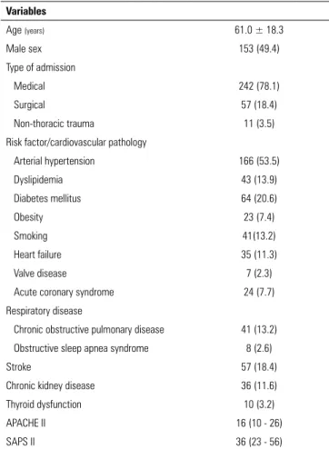
The mean age of the patients was 61.0 ± 18.3 years, and 49.4% (n = 153) were male. Table 1 summarizes the main demographic and clinical characteristics of our sample.
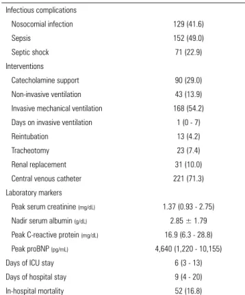
Table 2 summarizes the outcomes, complications (nosocomial infection, sepsis, septic shock, and death), the interventions performed during hospitalization, and the values of the laboratory markers studied.
During the study period, 73 patients with de novo AF (23.5%; 95%CI 18.9 - 28.7) were recorded. The incidence rates of de novo AF were 24.2% in males and 22.9% in females (p = 0.894). De novo AF occurred in 15.3% of
Figure 1 - Flowchart of patient inclusion in the study. AF - atrial fibrillation; ICU - intensive care unit.
Table 1 - General population characteristics (n = 310)
Variables
Age (years) 61.0 ± 18.3
Male sex 153 (49.4)
Type of admission
Medical 242 (78.1)
Surgical 57 (18.4)
Non-thoracic trauma 11 (3.5)
Risk factor/cardiovascular pathology
Arterial hypertension 166 (53.5)
Dyslipidemia 43 (13.9)
Diabetes mellitus 64 (20.6)
Obesity 23 (7.4)
Smoking 41(13.2)
Heart failure 35 (11.3)
Valve disease 7 (2.3)
Acute coronary syndrome 24 (7.7)
Respiratory disease
Chronic obstructive pulmonary disease 41 (13.2)
Obstructive sleep apnea syndrome 8 (2.6)
Stroke 57 (18.4)
Chronic kidney disease 36 (11.6)
Thyroid dysfunction 10 (3.2)
APACHE II 16 (10 - 26)
SAPS II 36 (23 - 56)
SD - standard deviation; APACHE II - Acute Physiology and Chronic Health Evaluation II; SAPS II - Simplified Acute Physiology Score. Values are expressed as the means ± standard deviations, n (%), or medians (interquartile ranges).
medical admissions, 15.8% of surgical admissions, and 9.1% of admissions due to non-thoracic trauma.
Table 3 summarizes the general characteristics of the population, according to the presence or not of de novo AF (univariate analysis). Upon admission, patients with
de novo AF were significantly older (70.1 ± 14.7 years
versus 58.1 ± 18.5 years; p < 0.001) and had higher
Table 2 - Outcomes, complications, interventions performed, and laboratory markers (n = 310)
Infectious complications
Nosocomial infection 129 (41.6)
Sepsis 152 (49.0)
Septic shock 71 (22.9)
Interventions
Catecholamine support 90 (29.0)
Non-invasive ventilation 43 (13.9)
Invasive mechanical ventilation 168 (54.2)
Days on invasive ventilation 1 (0 - 7)
Reintubation 13 (4.2)
Tracheotomy 23 (7.4)
Renal replacement 31 (10.0)
Central venous catheter 221 (71.3)
Laboratory markers
Peak serum creatinine (mg/dL) 1.37 (0.93 - 2.75)
Nadir serum albumin (g/dL) 2.85 ± 1.79
Peak C-reactive protein (mg/dL) 16.9 (6.3 - 28.8)
Peak proBNP (pg/mL) 4,640 (1,220 - 10,155)
Days of ICU stay 6 (3 - 13)
Days of hospital stay 9 (4 - 20)
In-hospital mortality 52 (16.8)
proBNP - pro-B type natriuretic peptide; ICU - intensive care unit. Values are expressed as n (%), medians (interquartile ranges), or means ± standard deviations.
(8.2% versus 1.7%; p = 0.007). All 6 patients with de novo AF and thyroid dysfunction had hypothyroidism. The median APACHE II scores (21 points versus 15 points) and SAPS II scores (47 points versus 34 points) were also significantly higher in patients with de novo AF (p = 0.004 and p < 0.001, respectively).
Table 4 summarizes the complications, interventions performed during hospitalization, and values of the laboratory markers studied, according to the presence or not of de novo AF (univariate analysis). Patients with de novo AF had a higher prevalence of septic shock (37% versus 18.6%; p = 0.007) and a greater need for catecholamine support (41.1% versus 25.3%; p = 0.012) and central venous catheterization (84.9% versus 67.1%; p = 0.003). The median peak values of serum creatinine (1.84mg/dL
versus 1.22mg/dL) and peak proBNP (9,461pg/mL versus
1,652pg/mL) were significantly higher in patients with de novo AF (p = 0.002 and p < 0.001, respectively).
Patients with prior HF (n = 35, 11.3%) had significantly higher levels of proBNP than those without HF (median 9,017pg/mL versus 2,130pg/mL; p < 0.001). Considering the subgroup with de novo AF, the proBNP levels were
Table 3 - Sample characteristics according to the presence or absence of de novo
atrial fibrillation
De novo AF (n = 73)
No de novo AF
(n = 237)
p value
Age in years 70.1 ± 14.7 58.1 ± 18.5 < 0.001
Male sex 37 (50.7) 116 (48.9) 0.894
Type of admission
Medical 63 (86.3) 179 (75.5) 0.290
Surgical 9 (12.3) 48 (20.3)
Non-thoracic trauma 1 (1.4) 10 (4.2)
Risk factors/cardiovascular pathology
Arterial hypertension 50 (68.5) 116 (48.9) 0.005
Dyslipidemia 12 (16.4) 31 (13.1) 0.446
Diabetes mellitus 12 (16.4) 52 (21.9) 0.408
Obesity 4 (5.5) 19 (8.0) 0.613
Smoking 8 (11.0) 33 (13.9) 0.693
Heart failure 19 (26.0) 16 (6.8) < 0.001
Valve disease 6 (8.2) 1 (0.4) 0.001
Acute coronary syndrome 2 (2.7) 4 (1.7) 0.629
Respiratory disease
Chronic obstructive pulmonary disease 9 (12.3) 32 (13.5) 1.000 Obstructive sleep apnea syndrome 3 (4.1) 5 (2.1) 0.398
Stroke 20 (27.4) 37 (15.6) 0.037
Chronic kidney disease 10 (13.7) 26 (11.0) 0.534
Thyroid dysfunction 6 (8.2) 4 (1.7) 0.007
APACHE II 21 (12 - 28) 15 (10 - 24) 0.004
SAPS II 47 (33 - 65) 34 (22 - 51) < 0.001
AF - atrial fibrillation; APACHE II - Acute Physiology and Chronic Health Evaluation II; SAPS II - Simplified Acute Physiology Score. Values are expressed as the means ± standard deviations, n (%), or medians (interquartile ranges).
not significantly different between patients with and those
without HF (median 11,068pg/mL versus 7,875pg/mL;
p = 0.222).
After the selection of the significant predictors in the univariate analysis and their application in the multivariable model (Table 5), the presence of stroke (OR = 10.09; 95%CI 1.54 - 66.27; p = 0.016) and elevated proBNP values (OR = 1.28; 95%CI 1.086 - 1.520; p = 0.004, for each 1,000pg/mL increment) were identified as independent predictors of de novo AF.
Table 4 - Complications, interventions performed, and laboratory markers according to the presence or absence of de novo atrial fibrillation
De novo AF (n = 73)
No de novo AF
(n = 237)
p value
Infectious complications
Nosocomial infection 37 (50.7) 92 (38.8) 0.079
Sepsis 41 (56.2) 111 (46.8) 0.182
Septic shock 27 (37.0) 44 (18.6) 0.002
Interventions performed
Catecholamine support 30 (41.1) 60 (25.3) 0.012
Non-invasive ventilation 13 (17.8) 30 (12.7) 0.332
Invasive mechanical ventilation 44 (60.3) 124 (52.3) 0.283 Days on invasive ventilation 2 (0 - 10) 1 (0 - 6) 0.082
Reintubation 6 (8.2) 7 (3.0) 0.086
Tracheotomy 8 (11.0) 15 (6.4) 0.205
Renal replacement 11 (15.1) 20 (8.4) 0.118
Central venous catheter 62 (84.9) 159 (67.1) 0.003
Laboratory markers
Peak serum creatinine (mg/dL) 1.84
(1.09 - 3.65)
1.22
(0.89 - 2.41) 0.002
Nadir serum albumin (g/dL) 1.94
(1.55 - 2.42)
2.15
(1.65 - 2.64) 0.140
Peak C-reactive protein (mg/dL) 18.8
(9.81 - 28.8)
16.2
(5.8 - 28.8) 0.422
Peak proBNP (pg/mL) 9,461
(2,951 - 17,882)
1,652
(535 - 5,289) < 0.001
AF - atrial fibrillation; proBNP - pro-B type natriuretic peptide. Values are expressed in n (%) or medians (interquartile ranges).
Table 5 - Multivariate model for prediction of de novo atrial fibrillation
Multivariate model* B OR 95%CI for OR p value
Age (per 1-year increment) 0.028 1.028 0.965 - 1.095 0.394
High blood pressure 0.936 2.550 0.482 - 13.487 0.271
Heart failure 0.997 2.711 0.447 - 16.438 0.278
Valve disease 19.818 4.04 x 10⁸ 0 0.999
Stroke 2.311 10.087 1.535 - 66.271 0.016
Thyroid dysfunction 2.407 11.105 0.784 - 157.2 0.075
APACHE II (per point increment) 0.140 1.150 0.990 - 1.336 0.067
SAPS II (per point increment) 0.062 1.064 0.987 - 1.146 0.104
Septic shock 1.584 0.940 0.872 - 1.013 0.162
Catecholamine support 0.528 1.696 0.247 - 11.624 0.591
Central venous catheter 0.239 1.269 0.157 - 10.292 0.823
Peak serum creatinine (per 1 mg/dL increment) 0.230 1.259 0.850 - 1.864 0.250
Peak proBNP (per 1,000 pg/mL increment) 0.250 1.284 1.086 - 1.520 0.004
B - coefficient B; OR - odds ratio; 95% CI - 95% confidence interval; APACHE II - Acute Physiology and Chronic Health Evaluation II; SAPS II - Simplified Acute Physiology Score; proBNP - pro-B type natriuretic peptide. * Only variables with p < 0.05 were included in the multivariable analysis.
Patients with de novo AF had significantly longer stays in the hospital (14 [7 - 23] days versus 8 [4 - 19] days; p = 0.002) and ICU (8 [4 - 16] days versus 6 [3 - 12] days; p = 0.031).
There were no significant differences in in-hospital mortality between patients with and those without de novo AF (20.5 versus 15.6%; p = 0.370).
DISCUSSION
Predictors of de novo atrial fibrillation: the role of proBNP
In our population, the presence of previous stroke and an elevated proBNP value were independent predictors
of de novo AF. The existence of previously documented
paroxysmal AF is one of the possible explanations for the high prevalence of prior stroke in this subgroup with
de novo AF. Such individuals presented sinus rhythm on
admission, though they may have had previous paroxysmal AF that manifested de novo during hospitalization.
In turn, proBNP was found to be a marker with good performance in predicting de novo AF in the ICU. To the best of our knowledge, there are no previous studies demonstrating this role of proBNP in general ICUs. A recent study by Chokengarmwong et al.(24) performed
Figure 2 - Receiver operating characteristic curve of peak pro-peptide natriuretic type B in the prediction of de novo atrial fibrillation. proBNP - pro-B type natriuretic peptide; AUC - area under the curve; 95%CI - 95% confidence interval.
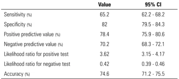
Table 6 - Performance of pro-B type natriuretic peptide > 5,666 pg/mL in the prediction of de novo atrial fibrillation
Value 95% CI
Sensitivity (%) 65.2 62.2 - 68.2
Specificity (%) 82 79.5 - 84.3
Positive predictive value (%) 78.4 75.9 - 80.6
Negative predictive value (%) 70.2 68.3 - 72.1
Likelihood ratio for positive test 3.62 3.15 - 4.17 Likelihood ratio for negative test 0.42 0.39 - 0.46
Accuracy (%) 74.6 71.2 - 75.5
admission is a predictor of de novo AF in the first 3 days of hospitalization in a surgical and trauma ICU. In our study, proBNP > 5,666pg/mL showed good specificity and reasonable sensitivity in the prediction of de novo AF. However, the pathophysiological relationship between AF and proBNP still needs to be explained and may be attributed to atrial dilation, atrial fibrosis, or even decompensation of the underlying disease.(25) However,
it seems more likely that proBNP, like troponin, is a consequence rather than a cause of stress and/or injury. Regardless of the type of pathophysiological relationship between AF and proBNP, elevated values of the latter allow the identification of patients at risk for AF. In turn,
the early identification of these patients allows establishing early strategies for the prevention of AF.
High incidence of de novo atrial fibrillation in the general intensive care unit
The incidence of de novo AF observed in our medical non-cardiac surgical ICU was 23.5%, which is considered high in this type of ICU. Although several previous studies focused on cardiac and surgical populations,(10-14,26)
our data suggest that de novo AF is also a fairly frequent problem in the polyvalent ICU. Previous studies on the
incidence of de novo AF in general ICUs have shown
that the frequency of these events can reach 7 to 15%. However, some of these studies focused on the incidence of supraventricular tachyarrhythmias, regardless of the type of arrhythmia;(4,17) in these studies, the incidence of
AF may be lower.
The increased proportion of septic patients with nosocomial infection in the ICU during the period of our study may explain the high incidence of AF. In fact, inflammation is a common process in critically ill patients and may be a mechanism in the genesis of AF.(27)
In critically ill patients, in addition to the infectious pathology, respiratory and cardiac pathologies, invasive procedures, and the use of mechanical ventilation and catecholamine support may be triggers of AF.(15)
Prognosis and prevention strategies
Previous studies have shown that AF is associated with higher in-hospital mortality in critically ill patients, especially in those with advanced age.(28) Although there
were no significant differences in in-hospital mortality between patients with and those without de novo AF in our cohort, the median days of hospital and ICU stay were significantly higher in the latter. To a certain extent, prolonged hospitalization in patients with AF may be associated with increased morbidity and higher health costs. Thus, the prevention of AF plays a central role in critically ill patients at increased risk (here identified by elevated proBNP). Several prophylactic AF strategies have been described,(29,30) most of which are described in
critically ill patients after thoracic surgery.
predictors. Recording the type and dose of catecholamines administered was not part of the study protocol, and these data may have a relevant impact on the prediction of AF. Data regarding the position of the central venous catheter and the possible rapid volume expansion phases may play relevant roles in both the proBNP levels and the prediction of AF; however, these data were not evaluated in the present study. Although the diagnostic sensitivity of proBNP should be not be considered a strong effect, this limitation is compensated at least partly by the considerable specificity of proBNP in detecting de novo AF in this population. Only a small proportion of patients had available echocardiographic parameters; therefore,
these data were excluded from the analysis. However, proBNP has the advantage of being an easily accessible marker in non-cardiac ICUs.
CONCLUSIONS
History of previous stroke and elevated proBNP on admission were independent predictors of de novo atrial fibrillation in the polyvalent intensive care unit. ProBNP can be a useful and easily and quickly accessible tool to stratify the risk of atrial fibrillation. The high incidence of de novo atrial fibrillation in the polyvalent non-cardiac intensive care unit emphasizes the importance of timely recognition of this pathology.
Objetivo: Avaliar quais os preditores de fibrilação atrial de
novo em doentes de uma unidade de cuidados intensivos não
cardíaca.
Métodos: Foram analisados 418 doentes internados entre janeiro e setembro de 2016 em uma unidade de cuidados inten-sivos não cardíaca. Registaram-se as características clínicas, as intervenções efetuadas e os marcadores bioquímicos durante a internação. Avaliaram-se ainda a mortalidade hospitalar e o tem-po de internação hospitalar e na unidade de cuidados intensivos.
Resultados: Foram incluídos 310 doentes, com média de idades de 61,0 ± 18,3 anos, 49,4% do sexo masculino, 23,5%
com fibrilação atrial de novo. O modelo multivariável
identifi-cou acidente vascular cerebral prévio (OR de 10,09; p = 0,016) e valores aumentados de proBNP (OR de 1,28 por cada aumento em 1.000pg/mL; p = 0,004) como preditores independentes de
fibrilação atrial de novo. A análise por curva Característica de
Operação do Receptor do proBNP para predição de fibrilação
atrial de novo revelou área sob a curva de 0,816 (p < 0,001), com
sensibilidade de 65,2% e especificidade de 82% para proBNP > 5.666pg/mL. Não se verificaram diferenças na mortalidade (p = 0,370), porém a duração da internação hospitalar (p = 0,002) e na unidade de cuidados intensivos (p = 0,031) foi superior nos
doentes com fibrilação atrial de novo.
Conclusões: História de acidente vascular cerebral prévio e proBNP elevado em internação constituíram preditores
inde-pendentes de fibrilação atrial de novo na unidade de cuidados
intensivos polivalente. O proBNP pode constituir ferramenta útil, de fácil e rápido acesso na estratificação do risco de fibrila-ção atrial.
RESUMO
Descritores: Fibrilação atrial/epidemiologia; Incidência; Cui-dados intensivos
REFERENCES
1. Furberg CD, Psaty BM, Manolio TA, Gardin JM, Smith VE, Rautaharju PM. Prevalence of atrial fibrillation in elderly subjects (the Cardiovascular Health Study). Am J Cardiol. 1994;74(3):236-41.
2. Reinelt P, Karth GD, Geppert A, Heinz G. Incidence and type of cardiac arrhythmias in critically ill patients: a single center experience in a medical-cardiological ICU. Intensive Care Med. 2001;27(9):1466-73.
3. Kirchhof P, Benussi S, Kotecha D, Ahlsson A, Atar D, Casadei B, Castella M, Diener HC, Heidbuchel H, Hendriks J, Hindricks G, Manolis AS, Oldgren J, Popescu BA, Schotten U, Van Putte B, Vardas P; ESC Scientific Document Group. 2016 ESC Guidelines for the management of atrial fibrillation developed in collaboration with EACTS. Eur Heart J. 2016;37(38):2893-2962.
4. Ommen SR, Odell JA, Stanton MS. Atrial arrhythmias after cardiothoracic surgery. N Engl J Med. 1997;336(20):1429-34. Erratum in: N Engl J Med 1997;337(3):209.
5. Brathwaite D, Weissman C. The new onset of atrial arrhythmias following major noncardiothoracic surgery is associated with increased mortality. Chest. 1998;114(2):462-8.
6. Makrygiannis SS, Margariti A, Rizikou D, Lampakis M, Vangelis S, Ampartzidou OS, et al. Incidence and predictors of new-onset atrial fibrillation in noncardiac intensive care unit patients. J Crit Care. 2014;29(4):697.e1-5.
8. Freedman B, Camm J, Calkins H, Healey JS, Rosenqvist M, Wang J, Albert CM, Anderson CS, Antoniou S, Benjamin EJ, Boriani G, Brachmann J, Brandes A, Chao TF, Conen D, Engdahl J, Fauchier L, Fitzmaurice DA, Friberg L, Gersh BJ, Gladstone DJ, Glotzer TV, Gwynne K, Hankey GJ, Harbison J, Hillis GS, Hills MT, Kamel H, Kirchhof P, Kowey PR, Krieger D, Lee VW, Levin LÅ, Lip GY, Lobban T, Lowres N, Mairesse GH, Martinez C, Neubeck L, Orchard J, Piccini JP, Poppe K, Potpara TS, Puererfellner H, Rienstra M, Sandhu RK, Schnabel RB, Siu CW, Steinhubl S, Svendsen JH, Svennberg E, Themistoclakis S, Tieleman RG, Turakhia MP, Tveit A, Uittenbogaart SB, Van Gelder IC, Verma A, Wachter R, Yan BP; AF-Screen Collaborators. Screening for Atrial Fibrillation: A Report of the AF-SCREEN International Collaboration. Circulation. 2017;135(19):1851-67.
9. Maisel WH, Rawn JD, Stevenson WG. Atrial fibrillation after cardiac surgery. Ann Intern Med. 2001;135(12):1061-73.
10. Bender JS. Supraventricular tachyarrhythmias in the surgical intensive care unit: an under-recognized event. Am Surg. 1996;62(1):73-5. 11. Knotzer H, Mayr A, Ulmer H, Lederer W, Schobersberger W, Mutz N, et
al. Tachyarrhythmias in a surgical intensive care unit: a case-controlled epidemiologic study. Intensive Care Med. 2000;26(7):908-14.
12. Seguin P, Signouret T, Laviolle B, Branger B, Mallédant Y. Incidence and risk factors of atrial fibrillation in a surgical intensive care unit. Crit Care Med. 2004;32(3):722-6.
13. Artucio H, Pereira M. Cardiac arrhythmias in critically ill patients: epidemiologic study. Crit Care Med. 1990;18(12):1383-8.
14. Christian SA, Schorr C, Ferchau L, Jarbrink ME, Parrillo JE, Gerber DR. Clinical characteristics and outcomes of septic patients with new-onset atrial fibrillation. J Crit Care. 2008;23(4):532-6.
15. Heinz G. Arrhythmias in the ICU: what do we know? Am J Respir Crit Care Med. 2008;178(1):1-2.
16. Conen D, Osswald S, Albert CM. Epidemiology of atrial fibrillation. Swiss Med Wkly. 2009;139(25-26):346-52.
17. Annane D, Sébille V, Duboc D, Le Heuzey JY, Sadoul N, Bouvier E, et al. Incidence and prognosis of sustained arrhythmias in critically ill patients. Am J Respir Crit Care Med. 2008;178(1):20-5.
18. Trappe HJ, Brandts B, Weismueller P. Arrhythmias in the intensive care patient. Curr Opin Crit Care. 2003;9(5):345-55.
19. Knaus WA, Draper EA, Wagner DP, Zimmerman JE. APACHE II: a severity of disease classification system. Crit Care Med. 1985;13(10):818-29.
20. Le Gall JR, Lemeshow S, Saulnier F. A New Simplified Acute Physiology Score (SAPSII) based on a European/North American multicenter study. JAMA. 1993;270(24):2957-63.
21. Vogelmeier CF, Criner GJ, Martinez FJ, Anzueto A, Barnes PJ, Bourbeau J, et al. Global Strategy for the Diagnosis, Management, and Prevention of Chronic Obstructive Lung Disease 2017 Report. GOLD Executive Summary. Am J Respir Crit Care Med. 2017;195(5):557-82.
22. Levey AS, Coresh J, Balk E, Kausz AT, Levin A, Steffes MW, Hogg RJ, Perrone RD, Lau J, Eknoyan G; National Kidney Foundation. National Kidney Foundation practice guidelines for chronic kidney disease: evaluation, classification, and stratification. Ann Intern Med. 2003;139(2):137-47. 23. Rhodes A, Evans LE, Alhazzani W, Levy MM, Antonelli M, Ferrer R, et al.
Surviving Sepsis Campaign: International Guidelines for Management of Sepsis and Septic Shock: 2016. Crit Care Med. 2017;45(3):486-552. 24. Chokengarmwong N, Yeh DD, Chang Y, Ortiz LA, Kaafarani HM, Fagenholz
P, et al. Elevated admission N-terminal pro-brain natriuretic peptide level predicts the development of atrial fibrillation in general surgical intensive care unit patients. J Trauma Acute Care Surg. 2017;83(3):485-90. 25. Svennberg E, Lindahl B, Berglund L, Eggers KM, Venge P, Zethelius B, et al.
NT-proBNP is a powerful predictor for incident atrial fibrillation - Validation of a multimarker approach. Int J Cardiol. 2016;223:74-81.
26. Seguin P, Laviolle B, Maurice A, Leclercq C, Mallédant Y. Atrial fibrillation in trauma patients requiring intensive care. Intensive Care Med. 2006;32(3):398-404.
27. Chung MK, Martin DO, Sprecher D, Wazni O, Kanderian A, Carnes CA, et al. C-reactive protein elevation in patients with atrial arrhythmias: inflammatory mechanisms and persistence of atrial fibrillation. Circulation. 2001;104(24):2886-91.
28. Alves GC, Silva Júnior GB, Lima RS, Sobral JB, Mota RM, Abreu KL, et al. Risk factors for death among critically ill elderly patients. Rev Bras Ter Intensiva. 2010;22(2):138-43.
29. Riber LP, Larsen TB, Christensen TD. Postoperative atrial fibrillation prophylaxis after lung surgery: systematic review and meta-analysis. Ann Thorac Surg. 2014;98(6):1989-97.