I
Sandra dos Santos Carvalho
Decoding E-cadherin glycans functions in cancer: from functional
glycomics to clinical applications.
Tese de Candidatura ao grau de Doutor em Ciências Veterinárias submetida ao Instituto de Ciências Biomédicas Abel Salazar da Universidade do Porto.
Orientador – Professora Doutora Salomé Soares de Pinho Marcos Pinto
Categoria – Professor Auxiliar Convidado
Afiliação – Instituto de Ciências Biomédicas Abel Salazar da Universidade do Porto (ICBAS-UP).
Co-orientador: Professor Doutor Celso Albuquerque Reis Categoria – Professor Auxiliar Convidado
Afiliação – Instituto de Ciências Biomédicas Abel Salazar da Universidade do Porto (ICBAS-UP).
Co-orientador: Professora Doutora Maria Raquel Campos Seruca
Categoria – Professor Afiliado
Afiliação – Faculdade de Medicina da Universidade do Porto (FMUP)
III
Funding * Financiamento
PhD Fellowship (SFRH/BD/77386/2011) provided by the Portuguese Foundation for Science and Technology (FCT) of the Portuguese Ministry of Science, Technology and Higher Education.
Bolsa Individual de Doutoramento (SFRH/BD/77386/2011) da Fundação Portuguesa para a Ciência e a Tecnologia (FCT) do Ministério da Ciência, Tecnologia e Ensino Superior.
V
The author of this thesis declares that, in accordance with “nº 2, alinea a, do Art.º 31º do Decreto-Lei nº 230/2009”, afforded a major contribution to the design and technical execution of the work, interpretation of the results and manuscript preparation resulting in the following accepted and in preparation articles:
Scientific publications
Julio Cesar Madureira de-Freitas-Junior
*
, Sandra Carvalho*
, Ana M. Dias, Patricia Oliveira, Joana Cabral, Raquel Seruca, Carla Oliveira, José Andrés Morgado-Diaz, Celso A. Reis, Salomé S. Pinho (2013). Insulin/IGF-I Signalling Pathways Enhances Tumour Cell Invasion through Bisecting GlcNAc N-glycans Modulation. An Interplay with E-cadherin. PLoS One. Nov 25; 8 (11):e811579. * These authors contributed equally to this work.S Carvalho, TA Catarino, AM Dias, M Kato, A Almeida, B Hessling, J Figueiredo, F Gärtner, JM Sanches, T Ruppert, E Miyoshi, M Pierce, F Carneiro, D Kolarich, R Seruca, Y Yamaguchi, N Taniguchi, CA Reis and SS Pinho (2015). Preventing E-cadherin aberrant N-glycosylation at Asn-554 improves its critical function in gastric cancer. Oncogene. Jul 20. doi: 10.1038/onc.2015.225.
Sandra Carvalho*, Tiago Oliveira*, Markus Bartels, Eiji Miyochi, Michael Pierce, Naoyuki Taniguchi, Fátima Carneiro, Raquel Seruca, Sabine Strahl, Celso A. Reis, Salomé S. Pinho (2015) O-mannosylation and N-glycosylation: two coodinated mechanisms regulating critical functions of E-cadherin in cancer. (In preparation). * These authors contributed equally to this work.
Pinho SS, Carvalho S, Marcos-Pinto R, Magalhães A, Oliveira C, Gu J, Dinis-Ribeiro M, Carneiro F, Seruca R, Reis CA. (2013) Gastric cancer: adding glycosylation to
the equation. Trends Mol Med. 2013 Nov; 19(11):664-76. doi:
VII
À minha orientadora, Doutora Salomé Pinho, por ter confiado em mim e nas minhas capacidades para iniciar este projeto. Pelo seu interesse incondicional, entusiamo contagiante e disponibilidade que sempre manifestou no decorrer dos trabalhos. As suas sugestões e a sua orientação foram indispensáveis para a realização deste trabalho.
Ao Professor Doutor Celso Reis, pelo acompanhamento em todas as etapas do projeto e pelas questões pertinentes bem como preciosas sugestões colocadas durante o decurso dos trabalhos.
À Professora Doutora Raquel Seruca, pelas sugestões e discussões dos trabalhos desenvolvidos e pelo entusiasmo pela investigação.
À Professora Doutora Fátima Gärtner, pela sua confiança em mim, e pela incansável disponibilidade que sempre manifestou.
À Professora Doutora Fátima Carneiro, por toda a sua disponibilidade, disponibilidade de recursos para a concretização de parte deste trabalho, e pela opinião especializada.
Ao Professor Doutor Sobrinho Simões, pelas excelentes condições de acolhimento no IPATIMUP e pelo entusiasmo genuíno à Ciência.
To all the co-authors of the publications for their important suggestions and contributions.
Ao grupo Glycobiology in Cancer, às “mucinas” e muitas outras pessoas do instituto, pela amizade, apoio e por todos os momentos que foram partilhados. Tornaram este longo caminho mais fácil de percorrer. À Catarina, Vânia e Ritinha pelo vosso apoio e ajuda.
À Joana Cabral, por me ter “acolhido” na fase inicial deste projeto e a quem devo toda a aprendizagem inicial das técnicas da glicobiologia. Pela pessoa genuína e bondosa que és. Foi e será sempre um enorme prazer conviver contigo.
VIII
acreditarem sempre em mim.
Ao Zé Carlos, pelo apoio, incentivo e por acreditar sempre que seria capaz. Por seres quem és…
À primeira pessoa que me incentivou a iniciar este percurso e que acreditou que chegaria à meta, ao meu pai. Sei que estarás lá presente.
IX
Cancro gástrico é uma doença agressiva com um elevado impacto na saúde mundial, devido ao diagnóstico tardio dos pacientes. Os eventos moleculares subjacentes ao processo de carcinogénese gástrica têm vindo a ser abordados de modo a identificar biomarcadores moleculares específicos para a deteção precoce do carcinoma gástrico. O cancro gástrico do tipo difuso, caracterizado por uma reduzida adesão intercelular, tem vindo a ser descrito como estando estritamente relacionado com a desregulação da proteína supressora de invasão, E-caderina. O comprometimento das funções biológicas da E-caderina resulta na perda da adesão celular e no aumento do potencial de invasão e metastização. E-caderina é uma glicoproteína que é modificada por glicosilação pós-translacionalmente e cujas modificações têm forte efeito na doença neoplásica. De facto, a transformação maligna está fortemente associada com padrões alterados de glicosilação na superfície das células tumorais.
A N-glicosilação da E-caderina mediada por GnT-III confere-lhe uma estabilidade na membrana celular o que conduz às suas funções adesivas adequadas. Esta glicoforma da E-caderina também regula a atividade da GnT-III por um mecanismo bidirecional o que contribui para a supressão de invasão e metastização. Neste estudo, demonstramos que este mecanismo é comprometido com a ativação da via de sinalização insulina / IGF-I no processo de invasão de células tumorais. Por outro lado, E-caderina pode também ser sujeita a N- glicosilação mediada por GnT-V o que compromete a adesão célula-célula. Pacientes com cancro gástrico exibem uma expressão aberrante da E-caderina especificamente modificada com estruturas β1,6 GlcNAc. Demonstramos que estes N-glicanos deletérios estão presentes especificamente no local de N-glicosilação Asn-554 da E-caderina, sendo o local chave para a sua desregulação funcional nas células tumorais gástricas. Além disso, também verificamos que o perfil de O-manosilação da E-caderina, descrito recentemente como sendo crucial para a adesão célula-célula, encontra-se comprometido num contexto de cancro gástrico com implicações nas suas funções biológicas. O impedimento da ocupação do local Asn-554 da E-caderina com N-glicanos β1,6 GlcNAc potencializa o perfil de O-manosilação da E-caderina, estando assim associado com a recuperação das suas funções biológicas. Adicionalmente, verifica-se a existência de um mecanismo coordenado entre o perfil de O-manosilação proteico e a N-glicosilação mediada por GnT-V em pacientes com cancro gástrico do tipo difuso. Assim sendo, a descrição do perfil de glicosilação da E-caderina em cancro gástrico pode contribuir para potenciais glico- biomarcadores de diagnóstico precoce do cancro gástrico do tipo difuso, e para o desenvolvimento de potenciais alvos terapêuticos.
XI
Gastric cancer is an aggressive disease with a high impact on global health due to the late diagnosis of gastric cancer patients. The molecular events underlying gastric cancer process have been addressed in order to identify molecular biomarkers specifics for the early detection of gastric carcinoma. Diffuse gastric cancer, characterized by a reduced intercellular adhesiveness, has been strongly correlated with the dysregulation of the invasion suppressor protein, E-cadherin. The impairment of E-cadherin biological functions results in loss of cell adhesion and increased cell invasion and metastatic potential. E-cadherin is a glycoprotein that is post-translationally modified by glycosylation which may exert a powerful effect on the outcome of neoplastic disease. In fact, malignant transformation is strongly associated with altered glycosylation patterns on the surface of cancer cells.
E-cadherin N-glycosylation mediated by GnT-III confers a stability to the E-cadherin at the cell membrane leading to the proper adhesive functions. Moreover, this E-cadherin glycoform also regulates the GnT-III activity by a bidirectional crosstalk which contributes to the suppression of tumour invasion and metastasis. In this study, we have shown that this interplay is compromised with the activation of the insulin/IGF-I signalling pathway in the process of tumour cell invasion. E-cadherin may also undergo GnT-V-mediated N-glycosylation that compromises the E-cadherin-mediated cell-cell adhesion. Human gastric cancer patients display an aberrant expression of E-cadherin specifically modified with the β1,6 GlcNAc branched glycans. Here, we have demonstrated that these deleterious N-glycans are present specifically in the E-cadherin N-glycosylation site Asn-554, being the key site for the functional dysregulation of E-cadherin in gastric tumour cells. Furthermore, we have shown that the O-mannosylation profile of E-cadherin, recently described crucial in cell-cell adhesion, is impaired in a gastric cancer context with implications in its biological functions. Precluding the occupancy of E-cadherin Asn-554 with β1,6 GlcNAc branched N-glycans potentiates the O-mannosylation profile of E-cadherin being associated with the recovery of the proper E-cadherin biological functions. In addition, we also have demonstrated the existence of a coordinated interplay between protein O-mannosylation and GnT-V-mediated N-glycosylation in human diffuse gastric cancer patients. Hence, the disclosure of the E-cadherin glycosignature in gastric cancer may contribute to potential glycobiomarkers for the early diagnosis of diffuse gastric cancer, and to the development of potential therapeutic targets.
XIII
a.a – amino acids
ALG - asparagine linked glycosylation Asn - asparagine
CA19-9 – cancer antigen 19-9 CBD - Catenin binding domain CKII - Casein kinase II
CMD - congenital muscular dystrophies CNX - calnexin
CRT - calreticulin
Dol-P - dolichol phosphate
Dpm1 - GDP-Man: Dol-P mannosyltransferase EBV - Epstein-Barr virus
E-cadherin- epitelial cadherin ECM - extracellular matrix
EMT- Epithelial-mesenchymal transition ER - Endoplasmic reticulum
ERAD - ER- associated degradation Fuc – fucose FUT8 - α1,6-fucosyltransferase Gal - galactose Gal - galactose GalNAc - N-acetylgalactosamine Glc - glucose GlcNAc - N-acetylglucosamine
GlcNAcT-I (or GnT-I) - N-acetylglucosaminyltransferase I GlcNAcT-II (or GnT-II) - N-acetylglucosaminyltransferase II GlcNAcT-III (or GnT-III) - N-acetylglucosaminyltransferase III GlcNAcT-IV (or GnT-IV) - N-acetylglucosaminyltransferase IV GlcNAcT-V (or GnT-V) - N-acetylglucosaminyltransferase V GlcNAcT-VI (or GnT-VI) - N-acetylglucosaminyltransferase VI GnT-IX/ GnT-Vb - β1,6-N-acetylglucosaminyltransferase-IX/Vb GSK-3β - glycogen synthase kinase-3β
H. pylori - Helicobacter pylori HAV - Histidine-Alanine-Valine
XIV
HDGC - Hereditary diffuse gastric carcinoma syndrome IARC - International Agency for Research on Cancer IGF-IR - IGF-I receptor
JMD - Juxtamembrane domain LOH - Loss of heterozygosity Man - mannose
MEB - Muscle-eye-brain
MGAT5 - mannoside acetylglucosaminyltransferase 5 gene miRNAs - MicroRNAs
Neu5Ac- sialic acid
OST - oligosaccharyltransferase
PIPKIγ - type I gamma phosphatidylinositol phosphate kinase PMTs - protein O-mannosyltransferases
polyLacNAc - poly-N-acetyllactosamine
POMGnT1 - β-1,2-N-acetylglucosaminyltransferase 1 POMGnT2 - β-1,4-N-acetylglucosaminyltransferase 2 POMK - protein O-mannose kinase
POMT1 - protein O-mannosyltransferase 1 POMT2 - protein O-mannosyltransferase 2 PP2A - protein phosphatase 2A
RPTPβ - receptor tyrosine phosphatase β RTK - receptors tyrosine kinase
Ser - serine
TGN - trans-Golgi network Thr - threonine
TJ - tight junctions
UGGT - UDP-Glc: glycoprotein glucosyltransferase UPR - unfolded protein response
WHO - World Health Organization WWD - Walker-Warburg syndrome α-DG - α- dystroglycan
XV
Chapter I ... 17
General Introduction ...19
Gastric Cancer ...19
Epidemiology and Aetiology ...19
Histological classification ...20
E-cadherin ...21
Extracellular domain of E-cadherin ...23
Intracellular domain of E-cadherin ...23
Regulation of E-cadherin-mediated cell- cell adhesion ...25
E-cadherin dysregulation in cancer ...25
Protein Glycosylation ...27
N-glycosylation ...28
O-Mannosylation...33
Glycosylation alterations associated with cancer ...36
N-linked glycans in cancer ...37
E-cadherin Post-translational modifications. Implications in cancer...39
References ...43
Rational and Aims ...61
Specific aims ...61
Chapter II ... 65
Insulin/IGF-I Signalling Pathways Enhances Tumour Cell Invasion through Bisecting GlcNAc N-glycans Modulation. An Interplay with E-cadherin. ...65
Chapter III ... 81
Preventing E-cadherin aberrant N-glycosylation at Asn-554 improves its critical function in gastric cancer ...81
XVI
O-mannosylation and N-glycosylation: two coordinated mechanisms regulating the
critical functions of E-cadherin in cancer ... 97
Chapter V ... 129
General Discussion ... 131 Concluding Remarks ... 140 Future Perspectives ... 142 References ... 143Chapter VI ... 149
Gastric Cancer: adding glycosylation to the equation ... 149
Appendix ... 165
Appendix I ... 167
Appendix II ... 173
17
19
General Introduction
Gastric Cancer
Epidemiology and Aetiology
Gastric cancer is an aggressive disease with a daunting impact on global health. Gastric cancer remains the sixth most common type of cancer, and is the fourth leading cause of cancer-related death worldwide, after lung, breast and liver cancer [1]. Despite the decline in incidence over the past few decades, gastric cancer continues to present a major clinical challenge due to late diagnosis of most gastric cancers since survival is highly dependent on the stage at which the tumour is diagnosed [2].
Large differences in gastric cancer incidence exists between continents, being highest incident rates found in Asia, followed by Europe, Central and South America, North America and Africa [3, 4]. Even within a given geographical area, certain ethnic groups have significantly higher risk of disease [5]. The risk of developing gastric cancer increases with age [6]. The male-to- female ratio in incidence and mortality is about 2:1 [7].
Gastric cancer is a multifactorial disease: infectious, environmental, and host-related factors may interact favouring the development of tumour [8]. Chronic infection with Helicobacter pylori (H. pylori), classified as a class I carcinogen according the International Agency for Research on Cancer (IARC), has been implicated in gastric carcinogenesis [9-11]. It is estimated that 50% of the world´s population is infected with H. pylori [12]. Another infectious agent associated with gastric cancer is the Epstein-Barr virus (EBV) [13]. Regarding environmental and lifestyle factors, tobacco smoking [14], low socioeconomic status, alcohol and meat consumption [15], diet rich in salt and poor in fruits and vegetables [16, 17], and high body mass index [18] are closely linked to increased risk of gastric cancer. At last, molecules involved in adhesion of the bacteria [19] as well as host genetic polymorphisms from diverse molecular pathways [20] are also associated with progression of the gastric carcinogenesis pathway. Genome-wide association studies have been largely used to evaluate single nucleotide polymorphisms simultaneously opening new avenues in cancer research [21].
20
Histological classification
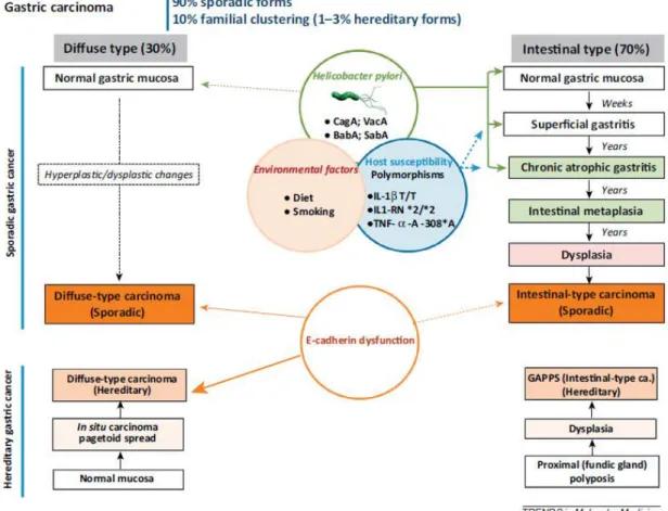
Gastric cancer is a complex and heterogeneous disease with different morphologies, histogenesis and molecular backgrounds [22, 23]. The major histological type of gastric cancer is adenocarcinoma, which originates from the glandular epithelium and accounts for 90% to 95% of all gastric malignancies [24]. Several gastric cancer classifications systems have been proposed over the past decades: World Health Organization (WHO) [25] and the Lauren´s classification [26]. The 2010 WHO system recognizes five major histologic patterns of gastric cancer: tubular, papillary, mucinous, poorly cohesive (including signet ring cell carcinoma), and mixed carcinomas [25, 27]. The Lauren´s criteria, which is the most commonly used classification, describes two main histological subtypes, the intestinal subtype and diffuse subtype gastric adenocarcinoma, which display distinct epidemiologic, morphological and molecular features [26, 28] (Figure 1).
The intestinal subtype of gastric cancer represents nearly 70% of the cases, being more frequently diagnosed in older male patients [29]. It is more likely to be sporadic than inherited [30]. Histologically, it is characterized by glandular architecture with cells displaying cohesiveness and various degrees of differentiation [31]. The main carcinogenic event associated with intestinal subtype cancer is H. pylori infection which leads to a sequence of histological lesions (known as Correa´s cascade) that culminate in a malignant lesion [32]. The cascade of events corresponds to a gradual and multistep progression from chronic gastritis to chronic atrophy to intestinal metaplasia to dysplasia and carcinoma [33]. At the molecular level, intestinal gastric cancer is associated with overexpression of c-met oncogene [34], K-ras mutations [35], loss-of-function of tumour suppressive genes TP53 [36] and APC [37], among others.
In contrast to the intestinal subtype, diffuse gastric cancer is generally diagnosed in younger patients [5]. The diffuse subtype of gastric cancer develops without precancerous lesions; it is characterized by poorly cohesive cells with little or no gland formation displaying a worse prognosis than the intestinal type [38, 39]. Diffuse gastric tumours have an important mechanism of carcinogenesis that is through defective intercellular adhesions, mainly resulting from E-cadherin dysregulation [38]. Abnormal E-cadherin expression can occur through biallelic inactivation of its related gene CDH1 via germline or somatic mutations [40], loss of heterozygosity (LOH) [41, 42], epigenetic silencing of gene transcription through CDH1 promoter hypermethylation [43, 44], transcriptional silencing that target CDH1 promoter [45] or by alteration of E-cadherin glycosylation [46, 47]. CDH1 inactivation occurs in early stages of diffuse type tumour development, whereas in intestinal type tumours it seem to take place in relatively late stages of carcinogenesis. Importantly,
21
CDH1 germline mutations characterizes the hereditary diffuse gastric carcinoma syndrome (HDGC) [48-50].
Figure 1 - Clinicopathological profiles and epidemiological settings of the two main histological subtypes of gastric cancer: intestinal subtype and diffuse subtype. Adapted from [47].
E-cadherin
Cadherins constitute a large superfamily of transmembrane glycoproteins that mediate specific cell-cell adhesion in a calcium-dependent manner [51]. The family is widespread in normal tissues but the individual members display pronounced tissue specificity [52]. The cadherin superfamily is mainly composed by “classical” cadherins of type 1, closely related cadherins of type II, desmossomal cadherins, protocadherins, and a variety of cadherin-related molecules [53, 54].
Classical cadherins were the first subtype of the cadherin superfamily identified in vertebrates [55-58]. Cadherins form primarily homophilic cell-cell interactions at the adherens junctions, and appear to modulate adhesion through dynamic interactions with the actin cytoskeleton [54, 59]. Epithelial cadherin (E-cadherin) is considered the
22
prototypical member of the cadherin superfamily, being identified in 1977 by Takeichi as a surface protein with Ca2+-dependent cell-cell adhesion properties [60]. E-cadherin is
expressed primarily in epithelial cells, where it localizes at the basolateral surface of the epithelial junctional complex- the adherens junctions [59].
The human CDH1 gene, encoding E-cadherin, is situated in the long arm of chromosome 16, within the locus 16q22.1 [61, 62]. CDH1 gene is organized into 15 introns and 16 exons, which are translated into protein comprising 882 aminoacids (a.a). The signal peptide corresponds to the first 27 a.a (exons 1-2), and is followed by the precursor peptide (exon 2-4) (Figure 2A) [42, 63, 64].
Figure 2 - Schematic representation of (A) different levels at which E-cadherin expression is regulated in human tumours, [42] (B) classical cadherin-catenin complex [65], and (C) three-dimensional structure of the extracellular domain (EC1–EC5) of E-cadherin combined with the representation of the four potential N-glycosylation sites [46].
23
The immature form of E-cadherin (the precursor protein) is a polypeptide composed by a propeptide sequence of about 130 a.a and a mature polypeptide of about 728 a.a (Figure 2A) [42]. The propeptide corresponds to a short signal sequence for import into the endoplasmic reticulum (ER) where undergoes cytoplasmic trimming. Following this trimming process, the mature E-cadherin is routed towards the basolateral surface of epithelial cells [64]. E-cadherin mature protein is organized in three major structural domains: an N-terminal ectodomain of about 550 a.a comprising five tandemly repeated subdomains (EC1-EC5), a single transmembrane domain, and a short cytoplasmic domain (C-terminal) of about 150 a.a [46, 64].
Extracellular domain of E-cadherin
The cell-cell adhesion mediated by E-cadherin is achieved through homophilic interactions of the extracellular domain of E-cadherin molecules. The N-terminal domain EC1 has been identified to correspond to the adhesive binding site [66]. EC1 contains a Histidine-Alanine-Valine (HAV) sequence which is thought to be essential for the process of cell-cell adhesion [67]. According to the literature, cadherins may form both lateral (cis) and adhesive (trans) dimers on the cell surface though distinct interactions involving EC1 or EC1-EC2 domains [68-70]. The cadherin molecules on the cell surface establish lateral or cis- interaction and then among adjacent cells form trans adhesive bonds, forming zipper-like structures [71]. Trans-interactions on opposing cell surfaces result in weak cell-cell adhesion, but strong cell-cell adhesion develops during lateral clustering of E-cadherin [72]. The five EC domains are rigidified by coordinating Ca2+ ions between any two consecutive
EC domains [66, 73] and this binding of Ca2+ is essential to confer resistance of the
extracellular region to proteolytic degradation [56, 74].
Intracellular domain of E-cadherin
Further strengthening of cell-cell adhesion requires subsequent linkage to the cytoskeleton that is accomplished through the interaction of the E-cadherin cytoplasmic domain with cytoplasmic proteins called catenins (Figure 2B) [66]. The cytoplasmic domain is subdivided into the juxtamembrane domain (JMD) that provides a specific binding site for p120-catenin and p120-related proteins [75, 76], and the catenin binding domain (CBD) which specifically binds to β-catenin and plakoglobin [77].
Interaction of β-catenin to E-cadherin is required for transportation of the newly synthesized E-cadherin protein from the ER to the cell membrane [78]. β-catenin binds the
24
C–terminal cytoplasmic domain of E-cadherin in a phospho-regulated manner [79]. Phosphorylation of the three serine residues located in the cadherin cytoplasmic domain (S684, S686, S692) by casein kinase II (CKII) and glycogen synthase kinase-3β (GSK-3β) leads to increase in affinity of E-cadherin and β-catenin interaction. In contrast, tyrosine phosphorylation of β-catenin at Y489 and Y654 weakens interaction with cadherin [80]. In addition, β-catenin plays an important role in cell signalling pathways, such as in Wnt signalling [81, 82]. β-catenin functions as a transcriptional co-regulator, moving from the cytoplasm into the nucleus and cooperating with TCF/LEF transcription in order to activate the expression of numerous genes involved in migration and proliferation [83, 84]. Furthermore, β-catenin plays a key role in cadherin-adhesive function by acting as an adaptor for a range of cytoplasmic proteins which in turn interact with the actin cytoskeleton [72]. The best known is α-catenin which exists in either a monomeric or homo-dimeric state: monomeric catenin binds to the N-terminal portion of β-catenin while homo-dimeric α-catenin binds to actin filaments [72, 85]. Phosphorylation of β-α-catenin at Y142 disrupts binding to α-catenin [80].
E-cadherin stabilization at the cell membrane also occurs through the association with p-120-catenin that interacts with the highly conserved juxtamembrane domain of E-cadherin [75]. E-E-cadherin-p120-catenin interaction prevents the entrance of E-E-cadherin into degrading endocytic trafficking pathways or accelerates the recycling of internalized cadherin back to the plasma membrane [86]. The phosphorylation of p120-catenin increases binding affinity to E-cadherin [72, 87]. However, the loss of E-cadherin-p120 catenin interaction destabilizes cadherin localization at the cell membrane, inhibiting its accumulation at cell borders [88]. Moreover, the weakness of E-cadherin-p120 catenin interaction promotes the ubiquitination-dependent endocytosis of E-cadherin by binding of E-cadherin to Hakai, an E3-ubiquitin ligase, in a Src-dependent manner [89, 90].
The E-cadherin trafficking may also be compromised by post-translational modifications of E-cadherin which have been reported to promote apoptosis [91]. O-GlcNAc glycosylation of cadherin cytoplasmic domain and incomplete processing arrest E-cadherin transport late in the secretory pathway by interfering the binding of E-E-cadherin with type I gamma phosphatidylinositol phosphate kinase (PIPKIγ), a protein required for E-cadherin recruitment to the adhesion sites [92].
25
Regulation of E-cadherin-mediated cell- cell adhesion
Dynamic regulation of cadherin-mediated cell-cell adhesion is associated with diverse morphogenetic processes [93]. The crucial role of such dynamism is evident during embryonic development. In fact, E-cadherin is expressed from the very early stages of development, at the two-cell stage [94]. E-cadherin is the first adhesion molecule expressed in the mouse embryo, and is reported to be essential during morula compaction and blastocyst formation [95, 96]. E-cadherin null embryos failed to form a blastocyst cavity, which emphasizes the crucial role of this molecule in tissue morphogenesis and developments. Defects in cell junctional and cytoskeletal organization resulted in failure to maintain a polarized and compacted state and also failure to form a trophectoderm epithelium [97].
In adult tissues, epithelial cells also display dynamic behaviours, such as rearrangement, movement and shape changes implicated in developmental growth, cell renewal, cell migration, and wound healing [54, 59]. The remodelling of adherens junctions, which comprise cadherin adhesion molecules associated to cytoplasmic proteins, has major roles in controlling these behaviours. Endocytosis is one of the mechanisms that modulates the adherens junctions [98]. It has been shown that endocytic trafficking of E-cadherin controls assembly, disassembly and stabilization of adherens junctions [99]. In addition, E-cadherin-mediated junction is also regulated by several signal transduction pathways, which transduce changes across the membrane to alter the state of the cadherin adhesive bond [54]. However, E-cadherin is not only a target for signalling pathways that regulate adhesion, but may itself send signals that regulate basic cellular processes, such as migration, proliferation, apoptosis and cell differentiation [100, 101].
E-cadherin dysregulation in cancer
Disruption of cell-cell contacts and loss of cellular adhesion constitute a key step in tumour development and progression. In fact, E-cadherin is the main suppressor of epithelial tumour invasion, since E-cadherin impairment results in loss of cell adhesion and alterations of epithelial morphology, increased invasiveness and acquisition of metastatic potential, ultimately contributing to malignancy [102]. E-cadherin has also been implied in the process of epithelial-mesenchymal transition (EMT), a process described to be important in cancer metastasis [103].
26
The downregulation or inactivation of E-cadherin is particularly evident and significant in gastric cancer. The presence of non-cohesive cells with reduced intercellular adhesiveness is a defining feature of diffuse gastric carcinomas that exhibit an aberrant expression or complete loss of E-cadherin expression [104]. Several molecular mechanisms have been described to underlie E-cadherin dysfunction in cancer (Figure 2A), including: LOH of chromosome 16q21-22 [41, 42], mutations of the E-cadherin gene CDH1 [40]; epigenetic silencing through promoter hypermethylation [43, 44]; transcriptional silencing that target CDH1 promoter [45]; microRNAs (miRNAs) [105], and endocytosis along with proteolytic processing of E-cadherin [64]. The absence or aberrant E-cadherin expression has been identified in sporadic diffuse/mixed gastric cancer cases due to the presence of CDH1 somatic mutations. Moreover, germline alterations of CDH1 gene characterize the HDGC. These germline mutations of the E-cadherin gene were first described in 1998 and since then, 68 families carrying germline CDH1 mutations have been identified worldwide including in Portugal [39, 106]. Furthermore, overexpression of E-cadherin transcriptional repressors has also been associated with E-E-cadherin dysfunction in several types of cancer. These transcription repressors include Snail, Slug, Twist and SIP/ZEB2 [107]. Another mechanism that leads to CDH1 downregulation is the hypermethylation of its promoter that occurs in a large CpG island in the 5’ proximal promoter region of CDH1 [108]. In addition, miRNAs have also been reported as another level of E-cadherin regulation [109]. Moreover, alterations in the endocytic/recycling pathway of E-cadherin can also lead to its dysfunction in cancer [89, 110]. Loss or delocalization of p-120 catenin can lead to E-cadherin endocytosis affecting its stability at the cell membrane [111]. In addition the recycling of E-cadherin can be impaired by the ectodomain shedding of E-cadherin by metalloproteinases and other proteases [112].
Despite those above mentioned genetic/epigenetic mechanisms underlying E-cadherin dysfunction in cancer, there is still a high percentage (around 70%) of human epithelial invasive cancers, including human sporadic gastric cancer cases (mainly diffuse-type) displaying E-cadherin dysfunction that is not explained by any of the aforementioned mechanisms of E-cadherin genomic alterations [113]. This gap of knowledge constitutes a concern in the clinical practice and it is therefore of paramount important to address the mechanisms linking E-cadherin dysfunction to the tumorigenesis of gastric cancer. In this regard, we have been proposed the existence of another mechanism of E-cadherin (dys) regulation in cancer that operates at the posttranslational level of E-cadherin, the glycosylation (Figure 2C) [46].
27
Protein Glycosylation
The molecular mechanisms of neoplastic progression continue to be a fundamental focus of biomedical research. Although genetic and epigenetic changes have been reported to drive the progression of neoplasia [114, 115], posttranslational glycosylation may exert an equally powerful effect on the outcome of neoplastic disease [116, 117]. In fact, analogous to genomics and proteomics, glycomics aims to define the structure and functional roles of glycans in complex biological systems [118, 119]. The mammalian glycome repertoire- the spectrum of all glycans structures- is estimated to be 10-104 times
larger than the proteome and far more complex than the genome and proteome [120-122]. Glycosylation consists in the covalent attachment of a carbohydrate to proteins and lipids producing different families of glycoconjugates [117] (Figure 3). Glycoprotein is a glycoconjugate in which a protein carries one or more glycans linked to a polypeptide backbone, usually via N- or O- linkage. N-linked glycans are attached to asparagine (Asn) residue of proteins in the consensus peptide sequence Asn- X- Ser/Thr, where X is any amino acid except proline [118]. O-glycans, particularly found on secreted or membrane-bound mucins, consist of O-linked glycan attached to serine (Ser) or threonine (Thr) residue which can be further extended resulting in different types of O-glycans structures [118, 123].
28
Protein glycosylation is not a random process but rather a non-template driven process [118], and characterized by the microheterogeneity phenomenon in which any single specific site on a protein may be occupied by a relatively limited number of glycan structures. The extent of microheterogeneity can vary from one glycosylation site to another, from glycoprotein to glycoprotein, and from cell to cell type. Several factors may affect glycan heterogeneity dictating the type of cell surface glycans present on a given glycoprotein: expression and localization of glycosyltransferases in the ER /Golgi complex, the ratio of their activities, their accessibility to substrate, the nucleotide sugar metabolism, as well as the Golgi pH [116, 117]. Such structural variation of glycans precludes the precise prediction of glycans structures in a given cell type.
Owing the complexity and dynamic nature of glycans, their biological roles span from nascent protein folding and intracellular trafficking to roles in molecular and cellular homeostasis [124], cell adhesion [125-127], cell-matrix [125] and host-pathogen interactions [47], immune modulation [128, 129], endocytosis, and signal transduction [124]. Therefore, minor alterations in glycan structure can significantly impact the structure and functions of glycoproteins by changing their conformation, stability, turnover, oligomerization, cell surface resident time, among other biological functions [117, 130].
N-glycosylation
The asparagine (N)-linked protein glycosylation occurs in all three domains of life: Bacteria, Archae and Eukarya [131]. In eukaryotic cells, about 90% of glycoproteins are likely to carry N-linked glycans with an average of 1,9 N-linked glycans per polypeptide chain [132]. The pathway initiates at the ER membrane where the glycan Glc3Man9GlcNAc2
is synthesized and covalently coupled to the polypeptide backbone, and subjected to further trimming [133]. Here, the glycans have a common role in promoting protein folding, quality control and some sorting events [134]. Once the glycoproteins have folded and oligomerized correctly, they move to the Golgi complex, where N-glycans are extensively modified acquiring a diverse spectrum of structures and novel functions [118].
The assembly of the core oligosaccharide is performed by a series of glycosyltransferases that are encoded by asparagine linked glycosylation (ALG) genes (Figure 4). The three carbohydrate building blocks of the core oligosaccharide substrate (N-acetylglucosamine- GlcNAc, mannose- Man, and glucose- Glc residues) emerge from the primary metabolism and enter the pathway as nucleotide activated sugars [118].
29
Figure 4 - Schematic representation of the N-linked oligosaccharide assembly. Adapted from [118].
The process starts on the cytoplasmic surface of the ER membrane by the transfer of GlcNAc-1-phosphate from UDP-GlcNAc to the lipid-like precursor dolichol phosphate (Dol-P), forming GlcNAc-P-P-Dol. A second GlcNAc and five Man residues are subsequently transferred in a stepwise manner from UDP-GlcNAc and GDP-Man, respectively. The Man5GlcNAc2-P-P-Dol is then translocated to the ER luminal side by a
bi-directional flippase [133]. Further addition of four Man residues from Dol-P-Man and three Glc residues donated by Dol-P-Glc to the partially synthesized N-glycans is carried out in a stepwise manner. Dol-P-Man and Dol-P-Glc donors are formed on the cytoplasmic face of the ER from GDP-Man and UDP-Glc through transfer of the respective sugar to Dol-P and “flipped” across the ER bilayer to the luminal face. At this point, the specific 14-residue oligosaccharide consisting of Glc3Man9GlcNAc2 is completely synthesized to be further
en-bloc transferred to the side-chain amide of Asn residues specified by the consensus sequence Asn-X-Ser/Thr site (where X is any amino acid except proline). This en bloc transfer is catalysed by the oligosaccharyltransferase (OST), a multisubunit protein complex associated with the translocon complex. OST binds to the membrane-anchored Dol-P-P-oligosaccharide and transfer the glycan to nascent protein by cleavage of GlcNAc-P bound, releasing Dol-P-P in the process [133]. The transfer of N-glycan to the innert side chain of Asn residues requires the formation of a loop so that the hydroxyl groups of Ser/Thr can contact the side-chain amide of Asn and render it more nucleophilic. The proline residue prevents the formation of such loop [135].
30
The hydrophilic carbohydrates attached to the protein alter the biophysical properties of the newly synthesized polypeptide increasing stability, solubility, and resistance to proteases [136-138]. In addition, the defined structures of the N-glycans serve as sorting signals creating a series of checkpoints to reflect the folding status of the glycoprotein [139]. Inhibition of glycosylation causes improper and incomplete folding of polypeptides and consequently failure to reach the native conformation. In this situation, these polypeptides are retained in the ER and targeted for degradation [140].
The N-glycan processing initiates immediately after the covalent attachment of the core glycan Glc3Man9GlcNAc2 to the protein in the lumen of the ER (Figure 5). Terminal
glucose residue is trimmed by α-glucosidase I generating the Glc2Man9GlcNAc2 N-glycan
which can associate with malectin. The possible functions of malectin include recruitment of α-glucosidase II for further processing and preventing aggregation of nascent polypeptides during the early synthesis [141].
31
The next glucose residue is then removed by α-glucosidases II generating the monoglucosylated intermediate Glc1Man9GlcNAc2, the ligand of the ER-resident lectin
chaperones calreticulin (CRT) and calnexin (CNX), members of the protein quality control. Both proteins, in conjugation with ERp57, an ER resident oxidoreductase, function as the major chaperone complex in the CNX/CRT complex. The binding of these non-stable glycoprotein intermediates with CNX/CRT/ERp57 complex assists the proper protein folding and also prevent the protein aggregation [140-142].
Removal of the last glucose residue by glucosidase II causes the glycoprotein release from CNX/CRT cycle. At this point, properly folded proteins are packaged in COPII-coated vesicles and transferred to the Golgi. However, if the protein remained incompletely folded, the enzyme called UDP-Glc: glycoprotein glucosyltransferase (UGGT) catalyses the re-glucosylation generating a monoglucosylated N-glycan on the glycoprotein and consequently promoting the re-association with CNX/CRT complex. The cycle is repeated until the protein reached its native conformation. However, if proper folding of the protein still cannot be achieved, the unfolded glycoprotein further undergoes extensive mannose trimming which removes three to four mannose residues, generating Man5-6GlcNAc2.This
intermediate will be then targeted to the ER- associated degradation (ERAD) pathway [139, 142].
After processing and trimming in the ER, the folded glycoproteins which arrive in the cis-Golgi are of the high mannose type containing usually eight or nine Man residues [143]. These high mannose-type N-glycans may remain unchanged during passage through the Golgi and be present on cell surface or secreted glycoproteins. However, further mannose trimming may occur in the cis compartment of the Golgi until to generate Man5GlcNAc2Asn, a key intermediate in the pathway to hybrid and complex N-glycans [118]
32
Figure 6 - Branching of complex N-glycans. Adapted from [118].
The Golgi membrane is covered with a spectrum of glycosyltransferases, glycosidases, and nucleotide sugar transporters that function together in a generally ordered manner from the cis-Golgi to the trans-Golgi network (TGN). Each Golgi glycosyltransferase transfers a sugar to a specific acceptor generated by preceding glycosyltransferases. Thus, Golgi glycosyltransferases must be appropriately localized in the cis-, medial-, trans- Golgi, or the TGN [143].
A set of α-mannosidases I in the cis-Golgi compartment removes mannose residues to generate the Man5GlcNAc2-Asn intermediate. After mannose trimming,
N-acetylglucosaminyltransferase I (GlcNAcT-I or GnT-I), localized to the medial Golgi, catalyses the addition of the first GlcNAc residue to Man5GlcNAc2-Asn. This biosynthetic
step is crucial for the conversion of high mannose to hybrid or complex-type N-glycans structures. Hybrid-type N-glycans keep the five mannose residues intact and extend the arm containing GlcNAc by addition of galactose (Gal), sialic acid (Neu5Ac) or other sugars. In complex-type N-glycans, the two outer mannose residues from GlcNAcMan5GlcNA2-Asn
are removed by action of α-mannosidase II (another resident of the medial-Golgi) to form GlcNAcMan3GlcNAc2-Asn [118, 143].
33
Afterwards a second GlcNAc residue is added to mannose α1-6 in the core by the action of N-acetylglucosaminyltransferase II (GlcNAcT-II or GnT-II) to yield the precursor for all bi-antennary complex glycans. Further branching may occurs: N-acetylglucosaminyltransferase IV (GlcNAcT-IV or GnT-IV) adds a GlcNAc residue to the Manα1-3 arm via β4 linkage initiating the synthesis of tri-antennary structures. Another enzyme N-acetylglucosaminyltransferase V (GlcNAcT-V or GnT-V) catalyses the transfer of GlcNAcβ1,6 branch to the Manα1-6 arms of bi-and tri- antennary substrates to form tri- and tetra-antennary N-glycans respectively. Then, N-acetylglucosaminyltransferase VI (GlcNAcT-VI or GnT-VI) may add the final branch to N-glycans to generate petra-antennary structures [118, 143].
Hybrid and complex N-glycans may carry a “bisecting” GlcNAc residue linked to the internal β-Man residue of the core by N-acetylglucosaminyltransferase III (GlcNAcT-III or GnT-III). The presence of a bisecting GlcNAc inhibits many of the otherwise possible elongation and branching reactions [118].
During subsequent terminal glycosylation, further sugar additions may occur in the trans-Golgi: 1) fucose (Fuc) residue addition to the GlcNAc residue adjacent to Asn (after GlcNAcT-I action); 2) elongation of branch GlcNAc residues of N-glycans (e.g. poly-N-acetyllactosamine or polyLacNAc); 3) and “capping” and “decoration” of elongated branches (addition of Neu5Ac, Fuc, Gal, N-acetylgalactosamine- GalNAc) [118].
O-Mannosylation
Protein O-mannosylation is a posttranslational process that is initiated at the ER by the covalent attachment of mannose structures to Ser or Thr residues of secretory and/or membrane proteins catalysed by the homologous protein O-mannosyltransferase 1 (POMT1) and 2 (POMT2). These O-mannose core structures may be further extended via different linkages originating distinct extended mannosylated structures [144]. The O-mannosylation in mammalian proteins has been demonstrated to play crucial roles in several biological mechanisms such as infections, cell adhesion, neuronal development [145, 146], and in cellular interactions-based pathologies, including congenital muscular dystrophies (CMD) [147-150] and cancer metastasis [151-153].
In mammals, O-linked glycans initiated by mannose attachment were first detected in a proteoglycan-enriched fraction of rat brain lysate, and were considered to be present on a limited number of glycoproteins from brain, nerve, and skeletal muscles [154]. In mammalian brain tissues, O-mannose glycans account for up to 30% of all O-linked glycans
34
to proteins [150, 155, 156] . Moreover, O-mannosyl glycans are the major modifications of secreted and cell wall proteins in yeast which makes the eukaryotic model yeast Saccharomyces cerevisiae as the best model to characterize O-mannosylation pathway and function of O-mannosyl glycans [144].
The most well characterized O-mannosylated mammalian protein is α- dystroglycan (DG), an integral glycoprotein of the dystrophin-glycoprotein complex [157, 158]. The α-DG links the extracellular matrix (ECM) to the actin cytoskeleton by interacting with ECM proteins in a glycosylation-dependent manner. Disruption of the O-mannosylation pathway causing the hypoglycosylation of α-DG results in the impairment of α-DG-mediated epithelial cell-basement membrane interaction, and underlies various forms of CMDs [159], as well as cancer metastasis [160]. Recent glycomics and glycoproteomic studies using mass spectrometry technology demonstrated a wide spectrum of known O-mannosylated proteins and their implications in cell biological functions and pathologies [161]. Several additional proteins were thus identified to be modified with O-mannosyl glycans, including CD24 [162], receptor tyrosine phosphatase β (RPTPβ) [163], neurofascin 186 [164], lecticans [165], cadherin’s and plexins [161]. However, the specific structures and the biological roles of O-mannosyl glycans in each proteins remain to be elucidated.
Beyond a structural role, O-mannosylation has been reported to be essential in ER protein quality control [166-170]. ER- stress situations trigger increased levels of protein O-mannosyltransferases (PMTs) by the unfolded protein response (UPR) [171]. Proteins which normally are not carriers of O-mannosyl glycans but failed to acquire the proper folding, undergo O-mannosylation in order to be targeted for degradation via proteasome-dependent ERAD pathway [168-170].
Evidences suggest that a single polypeptide translocating into the ER can undergo O- and/or N-glycosylation processes, and therefore competition between PMTs and OST enzyme complexes for acceptor proteins substrates was reported [144, 172]. Ecker et al demonstrated that N-glycosylation of cell wall protein Ccw5 only occurs in pmt4 mutant suggesting that O-mannosylation precedes and can prevents N-glycosylation [173]. Indeed, PMTs have the potential to alter N-glycosylation site occupancy: 24% of the identified glycopeptides specific N-glycans acceptor sequon were only used in pmt mutants. Nevertheless, Harty et al reported an opposite situation: N-glycans may introduce conformational changes which prevent O-mannosylation of N-glycosylated precursor [170]. Taken together, the potential crosstalk between O-mannosylation and N-glycosylation and its relevance in cell biology remain to be clarified, being a subject addressed on this PhD thesis.
35
The biosynthesis of O-mannosyl glycans is initiated at the cytosolic side of ER, where membrane dolichol is phosphorylated by the dolichol kinase Sec59, originating Dol-P [174]. The enzyme GDDol-P-Man: Dol-Dol-P mannosyltransferase (Dpm1) is then responsible for the transfer of mannose residue from GDP-Man to Dol-P, resulting Dol-P-Man, the only mannosyl donor in all eukaryotes [175, 176]. Dol-P-Man is then flipped from the cytosol to the luminal side of the ER [177]. In the ER lumen, protein O-mannosylation is initiated by the covalent attachment of mannose to Ser or Thr residues of secretory and membrane proteins [176] (Figure 7). The O-mannose core structures can be further extended via GlcNAcβ1-2Man, GlcNAcβ1-4Man, or GlcNAcβ1-6Man linkage [178-181]. Twenty-three different O-mannosylated structures have been characterized so far [182].
Figure 7 – Simplified O-mannosylation pathway. Adapted from [183].
The genes encoding PMTs, which initiates protein O-mannosylation, have been well characterized in the yeast Saccharomyces cerevisiae [144]. Saccharomyces cerevisiae comprises six PMTs grouped into three subfamilies, PMT1, PMT2, and PMT4 [184]. Two homologues, POMT1 and POMT2, have been identified in mammals [185] and classified as PMT4 and PMT2, respectively [186]. POMT1 and POMT2 are widely expressed in all mammalian tissues. These ER-resident proteins catalyse the initial step of O-mannosylation pathway- the transfer of mannose residue from Dol-P-Man to Ser and Thr residues of proteins in the secretory pathway via an α linkage [176, 187]. Formation of a POMT1 and POMT2 heterocomplex constitute a prerequisite for the proper O-mannosyltransferase activity in mammalian cells [187]. Knockout of POMT1 or POMT2 result in embryonic lethality and complete loss-of-function causing severe neuronal migration disorder, known as Walker-Warburg syndrome (WWD), the most severe phenotype of CMD [188, 189].
36
Concerning the O-Mannose elongation, one of the possible mannose-linkage structures existing in O-mannosyl glycans is GlcNAcβ1-2Man via protein O-Mannose β-1,2-N-acetylglucosaminyltransferase 1 (POMGnT1) activity [190]. This glycosyltransferase is expressed in a variety of mammalian tissues and is localized in the cis-Golgi [191]. After POMT1 and POMT2 actions, POMGnT1 catalyses the transfer of GlcNAc residue from UDP-GlcNAc into O-mannosyl-modified glycoprotein in a β2 linkage [192] (Figure 7). The muscle-eye-brain (MEB) disease is predominantly associated with POMGnT1 gene expression alterations [148, 193, 194].
An alternative elongation pathway implicates human protein O-mannose β-1,4-N-acetylglucosaminyltransferase 2 (POMGnT2) which is responsible for the transfer of GlcNAc from UDP-GlcNAc to O-mannose residue through β4-linkage [181]. POMGNT2 knockout mice exhibited abnormal neuronal migration and die in the first day of birth [195]. This pathways involves further extension by β3GalNAc-T2 and subsequent phosphorylation of the mannose residue at addition C6 position by protein O-mannose kinase (POMK) enzyme, generating GalNAcβ1-3GlcNAcβ1-4(P-6)Man structure [181]. POMGnT2, β3GalNAc-T2 and POMK are ER-resident enzymes and POMK-mediated phosphorylation requires the GalNAcβ1-3GlcNAcβ1-4Man structure generated by prior action of POMGnT2 and β3GalNAc-T2 [181] (Figure 7).
Finally, the last alternative extension pathway involves the action of β1,6-N-acetylglucosaminyltransferase-IX/Vb (GnT-IX/ GnT-Vb). The O-mannose glycan GlcNAcβ1-2(GlcNAcβ1-6)Man structure has been identified in α-DG glycoprotein from mammalian brain [179, 180]. The GnT-Vb activity requires the previous action of POMGnT1 to generate the GlcNAcβ1-2Man structure [196] (Figure 7). Other enzymes have been identified to be involved in further extension of the three types of O-mannosyl glycans core structures [182]. Mutations in known and putative glycosyltransferases involved in the biosynthesis of O-mannosyl glycans have been associated with defects in proper glycosylation of α-DG giving rise to human diseases.
Glycosylation alterations associated with cancer
Malignant transformation is strongly associated with altered glycosylation patterns on the surface of cancer cells, being a key event in the process of tumour development and progression [197]. In fact, expression of aberrant glycans have been implicated during the various steps of tumour progression, including proliferation, invasion, metastasis and
37
angiogenesis [130]. Altered expression and activity of glycosyltransferases and chaperones and mislocalization of glycosyltransferases are the major factors that affect the protein glycosylation in tumour cells [117]. Owing to the wide range of glycans with different structures and levels of expression present on tumour cells, compared with their non-transformed counterparts, cancer-associated aberrant glycosylation has been considered a hallmark of neoplastic cells [117]. Cancer-specific glycan epitopes have been presented themselves as potential cancer biomarkers for diagnostic, prognostic purposes and also as appealing therapeutic targets [117].
The major changes in glycan structures during malignancy encompass alterations both in O- and N-glycans structures which may occur both at early and late stages of cancer progression and metastasis. The most-widely occurring cancer-associated changes in glycosylation are: increased sialylation, truncated O-glycan, fucosylation, and branching N-glycans [116, 117, 198]. During neoplastic progression, many tumours exhibited high levels of the sialylated antigens SLea, SLex [199, 200] and STn [201, 202]. Furthermore, core
fucosylation have been reported to be related with cancer [203]. Upregulation of core fucosylation has been found in liver, ovarian and colon tumours tissues [203-205] and in the serum levels of hepatocellular carcinoma (HCC) and ovarian patients [205, 206].
N-linked glycans in cancer
The expression of complex β1,6-branched Asn-linked oligosaccharides structures have been directly associated with malignancy and metastatic potential of the neoplastic cells [207]. The β1,6-branched N-linked glycans results from the GnT-V activity, which is encoded by the mannoside acetylglucosaminyltransferase 5 (MGAT5) gene. During malignant transformation, MGAT5 is upregulated by the activation of the RAS-RAF-MAPK signalling pathway, which leads to the expression of branching in Asn-linked oligosaccharides mediated by GnT-V and to the promotion of tumour development and potential [208]. However, it is important to note that the occurrence of β1,6 branching in asparagine-linked oligosaccharides is dependent on tissue-specific regulation of GnT-V activity [209].
The role of GnT-V-mediated branched N-glycans in the process of tumour development and progression has been highlighted through various mechanisms of action. In fact, the GnT-V-dependent N-glycan modifications have been reported to impair epithelial contact inhibition in immortalized lung epithelial cell line, being associated with significantly increased cellular motility and tumour formation in athymic mice [210]. Likewise,
38
overexpression of GnT-V enhances the invasiveness of glioma and colon cancer cell lines [211, 212], as well as in mouse mammary carcinoma cells [209]. Furthermore, early events in breast carcinogenesis were found to be regulated by GnT-V through modulation of her-2-mediated signalling pathways [213]. In addition, transfection of GnT-V in mouse mammary cancer cell lines resulted in a significant induction of tumour growth and metastasis [214]. Accordingly, MGAT5-deficient mice displays significant suppression of mammary tumour growth and metastasis [215].
GnT-V-modified N-glycans have also been reported to modulate the cell signalling function of surface receptors, and consequently be implicated in cancer invasion and metastasis [205]. In fact, the extension of β1,6 GlcNAc branched glycans with poly N-acetyllactosamine structures on the surface of glycoprotein receptors leads to their binding to the galectins, a family of carbohydrate-binding proteins, originating the lattices (galectin-glycan structures) [216]. In turn, the constitutive endocytosis of the glycoprotein receptors is prevented, which contributes ultimately to the increased cell motility and tumour formation [217, 218].
The β1,6 GlcNAc branched N-glycans structures also modulates the cell-ECM and cell-cell interactions [125]. The presence of these glycans on α5β1 and α3β1 integrins enhances the migration and invasion potential in human fibrosarcoma and melanoma cells, respectively [219, 220]. The branched N-glycans also regulates the biological functions of the adhesion molecule E-cadherin, thereby affecting cell-cell interaction (as described below). At the clinical point of view, the β1,6 GlcNAc branched N-glycans structures have been reported as a predictors of poor outcome of breast carcinoma [221, 222].
The bisecting GlcNAc structures, catalysed by GnT-III, have been shown to have an important role in a cancer context [223]. GnT-III activity has been reported to counteract the role of GnT-V in cancer, regulating cancer cell survival and progression. GnT-III, encoded by MGAT3, catalyses the addition of GlcNAc in a β1,4-linkage, suppressing additional processing and elongation of N-glycans such as the β1,6-GlcNAc-branched N-glycans. MGAT3 transfection into mouse melanoma B16 cells with high metastatic potential resulted in a significant suppression of lung metastasis in mice due to a reduction of β1,6 GlcNAc branching glycans through competition with GnT-V [223].
The crucial role of GnT-III in the suppression of tumour metastasis results from the impairment of the cell-ECM interaction and promotion of the cell-cell adhesion [224]. The overexpression of GnT-III on α5 subunit reduces the α5β1 integrin binding to fibronectin, compromising the α5β1 integrin-mediated cell migration [225]. A similar impact was also
39
reported in MKN45 gastric cancer cells on α3β1 integrin-mediated cell spreading on fibronectin [226]. In contrast, the enhancement of cell-cell adhesion is acquired by the stabilizing effect that GnT-III confers to E-cadherin (as described below) [47].
E-cadherin Post-translational modifications.
Implications in cancer
E-cadherin is a glycoprotein that is post-translationally modified by glycosylation. Glycans have been described to precisely regulate the tumour cell-cell-adhesion by directly interfering with E-cadherin biological functions. The human E-cadherin ectodomain comprises four potential N-glycosylation sites: two putative sites located at EC4 subdomain 554 and Asn-566) and the remaining two potential sites at the EC5 subdomain (Asn-618 and Asn-633) [46].
The pattern of E-cadherin N-glycosylation was described to occurs in cell-density dependent manner, being modified with complex type or hybrid/high mannose oligosaccharides, in sparse and dense cultures, respectively [227]. These dynamic changes in the E-cadherin N-glycosylation profile are biologically relevant, since sparse cells (mimicking proliferative conditions) are known to form immature adherens junctions while dense cultures (mimicking differentiated state) form mature adhesion belts [227, 228].
The N-glycan at Asn-633 was found to be required for E-cadherin expression, folding and trafficking [229]. E-cadherin unglycosylated at this specific site is arrested in ER as a misfolded protein being degraded via ERAD pathway [230]. In turn, N-glycosylation at Asn-554 and Asn-566 were described to be important for cell cycle progression [231]. Additionally, N-glycosylation have been reported to affect the adhesive function of E-cadherin through modifying the assembly and stability of adherens junctions. In fact, removal of N-glycans at Asn-554 and Asn-566 resulted in elevated tyrosine phosphorylation level of β-catenin and consequently a reduced β- and α-catenin expression at adherens junctions, thus impairing the adhesive function of E-cadherin [229].
Recent studies with the neural cadherin (N-cadherin) further suggest that initial EC1 cadherin-dependent trans adhesive bonds established between opposing cadherin monomers are followed by lateral cadherin interactions controlled by N-glycosylation [232]. Removal of β1,6-branched N-glycans at EC2 and EC3 resulted in an increased N-cadherin cis-dimerization capability [233].
40
Several reports highlighted that DPAGT1 gene expression and protein N-glycosylation are coordinated with canonical Wnt signalling and E-cadherin- mediated cell-cell adhesion via positive and negative feedback loops [234]. In fact, canonical Wnt signalling pathway regulates DPAGT1 transcription, which in turn affects N-glycosylation status of Wnt components to further promotes Wnt signalling- positive feedback [235]. Moreover, high expression levels of DPAGT1 induce extensive N-glycosylation of E-cadherin with complex N-glycans, inhibiting E-E-cadherin mediated cell-cell adhesion [236] and impairing stability of adherens junctions [237]. In turn, reduced expression of DPAGT1 leads to hypoglycosylation of E-cadherin which culminate in depletion of β- and γ-catenin from promoter of target genes, and thereby in inhibition of canonical Wnt signalling [228]. An additional positive feedback loop was described between E-cadherin and GnT-III expression where E-cadherin mediated cell-cell adhesion upregulates GnT-III expression and its products (bisecting N-glycans) [238, 239], which in turn downregulate the tyrosine phosphorylation of β-catenin, contributing to suppression of invasion and metastasis [240].
Besides affecting the composition and maturity of adherens junctions, N-glycosylation indirectly controls the assembly of tight junctions (TJ). Actually, reduced E-cadherin N-glycosylation promotes recruitment of protein phosphatase 2A (PP2A) to adherens junctions [241]. As result, ZO-1 and other components of TJ become phosphorylated and participate in the assembly of TJ [241]. Furthermore, N-glycosylation impacts adherens junctions’ interactions with the cytoskeleton. E-cadherin hypoglycosylation improves the interaction of E-cadherin-β-catenin complex with PP2A and dynein which in turn associate with microtubules [242]. In contrast, E-cadherin-γ-catenin complexes establish association with the actin cytoskeleton via α-catenin and vinculin [241, 242].
During malignant transformation, E-cadherin undergoes an extensive modification on its N-glycosylation profile [243]. We and others have been demonstrating that E-cadherin regulates the transcription of GnT-III that in turn through competition with GnT-V, can glycosylate E-cadherin and promote a membranous localization of E-cadherin [127], and improvement of the competence of adhesive complex [244]. The N-glycosylation mediated by GnT-III was also associated with inhibition of E-cadherin endocytosis [126] and a delayed turnover of E-cadherin at the cell surface. Furthermore, E-cadherin modified with bisecting GlcNAc N-glycans enhances intercellular adhesion by recruitment of catenins [126], and downregulates intracellular signalling pathways involved in cell motility, supporting its contribution to tumour suppression [231]. In addition, MGAT3 glycogene expression and GnT-III-mediated E-cadherin glycosylation also contributes to an epithelial phenotype that
41
prevents the EMT process [245, 246]. Interestingly from the clinical point of view, it was demonstrated that E-cadherin from human gastric carcinoma is modified with GnT-V mediated branching N-glycans playing an important role on E-cadherin-mediated tumour invasion and progression [126].
GnT-V is known to be upregulated in gastric carcinoma [247], contributing to cancer cell invasion and metastases [215]. Gastric cancer cells overexpressing GnT-V display E-cadherin mislocalization [126]. Furthermore, GnT-V-mediated glycosylation on E-E-cadherin was shown to interfere with β-catenin and p120 catenin recruitment, disturbing the stability of adherens junctions and compromising cell-cell adhesion. Moreover, β1,6GlcNAc branched N-glycans on cadherins also affect downstream signalling pathways [248], contributing to increased cell migration and invasion [226]. Interestingly, patients with gastric carcinoma displaying loss of E-cadherin function (not explained at the genetic or structural level) exhibit an increase in β1,6GlcNAc-branched N-glycans on E-cadherin [126]. Taken together, it is clear that E-cadherin suffers a profound alteration on its N-glycosylation profile that accompany malignant transformation. However, the specific role of each N-glycans (structure-function relationship) on E-cadherin function in cancer remained to be clearly elucidated and will be addressed in this PhD thesis.
E-cadherin can also undergoes to increased fucosylation in cancer context. Some reports regarding E-cadherin core fucosylation was shown to have a negative effect on the cell-cell adhesion [249], suggesting that core fucosylated E-cadherin can serve as a promising prognostic marker for lung cancer patients [250]. However, other study reported that transfection of α1,6-fucosyltransferase (FUT8) improves binding of E-cadherin to β-catenin, through reduction of tyrosine 654 phosphorylation β-catenin and its transcriptional activity [251].
Recently, E-cadherin was identified as a major target for O-mannosylation [161, 252]. Lommel et al. demonstrated that O-mannosylation of E-cadherin is required for the morula to blastocyst transition before implantation [252]. Interestingly, O-mannosylation has shown to be crucial for E-cadherin-mediated cell-cell adhesion in a normal context [252]. Absence of O-mannosylated structures at the EC2-5 domains led to a disruption of E-cadherin localization [161]. Nonetheless, unravelling the molecular role of O-mannosyl glycans on E-cadherin as well as their impact on the modulation of E-cadherin adhesive properties in cancer progression remains to be elucidated and will be assessed in this PhD thesis.
43
References
1. Ferlay, J., Soerjomataram, I., Dikshit, R., et al. (2015). Cancer incidence and mortality worldwide: sources, methods and major patterns in GLOBOCAN 2012. Int J Cancer, 136, E359-86.
2. Lordick, F., Allum, W., Carneiro, F., et al. (2014). Unmet needs and challenges in gastric cancer: the way forward. Cancer Treat Rev, 40, 692-700.
3. Ferro, A., Peleteiro, B., Malvezzi, M., et al. (2014). Worldwide trends in gastric cancer mortality (1980-2011), with predictions to 2015, and incidence by subtype. Eur J Cancer, 50, 1330-44.
4. Ferlay, J., Steliarova-Foucher, E., Lortet-Tieulent, J., et al. (2013). Cancer incidence and mortality patterns in Europe: estimates for 40 countries in 2012. Eur J Cancer, 49, 1374-403.
5. Piazuelo, M.B. and Correa, P. (2013). Gastric cancer: Overview. Colomb Med (Cali), 44, 192-201.
6. Anderson, W.F., Camargo, M.C., Fraumeni, J.F., Jr., et al. (2010). Age-specific trends in incidence of noncardia gastric cancer in US adults. JAMA, 303, 1723-8.
7. Yamaoka, M. and Nakajima, S. (2009). Prevalence of subjects at a high or very high risk of gastric cancer in Japan. Gut Liver, 3, 95-100.
8. McLean, M.H. and El-Omar, E.M. (2014). Genetics of gastric cancer. Nat Rev Gastroenterol Hepatol, 11, 664-674.
9. Peek, R.M., Jr. and Blaser, M.J. (2002). Helicobacter pylori and gastrointestinal tract adenocarcinomas. Nat Rev Cancer, 2, 28-37.
10. Lochhead, P. and El-Omar, E.M. (2007). Helicobacter pylori infection and gastric cancer. Best Pract Res Clin Gastroenterol, 21, 281-97.
11. Lochhead, P. and El-Omar, E.M. (2008). Gastric cancer. Br Med Bull, 85, 87-100. 12. Salih, B.A. (2009). Helicobacter pylori infection in developing countries: the burden for how long? Saudi J Gastroenterol, 15, 201-7.
13. Akiba, S., Koriyama, C., Herrera-Goepfert, R., et al. (2008). Epstein-Barr virus associated gastric carcinoma: epidemiological and clinicopathological features. Cancer Sci, 99, 195-201.
14. La Torre, G., Chiaradia, G., Gianfagna, F., et al. (2009). Smoking status and gastric cancer risk: an updated meta-analysis of case-control studies published in the past ten years. Tumori, 95, 13-22.
![Figure 2 - Schematic representation of (A) different levels at which E-cadherin expression is regulated in human tumours, [42] (B) classical cadherin-catenin complex [65], and (C) three-dimensional structure of the extracellu](https://thumb-eu.123doks.com/thumbv2/123dok_br/15589043.1050369/22.892.129.758.421.958/schematic-representation-different-expression-regulated-classical-dimensional-extracellu.webp)
![Figure 3 - Common classes of glycoconjugates in mammalian cells. Adapted from [117].](https://thumb-eu.123doks.com/thumbv2/123dok_br/15589043.1050369/27.892.138.773.675.986/figure-common-classes-glycoconjugates-mammalian-cells-adapted.webp)
![Figure 4 - Schematic representation of the N-linked oligosaccharide assembly. Adapted from [118]](https://thumb-eu.123doks.com/thumbv2/123dok_br/15589043.1050369/29.892.130.682.108.482/figure-schematic-representation-n-linked-oligosaccharide-assembly-adapted.webp)
![Figure 5 - Schematic representation of quality control of glycoprotein folding. Adapted from [118]](https://thumb-eu.123doks.com/thumbv2/123dok_br/15589043.1050369/30.892.102.586.506.1059/figure-schematic-representation-quality-control-glycoprotein-folding-adapted.webp)
![Figure 6 - Branching of complex N-glycans. Adapted from [118].](https://thumb-eu.123doks.com/thumbv2/123dok_br/15589043.1050369/32.892.117.762.104.525/figure-branching-complex-n-glycans-adapted.webp)
![Figure 7 – Simplified O-mannosylation pathway. Adapted from [183].](https://thumb-eu.123doks.com/thumbv2/123dok_br/15589043.1050369/35.892.222.689.409.709/figure-simplified-o-mannosylation-pathway-adapted.webp)