Characterization of Cytomegalovirus Resistant Strains
in Hematopoietic Stem Cell Transplanted Patients
MSc dissertation in Medical and Molecular Oncology submitted to the
FACULTY OF MEDICINE, UNIVERSITY OF PORTO
Ana Bela Saraiva de Campos
Faculty of Medicine, University of Porto
SUPERVISION
Hugo Manuel Lopes de Sousa, MD, Phd
Molecular Oncology Goup – IC, IPO Porto FG – EPE, Porto, Portugal Virology Service, IPO Porto FG – EPE, Porto, Portugal
CO-SUPERVISION
Joana Patrícia Costa Ribeiro, MSc
Molecular Oncology Goup – IC, IPO Porto FG – EPE, Porto, Portugal Virology Service, IPO Porto FG – EPE, Porto, Portugal
David Boutteleau, PharmD Phd
Sorbonne Universités, UPMC Université Paris 06, CR7, Centre d’Immunologie et des Maladies Infectieuses (CIMI-Paris), F-75013, Paris, France
INSERM, U1135, CIMI-Paris, F-75013, Paris, France AP-HP, Hôpitaux Universitaires Pitié-Salpêtrière – Charles Foix, Service deVirologie,
Page ii CHARACTERIZATION OF CYTOMEGALOVIRUS RESISTANT
Preface
This study was realized in the Molecular Oncology & Viral Pathology Group of the Portuguese Oncology Institute of Porto (IPO Porto) with the collaboration of the Faculty of Medicine of the University of Porto.
A review article was submitted to publication in Reviews in Medical Virology: Campos AB., Ribeiro J., Bouttoleau D., Sousa H., Human Cytomegalovirus
Drug-Resistance Mutations in Stem Cell Transplantation: Current State of the Art.
The results obtained in this study will be submitted to publication: Campos AB., Ribeiro J, Bouttoleau D, Sousa H., Characterization of Cytomegalovívus strains in
Page iv CHARACTERIZATION OF CYTOMEGALOVIRUS RESISTANT
Acknowledgments
I would like to start by offering my sincere gratitude to my supervisor, Professor Hugo Sousa, who guided me during these last months. Thank you for your support and for always being available to help me. Thank you for the constant motivation, for the critical attitude that you have always shown regarding my work and for making me grow as person and as scientist. I would also like to thank Joana Ribeiro and Professor David Boutteleau, my co-supervisors, for helping me with their vast knowledge, for their availability and for the dedication that they have always shown.
I am also grateful to all my colleagues and to all the elements of the Molecular Oncology & Viral Pathology Group of the Portuguese Oncology Institute of Porto. Thank you, Cláudia, Nádia, Inês and Katia Kholod for your support and encouragement, for your friendship, for all the laughs and for all the random conversations!
Thank you Professor Henrique Almeida for your sympathy and availability in clarifying all my doubts.
To all my friends who supported me during these tough months I must offer special thanks. Your friendship and companionship was incredibly important to my success.
Very special thanks are required to my parents, José and Maria, and to my boyfriend Bruno. I have no words to express how grateful I am to each one of you. Thank you. I also need to thank my grandparents, aunts, uncles and cousins for always being there for me.
Finally, I could not fail to thank the One who guided me throughout my all life, especially now. My sincere gratitude to God, thank you for making this journey with me and for always set me on the right path.
Page vi CHARACTERIZATION OF CYTOMEGALOVIRUS RESISTANT
Abstract
Human cytomegalovirus (HCMV) infection is recognized as the major cause of morbidity and mortality in patients who receive hematopoietic stem cell transplant (HSCT). Currently, four antiviral drugs, Ganciclovir, Valganciclovir, Foscarnet and Cidofovir are used to prevent and treat HCMV infection and diseases. However, prolonged antiviral therapy, which is often necessary, may lead to antiviral resistance associated with the development of mutations in the phosphokinase (UL97) and/or DNA polymerase (UL54) viral genes. The characterization of HCMV drug resistance mutations is an important issue, improving the management of patients with alternative treatments. This study aims to characterize HCMV UL97 and UL54 mutations in twenty two patients with HCMV infection submitted to allogeneic HSCT at the Portuguese Oncology Institute of Porto (IPO Porto). To characterize HCMV mutations, DNA fragments of both UL97 and UL54 were amplified by nested PCR and sequenced for genetic characterization. The genetic information was compared with the reference HCMV strains and described as resistance mutations, polymorphisms or unknown mutations. Resistance mutations were identified in seven patients (32%): five (23%) harbored HCMV resistance mutations in UL97 (C592G, A594V, L595W and C603W) and two (9%) harbored HCMV resistance mutations in UL54 (P522S and L957F). These UL97 resistance mutationsare amongst the most frequently detected mutations in HCMV. UL54 gene was less frequently mutated and its mutations were detected in the absence of UL97 mutations. The P522S mutation is one of the most frequent UL54 gene mutations while L957F mutation has only been reported on laboratory strains, until now. Several polymorphisms were also found in both genes in combination with the resistance mutations. Unknown mutations in UL97 and/or in UL54 were also found, which may play an important role in the emergence of antiviral resistance. Resistance mutations, which lead to an impaired response to the therapy, confer different resistance levels which are an important factor that influence patient’s outcome. Other factors rather than the level of resistance may have been important in the definition of this
Page viii
CHARACTERIZATION OF CYTOMEGALOVIRUS RESISTANT
Resumo
A infecção pelo citomegalovírus humano (HCMV) é reconhecida como a principal causa de morbilidade e mortalidade em doentes que recebem transplante de células estaminais hematopoéticas (HSCT). Atualmente, existem quatro fármacos antivirais (Ganciclovir, Valganciclovir, Foscarnet e Cidofovir) para prevenir e tratar a infecção e doenças causadas pelo HCMV. Contudo, uma terapia antiviral prolongada pode levar à resistência antiviral associada ao desenvolvimento de mutações nos genes virais fosfocinase (UL97) e/ou DNA polimerase (UL54). A caracterização das mutações de resistência aos fármacos anti-HCMV é importante no sentido de melhorar o tratamento dos doentes. Este estudo teve como objectivo caracterizar as mutações nos genes UL97 e UL54 em vinte e dois doentes com infecção HCMV, que foram submetidos a HSCT alogénico, no Instituto Portugês de Oncologia do Porto. Os genes UL97 e UL54 foram amplificados por nested PCR e sequenciados para caracterização genética. A informação genética foi comparada com estirpes HCMV de referência e descrita como mutações de resistência, polimorfismos ou mutações de fenótipo desconhecido. Foram identificadas mutações de resistência em sete doentes (32%): cinco (23%) continham mutações no UL97 (C592G, A594V, L595W e C603W) e dois (9%) continham mutações no UL54 (P522S e L957F). As mutações de resistência encontradas no gene UL97 estão entre as mutações mais frequentes nas estirpes resistentes. As mutações do UL54 foram detectadas na ausência de mutações no UL97 e numa frequência menor. A mutação P522S é uma das mais frequentes no UL54 e a mutação L957F apenas tem sido reportada em estirpes laboratoriais. Vários polimorfismos foram encontados em ambos os genes, em combinação com as mutações de resistência. Adicionalmente, também foram identificadas mutações de fenótipo desconhecido, as quais poderão ser importantes no aparecimento da resistência antiviral. As mutações de resistência, que conferem diferentes níveis de resistência, levaram a uma resposta inadequada à terapia com impacto no outcome dos pacientes. Outros factores podem também ter sido importantes para o outcome dos doentes, sendo que a presença de polimorfismos e mutações de fenótipo desconhecido poderão ter ajudado a modular o nível
Page x CHARACTERIZATION OF CYTOMEGALOVIRUS RESISTANT
Table of Contents
Abbreviations and Acronyms List ... xv
List of Figures ... xix
List of Tables ... xxi
1. Literature’s Review ... 1 1.1. Human Cytomegalovirus (HCMV) ... 1 1.1.1. General characteristics ... 1 1.1.2. Structural characteristics... 2 1.1.3. Genetic characteristics ... 5 1.2. HCMV Infection ... 7 1.2.1. Epidemiology ... 7 1.2.2. Source of Transmission ... 7
1.2.3. Infection and replication Cycle... 8
1.2.4. HCMV Infection and Disease... 11
1.3. HCMV and HSCT ... 12
1.3.1. Epidemiology and Characteristics ... 12
1.3.2. Risk Factors ... 13 1.3.2.1. Before Transplantation ... 14 1.3.2.2. After Transplantation ... 16 1.3.3. Clinical Features ... 17 1.4. HCMV infection management ... 18 1.4.1. Anti-HCMV Drugs ... 18
1.4.1.1. Ganciclovir and Valganciclovir ... 19
1.4.1.2. Foscarnet ... 20
1.4.1.3. Cidofovir ... 21
1.4.2. Current Strategies ... 24
1.5. HCMV drug resistance ... 26
Page xii CHARACTERIZATION OF CYTOMEGALOVIRUS RESISTANT
STRAINS IN HEMATOPOIETIC STEM CELL TRANSPLANTED PATIENTS
2. Objectives ... 37
2.1. Main Objective ... 37
2.2. Secondary Objectives ... 37
3. Patients and Methods ... 39
3.1. Type of Study and Population ... 39
3.2. HCMV infection monitoring ... 39
3.3. Management of post-allo-HSCT HCMV infection ... 40
3.4. Data collection and analysis ... 40
3.4.1. Variable definitions ... 41
3.5. HCMV genotyping ... 41
3.5.1. Sample processing and nucleic acids extraction ... 41
3.5.2. HCMV genotypic antiviral resistance ... 42
3.5.2.1. Amplification of HCMV full-length UL97 gene ... 42
3.5.2.2. Amplification of HCMV partial UL54 gene ... 43
3.5.2.3. Sequencing of HCMV UL97 and UL54 ... 44
3.6. HCMV sequence analysis ... 45
4. Results ... 47
4.1. Characteristics of the study population ... 47
4.2. HCMV Infection ... 49
4.3. Genotypic Analysis of UL97 and UL54 genes ... 50
4.3.1. UL97 sequencing results ... 50
4.3.2. UL54 sequencing results ... 52
4.4. Characteristics of patients with resistance mutations ... 54
4.5. Analysis of HCMV infection and treatment in patients with resistance mutations 56 4.5.1. Patient 1 ... 56 4.5.2. Patient 4 ... 57 4.5.3. Patient 10 ... 58 4.5.4. Patient 11 ... 59 4.5.5. Patient 12 ... 60 4.5.6. Patient 6 ... 61 4.5.7. Patient 21 ... 62 5. Discussion ... 65
5.1. Correlation of HCMV resistance mutations with antiviral treatment response .... 65 5.1.1. Patient 1 ... 65 5.1.2. Patient 4 ... 66 5.1.3. Patient 10 ... 67 5.1.4. Patient 11 ... 67 5.1.5. Patient 12 ... 68 5.1.6. Patient 6 ... 68 5.1.7. Patient 21 ... 69 5.2. Overall Discussion ... 69
6. Conclusions and Future Directions ... 75
References ... 77
Page xiv
CHARACTERIZATION OF CYTOMEGALOVIRUS RESISTANT
Abbreviations and Acronyms List
A
aa Amino acid ACV Acyclovir Ag Antigenemia aGVHD Acute GVHD allo-HSCT Allogeneic HSCTAIDS Acquired immune deficiency syndrome
ATG Anti-thymocyte globulin
ATP Adenosine triphosphate
auto-HSCT Autologous HSCT
B
BMT Bone Marrow Transplant
bp Basepairs
C
CDV Cidofovir
cGVHD Chronic GVHD
CNS Central nervous systems
CsA Cyclosporine
D
D Donor
DB Dense bodies
DNA Deoxyribonucleic Acid
DOI Duration of infection
dsDNA Double stranded DNA
E
E Early
EBV Epstein Barr Virus
EDTA Ethylenediaminetetraacetic acid
ER Endoplasmic reticulum
F
Page xvi
CHARACTERIZATION OF CYTOMEGALOVIRUS RESISTANT
STRAINS IN HEMATOPOIETIC STEM CELL TRANSPLANTED PATIENTS
G
gB Glycoprotein B gC Glycoprotein Complexes GCV Ganciclovir gH Glycoprotein H GI Gastrointestinal gL Glycoprotein L gM Glycoprotein M gN Glycoprotein N gO Glycoprotein O gp Glycoprotein GVHD Graft-versus-host diseaseH
HAART Highly active antiretroviral therapy
HCMV Human Cytomegalovirus
HHV-5 Human Herpesvirus 5
HHV6 Human Herpesvirus 6
HHV7 Human Herpesvirus 7
HIV Human immunodeficiency virus
HLA Human leukocyte antigen
HSCT Hematopoietic stem cell transplant
HSV-1 Herpes simplex virus type 1
I
IC50 Half Maximal Inhibitory Concentration
IE Immediate early
IgG Immunoglobulin G
IPO Porto Portuguese Institute of Oncology of Porto
IRL Inverted repeat long
IRS Inverted repeat short
IV Intravenous
IVIGs Intravenous Immune Globulin
K
kb Kilobase kDa KilodaltonL
L LastM
MIEA Major Immediate Early Antigen
MRA Mutation Resistance Analyzer
N
NA Nucleic Acid
NIEPS Noninfectious enveloped particles
O
ORFs Open reading frames
P
PCR Polymerase chain reaction
pH Potential of hydrogen
PMNL Polimorphonuclear leukocytes
R
R Recipient
Rb Retinoblastoma
RT-qPCR Real time quantitative polymerase
chain reaction
S
SOT Solid organ transplant
T
TRL Terminal repeat long
TRM Transplant-related mortalitiy
TRS Terminal repeat Short
TTI Time to infection
U
UL Unique long US Unique short UV UltravioletV
VGCV ValganciclovirW
Page xviii
CHARACTERIZATION OF CYTOMEGALOVIRUS RESISTANT
List of Figures
Figure 1: Structure of a mature HCMV virion.. ... 2
Figure 2: Structural components of the HCMV genome ... 5
Figure 3: Structure of the four HCMV genome isomers, which are produced by inversion of UL and US regions ... 5
Figure 4: Schematic diagram of the linear, double-stranded DNA of HCMV ... 7
Figure 5: HCMV Life Cycle ... 11
Figure 6: Conditions associated with the risk of developing HCMV infection, reactivation, or disease in each of the following phase of therapy in HSCT patients. ... 14
Figure 7: Action mechanisms of systemic antivirals approved for treatment of HCMV infection. ... 19
Figure 8: UL97 structure and functional domains. ... 28
Figure 9: UL54 structure and functional domains. ... 32
Figure 10: Second PCR product with 2288 bp indicates the presence of the amplification product of HCMV UL97 gene ... 43
Figure 11: Second PCR product with 2408 bp indicates the presence of the amplification product of HCMV UL54 gene.. ... 44
Figure 12: UL97 sequencing analysis of codon 1 until 707 in comparison to Merlin strain ... 46
Figure 13: UL54 sequencing analysis of codon 272 until 1069 in comparison to TB40/E strain. ... 46
Figure 14: HCMV monitoring and antiviral treatment of patient 1 ... 56
Figure 15: HCMV monitoring and antiviral treatment of patient 4 ... 57
Figure 16: HCMV monitoring and antiviral treatment of patient 10 ... 58
Figure 17: HCMV monitoring and antiviral treatment of patient 11 ... 60
Figure 18: HCMV monitoring and antiviral treatment of patient 12.. ... 61
Figure 19: HCMV monitoring and antiviral treatment of patient 6 ... 62
Page xx CHARACTERIZATION OF CYTOMEGALOVIRUS RESISTANT
List of Tables
Table I: Antiviral agents used to prevent/ treat HCMV infection and disease.. ... 23
Table II: Primers used for amplification and sequencing of HCMV full-length UL97 gene. ... 44
Table III: Primers used for UL54 gene amplification and sequencing. ... 45
Table IV: Characteristics of the study population. ... 48
Table V: Analysis of HCMV infection among allogeneic HSCT recipients. ... 49
Table VI: Resistance mutations, polymorphisms and unknown mutations detected in UL97 by Merlin and AD169 strains. ... 51
Table VII: Resistance mutations, polymorphisms and unknown mutations detected in UL54 by TB40/E and AD169 strains. ... 53
Page xxii
CHARACTERIZATION OF CYTOMEGALOVIRUS RESISTANT
Chapter 1
Literature’s Review
1.1. Human Cytomegalovirus (HCMV)
In 1881, Ribbert described the presence of large intranuclear inclusions in kidney sections of a stillborn infant with congenital syphilis, who thought at that time that were caused by protozoal infections [1, 2]. These typical morphological alterations, known as “cytomegalic inclusions”, were also reported by several authors during the early 1900s, and were attributed to either syphilitic or protozoan infection [3]. Nevertheless, it was only in 1921 that Goodpasture and Talbert have suggested that these “cytomegalic inclusions” could be caused by a viral agent [1, 4, 5]. In fact they suggested that the occurrence of these morphologic alterations in the liver and kidney without the presence of inflammatory signs appeared to indicate that the causal agent could be transported by the bloodstream [1, 4, 5]. Furthermore, in 1950, Smith and Vellios also showed that this infection could be spread in the utero [4].
Despite the evidences for a viral association, it was only in 1956 that Smith et al. and Rowe et al. followed by Weller et al., in 1957, were able to isolate human CMV strains from the human salivary gland [4]. Later in 1960, Weller et al. proposed the term “cytomegalovirus” [1, 4]. Since then, human cytomegalovirus (HCMV) has been recognized as one of the most common opportunistic pathogens, especially found in immunocompromised patients as human immunodeficiency virus (HIV)-infected or transplanted patients [2, 4].
1.1.1.
General characteristics
Page 2 CHARACTERIZATION OF CYTOMEGALOVIRUS RESISTANT
STRAINS IN HEMATOPOIETIC STEM CELL TRANSPLANTED PATIENTS
tegument layer, and an envelope containing embedded viral glycoprotein complexes), a double-stranded DNA genome, kinetics of viral gene expression, persistence for the lifetime of the host after primary infection, reactivation from latency, and reinfection [8-14]. Within Herpesviridae family, HCMV belongs to the betaherpesvirinae subfamily and is alternatively known as Human Herpesvirus 5 (HHV-5) [12]. It is considered the betaherpesvirus prototype due to the tropism for salivary glands, the restricted host range and the slow replication that leads to slow spread of infectious particles in cell culture [8, 15, 16]. Even though the immune system keeps HCMV infections under control, a total HCMV clearance is rarely achieved, and the viral genome remains in a latent state [13, 17]. In fact, members of betaherpesvirinae subfamily are characterized by latent infections which are maintained in myeloid lineage cells [6]. Especially in cases of immune suppression, the reactivation of HCMV from latency is a key step in the pathogenesis of HCMV infection [6, 10-12, 17].
1.1.2.
Structural characteristics
The mature HCMV virion particle has between 150 and 200 nm of diameter and is structurally more complex when compared to other members of the Herpesviridae family [1, 7, 18]. Structurally, a mature virion consists of a double-stranded linear DNA genome within a 100 nm diameter complex icosahedral nucleocapsid, surrounded by a lipid envelope containing embedded viral glycoprotein complexes [6, 18, 19]. Between the nucleocapsid and the envelope there is the tegument, an amorphous structure with some degree of organization which contains viral phosphoproteins – Figure 1 [6, 18, 20].
Figure 1: Structure of a mature HCMV virion. dsDNA – double stranded DNA is packaged in an nucleocapsid, surrounded by the
HCMV-infected cell cultures are characterized by the production of infectious virions and two other types of particles: noninfectious enveloped particles (NIEPS) that are very similar to infectious virions but lack viral genomes packaged within the icosahedral nucleocapsid; and dense bodies (DB) that are composed by several tegument proteins, but lack an assembled nucleocapsid and viral genome. Depending upon the viral strain and the number of infectious cycles in culture, the ratios of the three forms of HCMV particles released from infected cells vary [8, 18, 20].
The virion structural proteins, mainly proteins of the capsid, have been described as having homology to other herpes virus [7]. The capsid is an icosahedral structure consisting of 12 pentons, 150 hexons, and 320 triplexes of proteins that self-assemble into an icosahedral structure [7]. Of the more than thirty viral proteins found in the complete infectious virion, at least four proteins constitute the capsid: major capsid protein (pUL86); minor capsid protein (pUL85); the minor capsid protein-binding protein (pUL46); and the small capsid protein (pUL48-49) [7, 18, 21]. The pUL86 is the most abundant protein component of the capsid (960 copies) and forms the penton and hexons of the icosahedral capsid. The pUL85 (two copies) and pUL46 (one copy), form the triplexes that are located between the pentons and hexons. The smallest capsid protein (pUL48-49) has been shown to decorate the hexons of the capsid and is essential for the assembly of infectious virions, perhaps through interactions with tegument proteins [7, 22].
Among herpesviruses, HCMV has the largest coding capacity for glycoproteins. At least 57 potential glycoproteins are encoded by AD169 laboratory strain, while clinical isolates and Toledo laboratory strain, contain an additional 13 open reading frames (ORFs) that may also encode glycoproteins [23-25]. While some glycoproteins are proposed to play specialized functional roles tailored to replication and pathogenic features in the biology of HCMV infection, others are likely functionally redundant [24, 25]. The phospholipid envelope contains 6 genes encoded glycoproteins: gpUL55 (gB), gpUL73 (gN), gpUL74 (gO), gpUL75 (gH), UL100 (gM), and gpUL115 (gL). These glycoproteins play essential roles in virus entry into host cells, cell-to-cell interaction and virion maturation [25-27]. Studies have revealed that the gB, gN, gM, gL and gH glycoproteins have an essential role for the production of infectious virus, in which an disruption of ORFs results in the failure to produce infectious progeny [7, 21]. Only gO is dispensable
Page 4 CHARACTERIZATION OF CYTOMEGALOVIRUS RESISTANT
STRAINS IN HEMATOPOIETIC STEM CELL TRANSPLANTED PATIENTS
encoding genes, associate to form three glycoprotein complexes: gCI, composed of homodimeric gB molecules linked by disulfide bonds; gCII, comprises gM, a type III membrane protein, and gN, a type I membrane protein; and gCIII, composed of gH heterotrimer plus gO and gL [7, 23-25, 28]. These glycoprotein complexes are highly conserved within herpesviruses and function as membrane glycoproteins. At least two glycoprotein complexes (gCI and gCIII) are required for viral entry into cells [27]. While gCI appears to be the primary glycoprotein involved in attachment to the cell, gCIII is implicated in HCMV and host cell membrane fusion [21]. Very little is known about the role of components of the gCII complex, which have been identified as heparin binding proteins of the envelope [23].
The remaining 20-25 structural proteins are enclosed in the tegument layer of HCMV virions, which is characterized by an amorphous layer between the nucleocapsid and the envelope [18, 20]. The tegument appears to be formed by the sequential addition of proteins collected from the nucleus and the cytoplasm of infected cells. Some are expressed only in the nucleus (ppUL69), others only in the cytoplasm (ppUL32/pp150 or ppUL99/pp28), and others (ppUL53 and ppUL83/pp65) are expressed in the nucleus but are often found in the cytoplasm [29].
Most tegument proteins are phosphorylated and are highly immunogenic [18, 20]. There are five predominant proteins in the tegument: the basic phosphoprotein ppUL32, the high molecular weight protein ppUL48, the high molecular weight-binding protein ppUL47, the upper matrix protein ppUL82 and the lower matrix protein ppUL83 [30], while more than 20 proteins have been identified at lower concentrations, including gene products of UL99, UL97, UL26, UL35 and UL88 [31]. Due to its large amounts, pp65 is the target antigen in antigenemia assays for rapid diagnosis of HCMV clinical infections [21]. The tegument proteins are thought to be involved in the maturation of virions, influence viral and cellular events in the early stages of infection, such as release of viral DNA from disassembling virus particles or the regulation of viral and cellular gene promoters [7, 18, 20, 21]. The tegument ppUL99/pp28 protein appears to be essential for virus replication by providing an essential function for virion envelopment, while pUL94 contributes for the secondary envelopment of virions [32, 33]. Furthermore, ppUL69 and ppUL82/pp71 are transactivators of both viral and cellular gene expression contributing for the dysregulation of cycle progression [7, 20]. pp71 has been shown to transactivate immediate early viral promoters as well as target cellular retinoblastoma (Rb) family
members for degradation and ppUL69 has been shown to restrict cell cycle progression [21, 34, 35].
1.1.3.
Genetic characteristics
The HCMV genome is the largest of all herpesviruses with approximately 230 000 basepairs (bp) [1, 19, 36, 37]. The organization of the viral genome consists of two covalently linked segments, (UL as unique long and US as unique short) bounded by repeat sequences located internally (internal repeat sequences binding the long and short segments, IRL and IRS, respectively) and terminally (terminal repeat bounding the long and short segments, TRL and TRS, respectively) - Figure 2 [11, 19, 37-39].
Figure 2: Structural components of the HCMV genome. UL and US indicate unique long and unique short regions, respectively. Repeat
sequences are denoted by: TR - terminal repeat, IR – internal repeat. Positions of genes of interest are indicated by dotted lines. Arrows show the direction of the open reading frame (ORF) for each gene. Adapted from [19].
Like the genome of other herpes virus, both segments can undergo inversion, resulting in four isomers of the viral genome [1, 7, 21, 39, 40]. Inversion of UL and US regions is mediated by direct repeat elements (a, b, c) at the genome termini and by inverted repeat elements at the UL-US junction (a´, b´, c´) - Figure 3 [21, 39].
Page 6 CHARACTERIZATION OF CYTOMEGALOVIRUS RESISTANT
STRAINS IN HEMATOPOIETIC STEM CELL TRANSPLANTED PATIENTS
The HCMV genome contains a single origin of replication, and like all human herpesviruses encodes a complete package of genes required for viral DNA replication including the DNA polymerase gene (UL54) [19, 36].
Viral genes conserved between different betaherpesviruses are most frequently found in the UL region of the genome, whereas genes located within the US region in general, encode functions that are specific for betaherpesviruses, including the host restriction of HCMV [7]. A significant number of genes in the UL region encode proteins that likely play key roles in the tissue and cellular distribution of the virus in vivo, yet are dispensable for in vitro replication [7].
The number of virus genes encoded by the genome of HCMV is greater than in other herpesviruses, but it remains unclear how many ORFs are expressed [40]. In fact, depending on the virus strain and the used prediction method, the HCMV genome has been estimated to encode between 192 and 252 ORFs [41]. Studies have assigned functional roles to some HCMV ORFs, nevertheless, the products of more than 50 HCMV ORFs have been considered dispensable for productive replication in vitro [21, 42]. Approximately 25% of the herpesvirus-conserved ORFs appear to encode functions related to viral DNA metabolism and replication, whereas the remaining 75% are thought to be involved in the maturation and structural organization of virions [21].
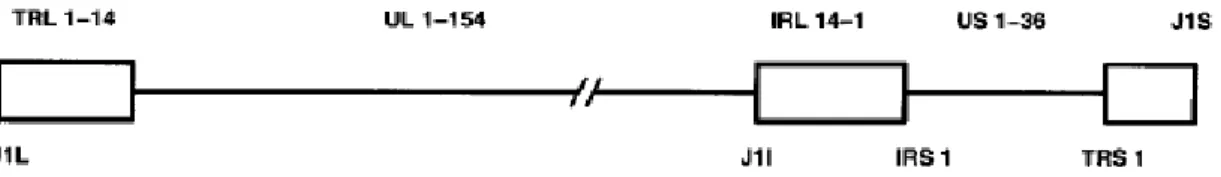
The AD169 laboratory strain was the first and the only completely sequenced HCMV strain, nevertheless it seems to have shorter genome than do many clinical isolates [36]. Analysis of its genome has revealed that it encodes 225 ORFs of about 100 or more amino acids (aa) residues in length, named according to the region and the numerical order in which they occur [19, 42]. There have been described the existence of unique ORFs, UL1-154 (with some ORFs receiving fractional designations such as UL21.5) and US1-36; and also repeated ORFs, J1L/J1I/J1S, TRL1-14/IRL1-14; and IRS1 plus TRS1 - Figure 4 [42]. Additional ORFs have been identified in two laboratory strains (Towne and Toledo) and in clinical isolates [42]. The Toledo strain, as well as some clinical isolates contains an additional 15 kilobases (kb) of DNA that is absent in the genomes of AD169 and Towne strains [19, 36, 41]. This large block of DNA seems to contain 19 genes encoding for viral glycoproteins and other specialized functions [36, 41].
Figure 4: Schematic diagram of the linear, double-stranded DNA of HCMV. Repeated genes (J1L/J1I/J1S, TRL1-14/IRL1-14) and
partially repeated genes (IRS1 and TRS1) are represented by rectangles, whereas unique gene blocks (UL1-UL154 and US1-US36) are designated by a line [42].
1.2. HCMV Infection
1.2.1.
Epidemiology
HCMV is a common virus with unknown seasonal predominance and its epidemiology varies in different regions of the world and between socioeconomic and age groups [1, 43, 44]. Generally, the prevalence of HCMV infection is higher in developing countries and among persons of lower socioeconomic status in developed nations [1, 45]. HCMV infection is considered important in certain risk groups such as immunocompromised individuals and pregnant women [43, 45].
The incidence of HCMV infection in the general population ranges from 36 to 90%, but the overall seroprevalence in developed countries is estimated to be in the range of 30– 70% [44, 46]. HCMV infection gradually increases with age, showing the lifelong risk of acquiring HCMV infection [1, 43, 45]. The prevalence of HCMV seropositivity in younger age varies according the social standing, being higher in lower economic classes [41, 47]. Racial differences have also been described, with higher seroprevalence found in African Americans and Hispanics than in Caucasians [1, 45]. An increased seroprevalence of HCMV has also been described in women attending sexually transmitted disease clinics and in young homosexual males [1, 44].
1.2.2.
Source of Transmission
HCMV is easily transmitted orally, through sexual intercourse, breastfeeding, placental transfer, blood transfusion or transplantation [44, 46]. The unsuspecting host is thus able to spread the virus both vertically and horizontally [1, 21, 48], since it is excreted
Page 8 CHARACTERIZATION OF CYTOMEGALOVIRUS RESISTANT
STRAINS IN HEMATOPOIETIC STEM CELL TRANSPLANTED PATIENTS
through body fluids (urine, saliva, tears, semen, milk, and cervical secretions) for months to years. [12, 43, 48].
Vertical transmission of HCMV can occur in three different ways: transplacental, intrapartum or breastfeeding [1]. Transplacental transmission can occur both in women infected for the first time during pregnancy and those infected long before conception [21]. A primary infection in the first 16 weeks of pregnancy is associated with higher rate of damage in fetal development [43]. Infection during delivery is due to shedding from the vagina or cervix, followed by ingestion of infected secretions by the offspring [21]. Breastfeeding is considered the most common mode of transmission to children and plays an important role in the epidemiology of HCMV infection as the virus is reactivated during lactation in nearly every seropositive mother [1, 43, 48].
Epidemiologic studies support the classification of HCMV as a sexually transmitted infection, consistent with excretion of this virus in cervical secretions, vaginal fluid, and semen [1]. Furthermore, HCMV has also been described as capable of being transmitted through blood or transplants [1]. The association between the acquisition of HCMV primary infection and blood transfusion was first suggested in 1960 as it is assumed that the virus is latent in the blood cells of healthy donors and is reactivated following transfusion when they encounter an allogeneic stimulus [1, 21].
1.2.3.
Infection and replication Cycle
HCMV infection starts with the attachment of HCMV membrane glycoproteins to the host cell surface receptors by a pH-independent mechanism [1, 3, 20, 21, 37] followed by the uncoated of viral capsids and rapid translocation of viral DNA into the nucleus – Figure 5 [3, 21].
The mechanism by which HCMV entry proceeds is cell type-dependent and occurs via fusion with the plasma membrane (on fibroblasts) or by acid-mediated endocytosis (in epithelial and endothelial cells). Both pathways require the viral gH/gL complex, however further characterization indicates that while gCIII (gH/gO/gL) is involved in binding of HCMV to fibroblasts, a complex consisting of gH/gL/UL128-131 plays a significant role in attachment and entry into epithelial and endothelial cells [1, 7, 20, 49].
Virus attachment and penetration are fast and efficient in both permissive and nonpermissime cell types [3, 21, 49]. The viral glycoprotein B (gB), encoded by UL55
gene, is the primary viral ligand that interacts with two separate binding sites: the heparin sulfate proteoglycans and a non-heparin receptor [21]. During the initial virus-cell interactions, HCMV attaches to the cell surface by low-binding of gB to heparan sulfate proteoglycans. The subsequent interaction of gB with its non-heparin receptor increases the stability of HCMV bind to cell surface [21]. However, the fusion of the viral envelope with the cell membrane to allow viral penetration is thought to require a further event mediated by the heteroligomeric gCIII (gH/gL/gO) complex with unidentified receptors - Figure 5 [7, 27].
After translocation into the nucleus, viral mRNA is transcribed and the transcripts exported from the nucleus to be translated by cellular machinery [3, 50]. Nevertheless, while some translated viral gene products are solely cytoplasmic proteins, others may return to the nucleus to regulate viral replication and cellular control [3, 50]. DNA replication, capsid assembly and DNA packaging occurs in the nucleus and viral assembly, which includes tegumentation and envelopment of new viral capsids, occur in the cytoplasm- Figure 5 [3, 51].
Similar to other herpesviruses, HCMV DNA replication begins 16 to 24 hours after infection [37] involving temporally ordered viral gene expression [12, 19, 37, 38]. The first transcribed viral genes are the immediate early (IE or α) genes, which are mainly transcriptional regulators, and have the ability to be transcribed in the absence of de novo protein synthesis, and are assumed to carry out key regulatory functions in permissive as well as in latent infection [1, 3, 19, 37, 38]. Expression of these genes is required for the transcription of early genes (E or β) [3, 12, 19, 37, 38]. Early genes are divided into two subclasses (β1/E and β2/E-L) and encode multiple proteins required for synthesis, processing and repair of DNA, capsid assembly, encapsidation, and establishment of immune evasion in the productively infected cell [1, 8, 12, 19, 37, 38].HCMV replication occurs after circularization of DNA and DNA synthesis starts by rolling circle replication which can undergo genomic inversion [47]. HCMV genome, unlike other herpesviruses, does not encode deoxyribonucleotide (dNTPs) biosynthesis enzymes, thus it has developed strategies to stimulate the biochemical pathways involved in the biosynthesis of DNA precursors [21]. This feature is considered crucial for its productive replication in the quiescent or terminally differentiated non-dividing cells [21]. Several trans acting factors
Page 10 CHARACTERIZATION OF CYTOMEGALOVIRUS RESISTANT
STRAINS IN HEMATOPOIETIC STEM CELL TRANSPLANTED PATIENTS
herpesvirus-conserved ORFs that provide the core replication proteins for viral DNA replication [8]. Among them, the single-stranded DNA-binding protein ppUL57 prevents the reannealing of DNA strands, followed by the unwinding by the helicase-primase complex, composed by primase (pUL70), helicase (pUL102) and primase-associated protein (pUL105) encoded by UL70, UL102, and UL105, respectively. The DNA polymerase processivity factor pUL44, encoded by UL44, prevents the dissociation of DNA polymerase pUL54, which is encoded by UL54, from the template [8, 19, 38]. The amino acid sequences of all of these proteins are highly conserved among HCMV strains and pUL54 3’-5’exonuclease activity (proofreading) is responsible for the high fidelity of the replication, which results in a low mutation rate [19]. The Late (L or γ) proteins are the last class of gene products expressed during HCMV replication and can be divided into two classes: gamma 1/leaky-late (γ1) and gamma 2/true-late (γ2) [8, 38]. Proteins encoded by these genes have mainly structural roles and contribute to the assembly and morphogenesis of the virion [1, 3, 12, 19, 37, 38]. Their transcription begins more than 24 hours after infection and requires prior viral DNA replication [37]. The late genes UL94, UL99, and also UL32 tegument proteins are essential for late events in virion assembly in the cytoplasm [32, 52].
The growth cycle of HCMV is relatively slow and viruses are not released until 72 to 96 hours [37]. Despite the fact that the precise molecular events of capsid assembly and DNA packaging are not yet understood, these processes are thought to occur in the nucleus – Figure 5 [53]. Three distinct capsid types, termed A, B, and C, are found in HCMV-infected cells [53]. The packaging of the DNA genome leads to the egress of the capsid scaffold and gives rise to a mature capsid - Figure 5 [21, 51]. Errors in packaging can result in cytoplasmic accumulation of non-infectious enveloped viral particles containing capsids lacking scaffold or DNA and capsids with only scaffold protein cores [54]. In fact, experiments revealed an important role for UL97 kinase (pUL97) in capsid assembly [19, 55]. Optimal nuclear egress requires active pUL97 and its detection correlates with abnormal subcellular distribution of viral structural protein assembly sites, both in the nucleus and in perinuclear structures, and consequent reduction in viral yield [19, 55]. Capsids are initially assembly through budding at the nuclear membrane, where they acquire a primary envelope derived from its inner leaflet. Then, they cross the lumen, fuse with the outer leaflet of the nuclear membrane or the endoplasmic reticulum (ER) membrane, lose their primary envelope, and move into the cytoplasm, where HCMV virion
particles acquire their tegument - Figure 5 [21]. Final envelopment of tegument-coated HCMV capsids occurs into a Golgi-derived secretory vacuole specifically destined for the plasma membrane and not marked for degradation in lysosomes or endosomes [14, 21]. Finally, the egress of infectious viral particles occurs by the fusion of secretory vacuoles containing HCMV viral particles with the cell membrane after transport via the vesicle trafficking pathways - Figure 5 [14, 21].
Figure 5: HCMV Life Cycle. 1) HCMV attachment to the cell membrane with penetration via endocytosis or fusion at the plasma
membrane; 2) Virion contents are released into the cytoplasm; 3) nucleocapsids are translocated into the nucleus, where viral DNA is released; 4) Viral replication and maturation follow the stimulation and parallel accumulation of viral synthesis function; 5) the encapsulation of replicated viral DNA as capsids, which are then transported from the nucleus to the cytoplasm; 6) envelopment of tegument-coated HCMV capsids occurs into a Golgi-derived secretory vacuole in the cytoplasm; 7) egress process that leads to virion release by exocytosis at the plasma membrane. Adapted from [6].
1.2.4.
HCMV Infection and Disease
HCMV infection is in the majority of cases asymptomatic, since the virus is maintained in a state of latency or low level shedding that is clinically undetectable [13, 19, 46]. Symptomatic infections in immunocompetent individuals are rare [4]. However, the groups at higher risk of HCMV infection and associated disease development are individuals with compromised or immature immune systems, including those infected with
Page 12 CHARACTERIZATION OF CYTOMEGALOVIRUS RESISTANT
STRAINS IN HEMATOPOIETIC STEM CELL TRANSPLANTED PATIENTS
HCMV infection can lead to HCMV-associated disease resulting in significant morbidity and mortality [8, 16].
Infants infected in utero are at risk for several congenital abnormalities and sensorineural hearing loss [13, 36, 40, 45, 46]. In patients who have acquired immune deficiency syndrome (AIDS) most commonly developed HCMV retinitis that leads to vision loss [13, 36, 46]. In transplant recipients, HCMV has both direct effects, resulting from viral invasion of organ systems, and indirect effects on the immune systems [46, 56]. The direct effects of HCMV primary infection or reactivation in organs are the development of end-organ diseases such as pneumonia, hepatitis, pancreatitis, gastrointestinal disease, retinitis, encephalitis, colitis, esophageal ulcers and others [13, 21, 36, 56]. Regarding the indirect effects, they are often associated with increased risk of other infections, bacterial and fungal, and promote acute graft rejection [46]. In fact, HCMV has repeatedly been associated with rejection after solid-organ transplantation and with graft-versus-host disease (GVHD) after hematopoietic stem cell transplant (HSCT) [19, 36, 56].
1.3. HCMV and HSCT
1.3.1.
Epidemiology and Characteristics
HCMV infection is the leading viral cause of morbidity and mortality in patients who receive hematopoietic stem cell transplant (HSCT) or solid organ transplant (SOT) (including kidney, liver, heart, heart-lung) [36, 46].
Studies have described that HSCT recipients have a higher prevalence of HCMV infection and associated diseases than SOT recipients [16, 57]. The incidence of HCMV infection following allogeneic HSCT (allo-HSCT) ranges from 32% to 70%, varying with the serological status of the recipient (R) and donor (D) [21]. Its incidence in seronegative recipients with seropositive donors (D+/R-) is lower than in seropositive recipients with seropositive donors (D+/R+), suggesting a transfer of adoptive immunity from donor to recipient. Thus, the most critical event is reactivation of a latent virus in seropositive recipients [15, 21, 46, 58-60]. In seronegative recipients, transmission mostly occurs through larger quantities of blood products that they receive [21].
A recent study from Portugal showed that 60.3% of allogeneic HSCT patients developed HCMV infection, mainly viral reactivations rather than primary infections (96.2% vs 3.8%, respectively) [61]. Typically, HCMV infection/reactivation appears within the first 100 days after transplant, both in allogeneic and autologous recipients, and affects mainly the lungs and the gastrointestinal tract [60, 61]. In HSCT recipients, late-onset HCMV disease (disease occurring >100 days after transplant) is similar to that observed in SOT patients, but with higher mortality [60, 62, 63]. Nevertheless, the increase in late-onset HCMV infections may be due to effective antiviral prophylaxis or preemptive treatment during the first 100 days after transplantation [58, 63, 64], by inhibiting the development of HCMV-specific T-cell lymphocyte response [62, 64, 65]. In fact, while prophylaxis and pre-emptive therapy are effective for the prevention of the HCMV disease during the antiviral treatment, cessation of therapy can result in the emergence of late-onset HCMV disease [58, 63].
1.3.2.
Risk Factors
The risk of developing HCMV reactivation or disease in HSCT patients is associated with the type of transplant and its associated-complications [59, 61, 64, 65]. Indeed, not all individuals are at the same risk as it varies with age, underlying disease, source of stem cells, donor/recipient (D/R) HCMV serological status, type of immunosuppression, and occurrence of graft-versus-host disease (GVDH) [36, 58, 59, 61, 64-67]. Moreover, the risk factors for HCMV infection vary during the transplantation period and while some can be predicted prior the transplant, other are dependent on the outcome of the transplant - Figure 6 [58-60, 64, 65].
Page 14 CHARACTERIZATION OF CYTOMEGALOVIRUS RESISTANT
STRAINS IN HEMATOPOIETIC STEM CELL TRANSPLANTED PATIENTS
Figure 6: Conditions associated with the risk of developing HCMV infection, reactivation, or disease in each of the following phase of
therapy in HSCT patients. Adapted from [65].
1.3.2.1. Before Transplantation
The risk factors that can be used prior transplantation to predict the occurrence of a HCMV reactivation/disease may be divided in host factors and transplant-related factors - Figure 6. Considering the host factors, it has been demonstrated that older ages represents a risk factor for developing HCMV reactivation/disease and for transplant-related mortality (TRM) [59, 61, 65]. The underlying disease and its treatment has not been specifically studied as a risk factor for development of HCMV reactivation/disease after HSCT, nevertheless, it has been demonstrated that the disease stage at the time of transplant is a highly significant predictor of mortality and that a diagnosis of chronic myelogenous leukemia in patients receiving T cells depleted from a HCMV–positive donor could represent a negative prognostic factor [59, 61, 65]. Furthermore, HCMV serostatus is considered mandatory in all transplant recipients and donors to evaluate the risk of HCMV reactivation/disease [58, 61, 66, 68, 69]. In autologous HSCT (auto-HSCT) recipients the probability of HCMV infection has been reported to be nearly 60% in seropositive patients and 23% in seronegative patients, however, the risk of developing HCMV reactivation/disease is lower than in allogeneic HSCT recipients [36, 65]. In allogeneic HSCT recipients, the risk of developing HCMV reactivation/disease has been reported as
Before transplant
• Host factors • Age
• Underlying disease
• Seropositivy status (donor and recipient)
• Transplant-related factors • T-cell depletion
• Autologous vs allogeneic
• Human leukocyte antigen (HLA)-match vs non-(HLA)-match donors • Immnunosuppresion
• Source of stem cells
(peripheral blood cells, cord blood and bone marrow)
After transplant
• Immnunosuppresion
• Presence of graft-versus-host disease (GVHD)
• Immune reconstitution • Other viral infections
• Other Opportunistic infections: parasitic, bacterial and fungal
5% of seronegative patients with seropositive donors (D+/R-), 14% of seropositive patients with seronegative donors (D-/R+), and 12% of seropositive patients with seropositive donors (D+/R+) [17, 59, 60, 65, 69]. Thus, the reactivation of HCMV occurs in nearly 80% of HCMV-seropositive recipients and 28% of seronegative recipients who receive a graft from a seropositive donor [65, 68]. Ljungman et al. suggests that seronegative patients that received grafts from seropositive donors have improved outcome [70], and in contrast, seropositive patients who receive grafts from seronegative donors have an increased risk of both repeated HCMV reactivation and disease [17, 59, 60, 65, 69]. Furthermore, seronegative recipients receiving transplant from seronegative donors have a very low risk of primary infection and a lower HCMV-related mortality [58, 59, 68].
Regarding the transplant-related factors, the type of transplant, Human leukocyte antigen (HLA) status, immunosuppression regimen, source of stem cells and T-cell depletion are considered important in the definition of risk for HCMV reactivation or disease - Figure 6 [17, 58, 60, 62, 64, 65, 67, 68]. Recipients of T cell-depleted allografts are the patients predominantly affected by HCMV reactivation, a rapid onset of HCMV-related symptoms, and a higher rate of fatal infections that may occur during the first 30 days after HSCT [58, 64, 66]. The risk of HCMV reactivation/disease is higher in recipients of transplants from an unrelated donor than in recipients of a related donor. However, in the allogeneic HSCT patients the risk of HCMV reactivation or disease is higher compared with patients receiving autologous HSCT [58, 60, 65, 66]. In recipients of unrelated or mismatched donor transplants, the increased risk of HCMV reactivation/disease is associated with a higher risk for HCMV-associated death and TRM, with higher mortality occurring in HCMV-positive patients receiving a transplant from negative donors [65]. Ljungman et al. also showed that allogeneic HSCT from seropositive donors was associated with better survival than from seronegative donors when the donors were unrelated, but not when the donors were HLA-identical siblings [70]. More aggressive chemotherapy regimens in transplant candidates have led to an increased infection of HCMV before transplantation [62]. Recipients of non-myeloablative stem cell transplants have a reduced risk of early HCMV infection and disease compared with standard myeloablative regimens [68]. However, they are also at an increased risk of late HCMV disease, mostly related to the use of antithymocyte globulin (ATG) or
Page 16 CHARACTERIZATION OF CYTOMEGALOVIRUS RESISTANT
STRAINS IN HEMATOPOIETIC STEM CELL TRANSPLANTED PATIENTS
60, 62]. A randomized study has showed, somewhat unexpectedly, that peripheral blood stem cell transplantation has not been associated with less HCMV infection and disease when compared to marrow transplantation [71]. However, in a non-randomized study, has been observed a moderate reduction in HCMV disease [72]. Cord blood transplantation is associated with similar rates of HCMV infection and has generated new populations of patients at high risk for HCMV reactivation and disease [60, 62]. However, blood products transfusion represents the main risk factor for HCMV acquisition in HCMV-negative patients receiving bone marrow from a HCMV-negative donor [65].
1.3.2.2. After Transplantation
Amongst the post-transplant related risk factors for HCMV reactivation or disease are the immune reconstitution, the development of GVDH, immunosuppression regime and the development of other infections - Figure 6 [17, 59, 60, 62, 64, 65, 68]. The development of HCMV disease occurs especially in HSCT recipients submitted to highly immunosuppressive regimens used to prevent rejection of the transplant, in particular those that lead to prolonged lymphocytopenia such as the use of fludarabine or analogues as well as alemtuzumab [60, 62]. In fact, the severity of the end-organ disease caused by HCMV is related to the degree of immune suppression [36, 60]. Furthermore, the immune reconstitution is also a significant factor, since while in SOT the host has the immune system working very early, in HSCT recipients the time to reach immune reconstitution may vary a lot [17, 64, 65]. Furthermore, allogeneic HSCT recipients, in contrast to autologous HSCT patients, are at a much higher risk of active HCMV infection because of the delayed recovery of T- and B-cell functions [17, 64]. In fact, studies have found HCMV-specific cytotoxic T-lymphocytes regeneration is dependent on HCMV serologic status, with both CD4+ and CD8+ T lymphocytes to be required for complete restoration of immunity [65]. However, the inability to control HCMV reactivation after allogeneic Bone Marrow Transplant (BMT) has been mainly associated with impaired function of antigen-specific CD8+ T cells rather than an inability to recover a sufficient numbers of HCMV-specific T cells [65]. In addition, it has been demonstrated that CD4+ T-helper cells regenerate relatively slowly following allogeneic BMT with subsequent limited help for CD8+ T cells to control HCMV replication and have observed lower HCMV-specific CD8 T-cell numbers during viral replication [17]. Therefore, HCMV pneumonia during the
first 120 days after HSCT is much more severe and life-threatening than it is in a patient after renal transplantation [36].
Another risk factor for HCMV reactivation or disease is the development of GVHD after transplantation and has been shown that acute and chronic GVHD significantly increases the risk of HCMV infection [17, 64, 65]. Moreover, patients that develop chronic GVHD (cGVHD), which is characterized by a severe combined cellular and humoral immunodeficiency, and acute GVHD (aGVHD), are at a prolonged risk to develop late-onset HCMV disease [60, 62, 64, 65].
Other viral infections, such as Human herpesvirus 6 (HHV6), Human herpesvirus 7 (HHV7) and Epstein Barr virus (EBV) have been implicated as risk factors for progression from active HCMV infection to HCMV disease and with reactivation of HCMV by suppressing the development of HCMV-specific immune responses [65]. In addition to viral infections, other opportunistic infections such as parasitic, bacterial, and fungal also emerge as common causes of HCMV reactivation/disease [58].
1.3.3.
Clinical Features
HCMV end-organ disease is classified as of early onset (<100 days after transplant) or late onset (>100 days after transplant) [36, 60]. Pneumonia and gastrointestinal involvement are the most frequently described diseases caused by HCMV in HSCT recipients; nevertheless, there are other documented conditions such as hepatitis, retinitis, encephalitis, hemorrhagic cystitis, unexplained fever, endothelial damage, and thrombotic microangiopathy [17, 36, 55, 58, 60, 68].
The interstitial pneumonia caused by HCMV is the most common life-threatening infectious complication of allogeneic HSCT, defined by the presence of pulmonary disease, combined with HCMV found in bronchoalveolar lavage fluid or lung tissue samples. It is still a potentially fatal disease, which usually occurs within the first 120 days after transplantation, with decreasing incidence and severity after the initiation of routine antiviral prophylaxis or pre-emptive therapy after HSCT [17, 36, 65, 68]. The incidence of HCMV pneumonia ranges from 10% to 30% in allogeneic HSCT recipients and from 1% to 6% in autologous HSCT recipients. It has been described that 69% of cases occur early
Page 18 CHARACTERIZATION OF CYTOMEGALOVIRUS RESISTANT
STRAINS IN HEMATOPOIETIC STEM CELL TRANSPLANTED PATIENTS
factors: prolonged deficiency in HCMV-specific cytotoxic T-lymphocyte activity, recipient seropositivity, older age, presence of acute GVHD, use of cyclosporine (CsA) as GVHD prophylaxis, and the underlying disease [17, 36, 65]. Lymphocytopenia, male gender, and severe acute GVHD are also often referred as contributing for severe HCMV pneumonia [17, 36, 65].
Gastrointestinal (GI) HCMV disease has been described both in autologous and allogeneic HSCT as a combination of clinical symptoms in gastrointestinal tract, findings of macroscopic mucosal lesions at endoscopy, and the symptoms vary depending on the location of the disease [17, 36, 65]. It is characterized by an erosive and/or an ulcerative condition that can occur at any location in the GI tract, from mouth to rectum [36, 65]. The incidence rates for GI HCMV disease are of 2% at 2 years after HSCT, with higher frequency in allogeneic than in autologous HSCT recipients [17]. The risk factors for the development of GI HCMV disease include allogeneic transplantation, use of steroids, the presence of intestinal acute GVHD and the HCMV-seropositive recipients [17, 65].
1.4. HCMV infection management
1.4.1.
Anti-HCMV Drugs
Currently, four antiviral drugs are used to prevent/treat HCMV infection and diseases. These antiviral drugs act by inhibiting effective HCMV DNA synthesis and have been shown to be effective in the prevention and/or treatment of HCMV infection and diseases: Ganciclovir (GCV), Valganciclovir (VGCV), Cidofovir (CDV) and Foscarnet (FOS) - Figure 7 [40, 46, 73-76]. Despite their clinical utility being limited by the efficacy, limited oral bioavailability, development of resistance in clinical practice, and associated toxicities, these drugs have been used to treat many forms of HCMV disease in immunocompromised patients [40, 46].
Figure 7: Action mechanisms of systemic antivirals approved for treatment of HCMV infection. GCV/VGCV requires phosphorylation
by the phosphokinase (pUL97). After monophosphorylation by pUL97, the cellular kinases add two additional phosphates. GCV triphosphate is the active form of the drug incorporated into viral DNA by the viral DNA polymerase (pUL54). CDV is a monophosphate analog and does not require initial viral phosphorylation. Cellular kinases add additional phosphates to produce CDV diphosphate, which is incorporated into the viral DNA by pUL54 leading to termination of viral DNA replication. FOS is a pyrophosphate analog, which does not require activation and is not incorporated into the growing viral DNA chain. It blocks directly the release of pyrophosphate by pUL54 and therefore resulting in chain termination. Adapted from [19, 77].
1.4.1.1. Ganciclovir and Valganciclovir
Ganciclovir (GCV) or (9-[1, 3-dyhydroxy-2-propoxymethyl] guanine) was the first antiviral agent approved for the treatment of HCMV infection/disease, and remains the first-line treatment for HCMV infection/disease in immunocompromised patients [36, 46, 77, 78]. It is an inactive nucleoside analogue of guanosine and homologue of acyclovir (ACV) [10, 36, 40, 77]. GCV is converted to GCV triphosphate that is the active form of drug by a multistep process dependent on both viral and cellular enzymes - Figure 7 and Table I [19, 46, 78-80]. The two target viral proteins involved in GCV anabolism are: UL97 and UL54 [19, 40, 78]. The UL97 gene of HCMV encodes a viral protein kinase that phosphorylates GCV to GCV monophosphate [10, 36, 40, 46, 77-79, 81]. Two subsequent phosphorylation steps are performed by host cellular kinases that result in the formation of the GCV triphosphate metabolite, which is a competitive inhibitor of the natural substrate (deoxyguanosine triphosphate) for the viral DNA polymerase encoded by UL54 - Figure 7
Page 20 CHARACTERIZATION OF CYTOMEGALOVIRUS RESISTANT
STRAINS IN HEMATOPOIETIC STEM CELL TRANSPLANTED PATIENTS
HCMV DNA continue to be synthesized [36, 80], nevertheless its effects result from the ability to difficult and slow the elongation of viral DNA [36, 46, 81].
GCV has been shown to reduce the severity of HCMV retinitis, gastrointestinal disease and, to a lesser extent, pneumonia in SOT, HSCT and AIDS patients [21, 36, 40, 46]. GCV was initially approved by Food and Drug Administration (FDA) in 1989 for intravenous (IV) use [19, 46, 79]. Nevertheless, and despite its high bioavailability and therapeutic efficacy, IV use is limited since hospitalization is required for administration [46, 75]. Moreover, GCV use is limited by the occurrence of hematologic side effects, primarily neutropenia, and thrombocytopenia, mainly in the early phases of HSCT - Table I [17, 36, 46, 65, 68]. An oral capsule released in 1994 represented a major advance for treatment of HCMV retinitis, but could only be used as maintenance therapy, as the low bioavailability of the oral formulation was considered insufficient for induction therapy [19, 46, 79]. GCV poor oral bioavailability (5.6%) leads to the development of Valganciclovir (VGCV), a L-valyl ester prodrug which after oral administration is rapidly metabolized in the liver and intestinal wall - Table I [19, 36, 40, 46, 67, 77, 79]. The adverse effects of VGCV are similar to those of GCV, mainly, neutropenia and thrombocytopenia - Table I [36, 46, 67]. Nevertheless, VGCV has a much better bioavailability (60%) and is a suitable replacement for IV GCV in many clinical applications [19, 36, 40, 46, 67, 77, 79]. Thus, given the convenience to treat patients without hospitalization, VGCV tends to be widely used among transplant recipients, not only for prophylaxis, but also for preemptive and maintenance therapy [46, 67, 73].
Although GCV and VGCV have been effective in prevention and treatment of HCMV disease, the emergence of GCV/VGCV-resistant HCMV strains has posed a more significant threat due to an aggressive disease course and a greater mortality risk. Treatment options for these strains are limited, with FOS being recommended as the initial treatment option followed by CDV; nevertheless, these agents are known to have substantial side effects, the most notable of which is nephrotoxicity - Table I [83].
1.4.1.2. Foscarnet
Foscarnet (FOS) a pyrophosphate analogue, with the chemical name of phosphonoformic acid, which reversibly, and noncompetitively, inhibits the activity of the HCMV DNA polymerase - Table I [36, 40, 82]. FOS does not require intracellular
activation to exert its antiviral activity and is not incorporated into the growing viral DNA chain [19, 40, 77, 78, 80-82]. This noncompetitive inhibitor reversibly blocks the pyrophosphate binding site of the viral DNA polymerase and inhibits the cleavage of pyrophosphate from deoxynucleoside triphosphates - Figure 7 and Table I [19, 40, 46, 77-81]. FOS is administered as large volume intravenous solution since it must be present in high concentrations inside the cell to remain in contact with the viral DNA polymerase and inhibit DNA replication; hence, when the intracellular concentration decreases viral DNA synthesis resumes [10, 19, 36].
FOS was FDA approved in 1991 [10, 19] and despite its utility has been associated with nephrotoxicity and metabolic toxicity as well as renal failure, hypocalcemia, hypomagnesemia and hypophosphatemia - Table I [10, 36, 46]. Due to its side effects, FOS is considered a second-line therapy, preferred over GCV especially in patients with myelosupression or weak graft after HSCT, to treat patients with AIDS and HCMV reti-nitis who are failing GCV therapy due to viral resistance, or those who cannot be treated with GCV due to dose-limiting neutropenia or leucopenia [17, 36, 46, 68]. Furthermore, some studies refer that patients must be on long-term maintenance regimens with IV FOS to prevent the relapse or progression of HCMV disease [21, 36].
1.4.1.3. Cidofovir
Cidofovir (CDV) is a monophosphate nucleotide analogue, with the chemical name ([S]-1-[3-hydroxy-2-phosphonylmethoxypropyl] cytosine) [36, 40, 46, 78, 80]. Because a single phosphate-like group is already present in this monophosphate analog, CDV does not require initial phosphorylation by pUL97 kinase but is dependent on diphosphorylation by cellular kinases for activation - Figure 7 and Table I [19, 40, 80, 81]. Cellular kinases add the additional phosphate group to CDV, converting it into CDV diphosphate, an analog of deoxycytosine [46, 77, 81]. Similarly to GCV, the incorporation of CDV-diphosphate into viral DNA causes a slowing and subsequent cessation of HCMV DNA replication - Figure 7 and Table I [77, 78, 81]. Sequential incorporation of two CDV molecules completely inhibits further synthesis of HCMV DNA since it cannot be excised by pUL54 3-to-5 exonuclease activity [84].
Page 22 CHARACTERIZATION OF CYTOMEGALOVIRUS RESISTANT
STRAINS IN HEMATOPOIETIC STEM CELL TRANSPLANTED PATIENTS
CDV was FDA approved in 1996 as an IV formulation for the treatment of a broad- range of DNA viruses infections, including all the herpesviruses [40, 68]. CDV oral bioavailability is less than 5% [19, 46]; nevertheless it has a very long intracellular half-life when compared with GCV and FOS [36, 68]. Despite its efficacy as an anti-HCMV agent, due to the poor oral bioavailability and concerns about dose-related nephrotoxicity, lipid ester analogs, including as hexadecyloxypropyl-CDV (CMX001) and octadecyloxyethyl-CDV, are being tested against herpesviruses – Table I [46].
CDV has excellent activity against HCMV and has been reported to be effective in the treatment of HCMV retinitis in AIDS patients [40, 46] . CDV has also been studied for HCMV infection and disease in allogeneic HSCT [36], however, it has several side-effect such as nephrotoxicity and myelosuppression [17, 46, 68], and therefore it is considered a third-line agent for HCMV infection – Table I [17].


![Table I: Antiviral agents used to prevent/ treat HCMV infection and disease. Adapted from [17]](https://thumb-eu.123doks.com/thumbv2/123dok_br/15145182.1012262/45.892.151.767.132.1007/table-antiviral-agents-prevent-treat-infection-disease-adapted.webp)
![Figure 8: UL97 structure and functional domains. Adapted from [19, 55, 77, 95].](https://thumb-eu.123doks.com/thumbv2/123dok_br/15145182.1012262/50.892.126.751.371.740/figure-ul-structure-functional-domains-adapted.webp)
![Figure 9: UL54 structure and functional domains. Adapted from [19, 77, 95].](https://thumb-eu.123doks.com/thumbv2/123dok_br/15145182.1012262/54.892.143.733.608.1061/figure-ul-structure-functional-domains-adapted.webp)


