Skin Cancer – Clinical Investigations
Dermatology 2018;234:37–42Lentigo Maligna – Not Always a Face and
Neck Disease of the Elderly
Ana Filipa Duarte
a, bBernardo Sousa-Pinto
b–dAna Margarida Barros
aEckart Haneke
a, e–gOsvaldo Correia
a, c, daCentro de Dermatologia Epidermis, Instituto CUF, Porto, Portugal; bMEDCIDS – Department of Community
Medicine, Information and Health Decision Sciences, Faculty of Medicine, University of Porto, Porto, Portugal;
cCINTESIS – Center for Health Technology and Services Research, University of Porto, Porto, Portugal; dBasic and
Clinical Immunology Unit, Department of Pathology, Faculty of Medicine, University of Porto, Porto, Portugal;
eDepartment of Dermatology, Inselspital, University of Bern, Bern, Switzerland; fDermatology Clinic Dermaticum,
Freiburg, Germany; gDepartment of Dermatology, University of Ghent, Ghent, Belgium
Received: March 17, 2018 Accepted: April 17, 2018 Published online: June 12, 2018
Ana Filipa Duarte, MD © 2018 S. Karger AG, Basel
DOI: 10.1159/000489397
Keywords
Lentigo maligna · Extrafacial location · Early diagnosis
Abstract
Introduction: Lentigo maligna (LM) is a rare form of in situ
melanoma, frequently seen as a large patch in elderly pa-tients. The aim of this study was to assess clinical and dermo-scopic features of LM. Material and Methods: A retrospec-tive study of LM patients presenting to our center between July 2007 and July 2017 was performed. Demographic data, anatomical location, laterality, diameter, Clark level, Breslow stage, “ABCD” signs and dermoscopic features were regis-tered. Facial versus extrafacial LM were compared. Results: We found 21 LM, of which 12 had an extrafacial location and 9 a facial location. Half of the extrafacial lesions were located on an upper limb. The median age at diagnosis was 63 years (ranging from 38 to 84 years). Most LM cases were female (16/21) with phototype II (13/21). More than half of the pa-tients (11/21) had a history of a skin neoplasm or actinic ker-atosis. The median diameter found was 6 mm (interquartile range = 4.5 mm), ranging from 1 to 15 mm. Five lesions were invasive (median Breslow depth of 0.2 mm), and 4 of them
were extrafacial. Discussion: In this study LM was more fre-quently found in an extrafacial location and as a small patch with a 6-mm diameter medium. The epidemiology of LM/LM melanoma might be changing. Full body examination and dermoscopy are of the utmost importance for the diagnosis. Dermatologists should be aware and search for small lesions outside the face and neck, particularly in middle-aged fe-male patients with photo-damaged skin.
© 2018 S. Karger AG, Basel
Introduction
Lentigo maligna (LM) is relatively rare form of ma-lignant melanoma (MM), corresponding to 4–15% of all MM cases [1]. LM usually occurs in sun-damaged skin and is typically poorly circumscribed; its diagnosis is based on atypical junctional melanocytic hyperplasia combined with solar elastosis and epidermal atrophy [1, 2]. When LM invades the dermis, it is called lentigo maligna melanoma (LMM). The exact percentage of LM evolving to LMM is not clear, varying from 5 to 50% [1, 3].
LM occurs almost exclusively in middle-aged and el-derly Caucasians [2]; its incidence is increasing because of a higher cumulative exposure to ultraviolet radiation [4]. Classically, LM presents as a slowly enlarging patch with variable color and with ill-defined borders, most fre-quently located on the head and neck (86%), with predi-lection for the cheeks [5].
The clinical presentation may be subtle, particularly in early stages, and therefore, delayed diagnosis is common [4]. Because of its potential significant subclinical exten-sion, LM has a higher risk of local recurrence than other types of correctly treated melanoma. Once LM progresses to its vertically invasive form, its prognosis is similar to that of other types of MM. The diagnosis is a challenge in most
Patients with melanoma (n = 152) between 2007 and 2017 at our center Patients with lentigo maligna (n = 21) between 2007 and 2017 at our center Clinical features (n = 21)
Dermoscopic features (n = 20)
a
b c
Fig. 1. Flowchart of the Methods.
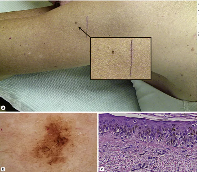
Fig. 2. Lentigo maligna. a Hyperpigmented patch, 3 mm in diameter, on the left arm, in a photo-damaged area,
of a woman in her sixties. b Dermoscopy (×30): atypical pigmented network, irregular striae. c Histopathology (×10): atypical melanocyte proliferation, small nests, pagetoid spread and light halo melanocytes.
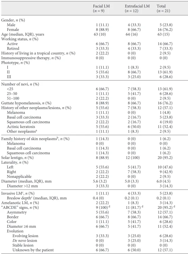
Table 1. Description of cases of facial and extrafacial lentigo maligna (LM) in 21 patients Facial LM (n = 9) Extrafacial LM(n = 12) Total(n = 21) Gender, n (%) Male 1 (11.1) 4 (33.3) 5 (23.8) Female 8 (88.9) 8 (66.7) 16 (76.2)
Age (median, IQR), years 63 (10) 64 (16) 63 (15) Working status, n (%)
Active 6 (66.7) 8 (66.7) 14 (66.7)
Retired 3 (33.3) 4 (33.3) 7 (33.3)
History of living in a tropical country, n (%) 2 (22.2) 0 (0) 2 (9.5) Immunosuppressive therapy, n (%) 0 (0) 0 (0) 0 (0) Phototype, n (%) I 1 (11.1) 1 (8.3) 2 (9.5) II 5 (55.6) 8 (66.7) 13 (61.9) III 3 (33.3) 3 (25.0) 6 (28.6) Number of nevi, n (%) <25 6 (66.7) 7 (58.3) 13 (61.9) 25–50 1 (11.1) 5 (41.7) 6 (28.6) 51–100 2 (22.2) 0 (0) 2 (9.5) Guttate hypomelanosis, n (%) 8 (88.9) 8 (66.7) 16 (76.2) History of other neoplasms/lesions, n (%) 5 (55.6) 7 (58.3) 12 (57.1)
Melanoma 1 (11.1) 0 (0) 1 (4.8)
Basal cell carcinoma 3 (33.3) 2 (16.7) 5 (23.8) Squamous cell carcinoma 2 (22.2) 2 (16.7) 4 (19.0) Actinic keratosis 5 (55.6) 6 (50.0) 11 (52.4) Other neoplasmsa 1 (11.1) 1 (8.3) 2 (9.5)
Family history of skin neoplasmsb, n (%) 1 (14.3) 0 (0) 1 (6.2)
Melanoma 0 (0) 0 (0) 0 (0)
Basal cell carcinoma 1 (14.3) 0 (0) 1 (6.2) Squamous cell carcinoma 1 (14.3) 0 (0) 1 (6.2) Solar lentigo, n (%) 8 (88.9) 12 (100) 20 (95.2) Laterality, n (%)
Left 5 (55.6) 5 (41.7) 10 (47.6)
Right 2 (22.2) 7 (58.3) 9 (42.9)
Nonapplicable 2 (22.2) 0 (0) 2 (9.5)
Diameter (median, IQR), mm 5.6 (3.2) 5.0 (3.3) 6.0 (4.5)
Diameter >12 mm 3 (33.3) 0 (0) 3 (14.3)
Invasive LMc, n (%) 1 (11.1) 4 (33.3) 5 (23.8)
Breslow depthc (median, IQR), mm 0.4 (0) 0.2 (0.1) 0.2 (0.1)
Amelanotic LM, n (%) 2 (22.2) 1 (8.3) 3 (14.3) “ABCDE” signs, n (%) 9 (100) d 11 (81.7) d 20 (95.2) d Asymmetry 5 (55.6) 7 (58.3) 12 (57.1) Border 6 (66.7) 8 (66.7) 14 (66.7) Color 1 (11.1) 5 (41.7) 6 (28.6) Diameter ≥6 mm 6 (66.7) 5 (41.7) 11 (52.4) Evolution Evolving lesion 3 (33.3) 3 (25.0) 6 (28.6) De novo lesion 0 (0) 3 (25.0) 3 (14.3) Stable lesion 0 (0) 0 (0) 0 (0)
cases. Suspicious lesions should undergo dermoscopic evaluation that may contribute to an early diagnosis [6].
The aim of this study was to characterize the clinical and dermoscopic features of LM/LMM in our center dur-ing the last decade.
Methods
For further details, see the online supplementary material (see www.karger.com/doi/10.1159/000489397 for all online suppl. ma-terial) (Fig. 1).
Results
Over the assessed 10-year period, we observed a total of 21 LM (out of 152 melanomas observed within the same period), of which 12 had an extrafacial location. Half of the latter were located on the upper limbs (upper
arm: n = 3; forearm: n = 2; hand: n = 1; Fig. 2), followed by the lower limbs (lower leg: n = 4; thigh: n = 1) and the upper back (n = 1). In all patients, LM was first suspected by a dermatologist. The median age at diagnosis was 63 years (ranging from 38 to 84 years).
Most LM cases were female (16/21) and occurred in patients with phototype II (13/21) and less than 25 nevi (13/21) (Table 1). More than half of the patients (11/21) had a history of a skin neoplasm or actinic keratosis. The median diameter for the assessed LM was 6 mm (inter-quartile range, IQR = 4.5 mm – ranging from 1 to 15 mm), corresponding to a median diameter of 5.1 mm among females (IQR = 5.3 mm) and 8.0 mm among males (IQR = 2.5 mm).
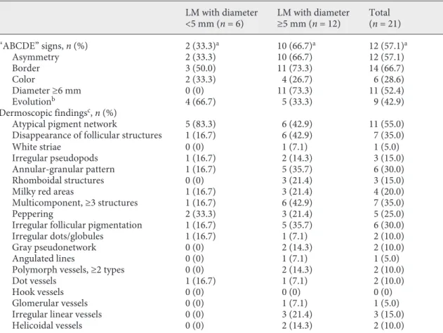
Of the “ABCD” signs, asymmetry, irregular border and diameter ≥5 mm were observed in more than half of the neoplasms; however, the presence of at least 3 ABCD signs was only present in 2 out of 6 cases of LM with di-ameter <5 mm (Table 2). The most common dermoscop-Table 1 (continued)
Facial LM
(n = 9) Extrafacial LM(n = 12) Total(n = 21) Dermoscopic findingse, n (%)
Atypical pigment network 1 (11.1) 10 (90.9) 11 (55.0) Disappearance of follicular structures 7 (77.8) 0 (0) 7 (35.0)
White striae 1 (11.1) 0 (0) 1 (5.0)
Irregular pseudopods 0 (0) 3 (27.3) 3 (15.0) Annular-granular pattern 6 (66.7) 0 (0) 6 (30.0) Rhomboidal structures 3 (33.3) 0 (0) 3 (15.0)
Milky red areas 2 (22.2) 2 (18.2) 4 (20.0)
Multicomponent, ≥3 structures 6 (66.7) 1 (9.1) 7 (35.0)
Peppering 2 (22.2) 3 (27.3) 5 (25.0)
Irregular follicular pigmentation 6 (66.7) 0 (0) 6 (30.0) Irregular dots/globules 0 (0) 2 (18.2) 2 (10.0)
Gray pseudonetwork 2 (22.2) 0 (0) 2 (10.0)
Angulated lines 1 (11.1) 0 (0) 1 (5.0)
Polymorph vessels, ≥2 types 2 (22.2) 0 (0) 2 (10.0)
Dot vessels 1 (11.1) 1 (9.1) 2 (10.0)
Hook vessels 0 (0) 0 (0) 0 (0)
Glomerular vessels 0 (0) 1 (9.1) 1 (5.0)
Irregular linear vessels 2 (22.2) 1 (9.1) 3 (15.0)
Helicoidal vessels 2 (22.2) 0 (0) 2 (10.0)
IQR, interquartile range. a Includes 1 patient with a history of thyroid cancer and another with a history of
leiomyoma. b Family history of melanoma is unknown for 6 patients, while family history of basal cell carcinoma
and squamous cell carcinoma is unknown for 5 patients. c All invasive LM were staged as Clark level II and
Bre-slow stage I. d Frequency of patients presenting with at least 1 “ABCDE” sign. For facial LM, 1/3 of patients only
presented with 1 “ABCDE” sign, while 2/3 of them presented with 2 or more criteria. For extrafacial LM, 17% of patients only presented with 1 sign, while 75% presented with 2 or more signs. e Dermoscopic information not
ic findings were atypical pigment network (11/20), dis-appearance of follicular structures (7/20) and multicom-ponent pattern (7/20) (Table 1). Five lesions were inva- sive – LMM – all of them staged as Clark level II, having a median Breslow depth of 0.2 mm (IQR = 0.1 mm).
Facial LM were observed for half of the female patients (8/16) but for only one fifth (1/5) of male patients. Results of the comparisons between facial versus extrafacial LM, and between neoplasms of <5 versus ≥5 mm diameter are presented in Table 1 and Table 2, respectively.
Discussion
Despite being a small retrospective series of a single center, this study has some interesting results, particu-larly regarding the location and median diameter of the assessed neoplasm, as well as the patients’ median age.
In fact, in our study, almost 60% of LM were located outside the face and neck. Extrafacial location had been reported by previous studies, but never to such an extent. In most studies, LM involving the head and neck account-ed for 75–85%, and only 15–25% were outside the face and neck. Patient age and gender distribution were also surprisingly different from those usually described in the literature [5, 7] in this study, the median age was lower than usually reported (63 vs. 70–75 years) [7]. In addition, more than three quarters of our patients were female, while most previous studies had reported LM to be more common among males [5, 7].
Changing patterns of sun exposure may contribute to the changing epidemiology of LM. The face is consistent-ly more exposed than other areas, but the relationship between sun exposure and LM/LMM might be complex. The regular use of sunscreens on the face may not totally explain the higher incidence of extrafacial lesions,
be-Table 2. Comparison of the frequency of “ABCDE” signs and dermoscopic findings in lentigo maligna (LM)
cases with <5 and ≥5 mm diameter
LM with diameter
<5 mm (n = 6) LM with diameter≥5 mm (n = 12) Total (n = 21) “ABCDE” signs, n (%) 2 (33.3)a 10 (66.7)a 12 (57.1)a Asymmetry 2 (33.3) 10 (66.7) 12 (57.1) Border 3 (50.0) 11 (73.3) 14 (66.7) Color 2 (33.3) 4 (26.7) 6 (28.6) Diameter ≥6 mm 0 (0) 11 (73.3) 11 (52.4) Evolutionb 4 (66.7) 5 (33.3) 9 (42.9) Dermoscopic findingsc, n (%)
Atypical pigment network 5 (83.3) 6 (42.9) 11 (55.0) Disappearance of follicular structures 1 (16.7) 6 (42.9) 7 (35.0)
White striae 0 (0) 1 (7.1) 1 (5.0)
Irregular pseudopods 1 (16.7) 2 (14.3) 3 (15.0) Annular-granular pattern 1 (16.7) 5 (35.7) 6 (30.0) Rhomboidal structures 0 (0) 3 (21.4) 3 (15.0) Milky red areas 1 (16.7) 3 (21.4) 4 (20.0) Multicomponent, ≥3 structures 1 (16.7) 6 (42.9) 7 (35.0)
Peppering 2 (33.3) 3 (21.4) 5 (25.0)
Irregular follicular pigmentation 1 (16.7) 5 (35.7) 6 (30.0) Irregular dots/globules 1 (16.7) 1 (7.1) 2 (10.0) Gray pseudonetwork 0 (0) 2 (14.3) 2 (10.0)
Angulated lines 0 (0) 1 (7.1) 1 (5.0)
Polymorph vessels, ≥2 types 0 (0) 2 (14.3) 2 (10.0)
Dot vessels 1 (16.7) 1 (7.1) 2 (10.0)
Hook vessels 0 (0) 0 (0) 0 (0)
Glomerular vessels 0 (0) 1 (7.1) 1 (5.0)
Irregular linear vessels 0 (0) 3 (21.4) 3 (15.0) Helicoidal vessels 0 (0) 2 (14.3) 2 (10.0)
cause females typically use more sunscreen all over the year than males (among whom we only found 1 facial le-sion).
The median diameter at diagnosis was very small (me-dian of 6 mm; 9 lesions with ≤5 mm). The bigger lesion observed had 15 mm and was smaller than the mean di-ameter found in a Spanish study (15.8 mm) [7]. Females had a smaller median diameter than males, suggesting a more delayed diagnosis among the latter.
Although the “ABCD” criteria have a good semiologi-cal value in the differential diagnosis of pigmented lesions [8], in this study only 2 cases of LM <5 mm had 3 signs of this rule, which does therefore not seem to be useful for the diagnosis of early small lesions.
Dermoscopy of facial pigmented lesions has been widely studied [4, 9, 10]. In our series, the most frequent feature for facial LM (particularly in lesions ≥5 mm) were disappearance of follicular structures, annular granular pattern and irregular follicular pigmentation, but also multicomponent pattern. Dermoscopic features of extra-facial LM include a combination of features of extra-facial LM and in situ superficial spreading MM [9]. None of the ex-trafacial cases of this series had a single feature of facial LM. Curiously, only 1 case of angulated lines was found; it concerned a facial LM, and not an extrafacial lesion, where this pattern has been previously reported [11].
Reflectance confocal microscopy has been described as an important complementary tool for diagnosis,
treat-ment guidance and follow-up of LM [12, 13], but it is a time-consuming and highly costly technique. Although more sensitive, it is less specific for the diagnosis of LM [14].
A routine full-body examination of the patients, and a regular photographic documentation with dermoscopy [15], associated with higher suspicions on patients with photo damage or a skin cancer history, may justify the small diameter of the lesions in this study. Clinical suspi-cion supported by complete history and close inspection assisted by dermoscopy are of most importance to recog-nize early LM.
Key Message
60% of lentigo maligna are located in photo-exposed areas out-side the head and neck.
Statement of Ethics
Subjects have given their informed consent for the study.
Disclosure Statement
The authors have no conflict of interest to declare.
References
1 McKenna JK, et al: Lentigo maligna/lentigo maligna melanoma: current state of diagnosis and treatment. Dermatol Surg 2006;32:493– 504.
2 Smalberger GJ, Siegel DM, Khachemoune A: Lentigo maligna. Dermatol Ther 2008;21: 439–446.
3 Weinstock MA, Sober AJ: The risk of progres-sion of lentigo maligna to lentigo maligna melanoma. Br J Dermatol 1987;116:303–310. 4 Pralong P, et al: Dermoscopy of lentigo ma-ligna melanoma: report of 125 cases. Br J Der-matol 2012;167:280–287.
5 Cox NH, Aitchison TC, MacKie RM: Extrafa-cial lentigo maligna melanoma: analysis of 71 cases and comparison with lentigo maligna melanoma of the head and neck. Br J Derma-tol 1998;139:439–443.
6 Schiffner R, et al: Improvement of early rec-ognition of lentigo maligna using dermatos-copy. J Am Acad Dermatol 2000;42:25–32. 7 Martinez-Leborans L, et al: Extrafacial lentigo
maligna: a report on 14 cases and a review of the literature. Actas Dermosifiliogr 2016; 107:e57–e63.
8 Thomas L, et al: Semiological value of ABCDE criteria in the diagnosis of cutaneous pig-mented tumors. Dermatology 1998;197:11– 17.
9 Lau YN, Affleck AG, Fleming CJ: Dermato-scopic features of extrafacial lentigo maligna. Clin Exp Dermatol 2013;38:612–616. 10 Stolz W, Schiffner R, Burgdorf WH:
Derma-toscopy for facial pigmented skin lesions. Clin Dermatol 2002;20:276–278.
11 Vanden Daelen A, et al: A digital dermoscopy follow-up illustration and a histopathologic correlation for angulated lines in extrafacial lentigo maligna. JAMA Dermatol 2016;152: 200–203.
12 Guitera P, et al: Improving management and patient care in lentigo maligna by mapping with in vivo confocal microscopy. JAMA Der-matol 2013;149:692–698.
13 Gomez-Martin I, et al: Histopathologic and immunohistochemical correlates of confocal descriptors in pigmented facial macules on photodamaged skin. JAMA Dermatol 2017; 153:771–780.
14 Cinotti E, et al: Dermoscopy vs reflectance confocal microscopy for the diagnosis of len-tigo maligna. J Eur Acad Dermatol Venereol 2018;17:14791.
15 Nufer KL, Raphael AP, Soyer HP: Dermos-copy and overdiagnosis of melanoma in situ. JAMA Dermatol 2018;154:398–399.