TNF-
a
inhibits SCF, ghrelin, and substance P
expressions through the NF-
k
B pathway activation
in interstitial cells of Cajal
Keyu Ren
1, Chunming Yong
2, Hao Yuan
1, Bin Cao
1, Kun Zhao
1and Jin Wang
3 1Department of Gastroenterology, The Affiliated Hospital of Qingdao University, Qingdao, China 2
Department of Emergency Medicine, The Affiliated Hospital of Qingdao University, Qingdao, China 3
Department of Pathology, School of Basic Medicine, Medical College of Qingdao University, Qingdao, Shandong, China
Abstract
Ulcerative colitis is a chronic inflammatory disease of the colon where intestinal motility is disturbed. Interstitial cells of Cajal (ICC) are required to maintain normal intestinal motility. In the present study, we assessed the effect of tumor necrosis factor-alpha (TNF-a) on viability and apoptosis of ICC, as well as on the expression of stem cell factor (SCF), ghrelin, and substance P. ICC were derived from the small intestines of Swiss albino mice. Cell viability and apoptosis were measured using CCK-8 assay and flow cytometry, respectively. ELISA was used to measure the concentrations of IL-1b, IL-6, ghrelin, substance P, and endothelin-1. Quantitative RT-PCR was used to measure the expression of SCF. Western blotting was used to measure the expression of apoptosis-related proteins, interleukins, SCF, and NF-kB signaling pathway proteins. TNF-ainduced inflammatory injury in ICC by decreasing cell viability and increasing apoptosis and levels of IL-1band IL-6. TNF-adecreased the levels of SCF, ghrelin, and substance P, but had no effect on endothelin-1. TNF-adown-regulated expressions of SCF, ghrelin, and substance P by activating the NF-kB pathway in ICC. In conclusion, TNF-adown-regulated the expressions of SCF, ghrelin, and substance P via the activation of the NF-kB pathway in ICC.
Key words: TNF-a; SCF; Ghrelin; Substance P; NF-kB; Interstitial cells of Cajal
Introduction
Ulcerative colitis is an idiopathic, chronic inflammatory disease of the colon, most commonly affecting people aged 30 to 40 years (1,2). The pathogenesis is multifactorial, involving dysregulated immune responses, genetic pre-disposition, epithelial barrier defects, and environmental factors (3). Ulcerative colitis is characterized by mucosal inflammation of the rectum and colon. Major symptoms include bloody diarrhea, fatigue, abdominal cramps, urgency, and fever (4–6). Ulcerative colitis is most prevalent in Europe (505 per 100.000 persons), followed by Canada (248 per 100.000 persons), and the USA (214 per 100.000 persons). The primary aim of medical management is to induce and maintain clinical and endoscopic remission (7). Treatments of ulcerative colitis include aminosalicylates for mild to moderate disease, topical and systemic steroids for disease flares, and immunosuppressants and bio-logical drugs for moderate to severe disease. Colectomy is required for patients with refractory disease or colonic neoplasia (3); however, effective control of ulcerative colitis still remains difficult.
Currently, studies on the pathogenesis of ulcerative colitis are mostly focused on immunity, heredity, infection, and intestinalflora factors (8–10); very few studies have focused on the changes of intestinal motility (11). Inter-stitial cells of Cajal (ICC), expression of stem cell factor (SCF), ghrelin, and endothelin-1 play important roles in intestinal motility (12–14). ICC are mesenchymal cells that are distributed along the gastrointestinal (GI) tract and func-tion as pacemaker or neuromediator cells between nerves and smooth muscle cells. ICC are needed for normal intestinal motility (15). ICC express the receptor tyrosine kinase, c-Kit, which is a recognized marker for ICC (16). It has been reported that the ultrastructure of ICC is dis-turbed in patients with ulcerative colitis (17).
The c-Kit/SCF signal is responsible for phenotypic maintenance, proliferation, and differentiation (18). SCF is decreased in mice models of ulcerative colitis, indicating that the pathogenesis of ulcerative colitis is related to c-Kit/SCF (19). Ghrelin is a gut-brain peptide that exerts endocrine effects (food intake control) and plays important
Correspondence: Jin Wang:<[email protected]>
roles in energy homeostasis and modulation of immune and inflammatory responses (20). Jung et al. (21) proposed that circulating ghrelin levels and obestatin/ghrelin ratio served as markers of activity in ulcerative colitis patients. Endothelins are peptides that exert various biological effects, including vasoconstriction and stimulation of cell proliferation (22). Murch et al. (12) reported high endothelin-1 immunoreactivity in Crohn’s disease and ulcerative colitis. Thus, it is confirmed that ICC, SCF, ghrelin, and endothelin-1 are associated with ulcerative colitis.
Cytokines, such as tumor necrosis factor-alpha (TNF-a) and interleukin (IL), play important roles in the inflammatory process of ulcerative colitis. Increasing evidence indicates a genetic association between TNF-aand ulcerative colitis. Additionally, increased TNF-a levels have been noted in patients with ulcerative colitis. TNF-ais an important con-stituent in the pathophysiology of ulcerative colitis, and thus, agents targeting TNF-a have been studied for ulcerative colitis (23). In the present study, we aimed to assess the effects of TNF-a on components related to intestinal motility using ICC, derived from mice.
Material and Methods
Cell culture and treatment
Animal care and studies were done according to the guidelines of the ethics committee of Qingdao University. Six Swiss albino mice (20–25 g) were used in this study. The small intestines of the mice were removed (from 1 cm below the pyloric ring to the cecum) and opened along the mesenteric border. Luminal contents were washed by Krebs-Ringer bicarbonate solution, and the tissues were pinned to the base of a Sylgard dish. Mucosa was removed by sharp dissection. Small tissue strips of intestinal muscle (consisting of both circular and longitudinal muscles) were equilibrated in nominally Ca2+-free solution (5.36 mM KCl, 125 mM NaCl, 0.34 mM NaOH, 0.44 mM NaHCO3, 10 mM glucose, 2.9 mM sucrose, and 11 mM HEPES; pH 7.4) for 30 min. The cells were then dispersed in a solution containing collagenase (1.3 mg/mL; Worthington Biochemical, USA), bovine serum albumin (BSA; 2 mg/mL; Sigma-Aldrich, USA), trypsin inhibitor (Sigma-Aldrich, 2 mg/mL), and ATP magnesium salt (0.27 mg/mL). The cells were placed onto sterile glass cover slips coated with murine collagen (2.5mg/mL; Falcon/BD, USA) in a 35-mm culture dish. The cells were then cultured at 37°C in an incubator with 95% O2-5% CO2 atmosphere in smooth muscle growth medium (SMGM; Clonetics, USA) sup-plemented with 2% antibiotics/antimycotics (Gibco, USA), and murine SCF (5 ng/mL; Sigma-Aldrich). ICC were identified using an immunological technique by anti-c-Kit antibody (Santa Cruz, USA) at a dilution of 1:50 for 20 min. ICC were morphologically different from other cell types in the culture, and were identified using phase contrast microscopy after verification with c-Kit anti-body. The cells were treated with different concentrations
(10 to 40 ng/mL) of TNF-a. SN50 (20mM) was used as nuclear factor-kappaB (NF-kB) inhibitor.
Cell counting kit-8 (CCK-8) assay
Cell viability was assessed using CCK-8 assay (Dojindo Molecular Technologies, USA). Cells were seeded on a 96-well plate, with 5000 cells/well. After stimulation, CCK-8 solution was added to the culture medium, and the cul-tures were incubated for 1 h at 37°C in humidified 95% air and 5% CO2. Absorbance was measured at 450 nm using Microplate Reader (Bio-Rad, USA).
Apoptosis assay
Flow cytometry analysis was done to identify and quan-tify apoptotic cells using Annexin V-FITC/PI apoptosis detection kit (Beijing Biosea Biotechnology, China). Cells (100,000 cells/well) were seeded in a 6-well plate. Treated cells were washed twice with cold phosphate buffered saline (PBS) and resuspended in buffer. The adherent and floating cells were combined and treated according to the manufacturer’s instructions and measured withflow cytom-eter (Beckman Coulter, USA) to differentiate apoptotic cells (Annexin-V positive and PI-negative) from necrotic cells (Annexin-V and PI-positive).
Enzyme-linked immunosorbent assay (ELISA)
Cells (100,000 cells/well) were plated onto 96-well plates and cultured at 37°C overnight. Then, after desired treatments, culture supernatant was collected for meas-urements using ELISA kits, according to the recom-mended protocols. Levels of ghrelin and endothelin-1 were assessed by kits from Abcam (UK). Level of sub-stance P was determined by kit from Cayman Chemical (USA). Concentrations of inflammatory cytokines were measured by kits from R&D Systems (UK).
Quantitative reverse transcription polymerase chain reaction (qRT-PCR)
Total RNA was isolated from transfected cells using Trizol reagent (Invitrogen, USA) and treated with DNaseI (Promega, USA). Reverse transcription was performed using MultiScribe Reverse Transcriptase (Thermo Fischer Scientific, USA) and random hexamers or oligo(dT). The reverse transcription conditions were 10 min at 25°C, 30 min at 48°C, and afinal step of 5 min at 95°C. Real-time PCR for evaluation of SCF mRNA level was performed using Powerup SYBR Green Master Mix (Thermo Fischer Scientific) following the protocol of supplier. Fold relative expression was calculated according to the 2-DDCtmethod (24), normalizing to GAPDH.
Western blotting
(Pierce, USA). The western blotting system was estab-lished using Bio-Rad Bis-Tris Gel system according to the manufacturer’s instructions. Proteins in the gels were transferred to polyvinylidene difluoride (PVDF) membranes, followed by blocking with 5% non-fat milk. Primary anti-bodies, such as anti-Bcl-2 (ab196495), anti-Bax (ab182733), anti-pro caspase-3 (ab115183), anti-cleaved caspase-3 (ab49822), anti-IL-1b (ab200478), anti-IL-6 (ab208113), anti-SCF (ab64677), anti-p65 (ab16502), anti-phospho (p)-p65 (ab86299), IkBa, inhibitor of nuclear factor kB a (IkBa; ab32518), p-IkBa(ab133462), GAPDH (ab181603, all Abcam), anti-pro caspase-9 (9504), and anti-cleaved caspase-9 (9509; both Cell Signaling Technology, USA) antibodies were incubated with the membrane at 4°C overnight, followed by washing and incubation with second-ary antibody marked by horseradish peroxidase (ab205718, Abcam) for 1 h at room temperature. After rinsing, the polyvinylidene difluoride membrane-carried blots and antibodies were transferred into Bio-Rad ChemiDoct XRS system, and then 200mL Immobilon Western Chemi-luminescent HRP substrate (Millipore, USA) was added to cover the membrane surface. The signals were captured using Image Labtsoftware (Bio-Rad).
Statistical analysis
All experiments were repeated three times. The results of multiple experiments are reported as means±SD. Statis-tical analyses were performed using SPSS 19.0 statisStatis-tical software (USA). P values for comparisons between two groups were calculated using unpaired two-tailed t-test,
and for three groups or more were calculated using one-way analysis of variance (ANOVA) with Holm-Sidak’spost hoc test. Po0.05 was considered to indicate a statistically significant result.
Results
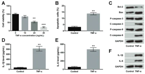
TNF-ainduced inflammatory injury in ICC
ICC were treated with TNF-a at 10, 20, or 40 ng/mL concentrations, and then cell viability was measured using CCK-8 assay and apoptosis was determined using flow cytometry. As shown in Figure 1A, TNF-a significantly decreased cell viability at 20 ng/mL (F(3,8)=38.82, Po0.01) and 40 ng/mL concentrations (F(3,8)=38.82, Po0.001). The concentration of TNF-afor subsequent experiments was 20 ng/mL. TNF-a significantly increased apoptosis compared to the control (Po0.01; Figure 1B). Western blotting analysis also confirmed thisfinding, where TNF-a decreased the expression of anti-apoptotic protein (Bcl-2) and increased the expressions of pro-apoptotic proteins (Bax and cleaved caspases 3 and 9) compared to the control (Figure 1C). Then, we measured the concentra-tions of pro-inflammatory cytokines, IL-1band IL-6, using ELISA. As shown in Figure 1D and E, TNF-a increased the levels of IL-1band IL-6 compared to the control (both Po0.01). Western blotting confirmed these findings as TNF-aincreased the expressions of IL-1band IL-6 (Figure 1F). Thesefindings indicated that TNF-a induced inflam-matory injury in ICC by decreasing cell viability and increas-ing apoptosis and secretion of pro-inflammatory cytokines.
Figure 1.TNF-ainduced inflammatory injury in interstitial cells of Cajal (ICC). ICC were treated with TNF-aat 10, 20, or 40 ng/mL con-centrations.A, Cell viability was measured using CCK-8 assay.B, Apoptosis was measured usingflow cytometry assay.C, Expression of apoptosis-associated proteins was determined by western blotting.D–F, Concentrations of IL-1band IL-6 were measured using
TNF-adecreased the levels of SCF, ghrelin, and
substance P
Next, we assessed the effect of TNF-aon SCF using qRT-PCR and western blotting analyses. Results showed that TNF-asignificantly decreased the expression of SCF compared to the control (Po0.05; Figure 2A). Then, we assessed the effect of TNF-a on the levels of ghrelin, substance P, and endothelin-1 using ELISA, and found that TNF-a significantly deceased the levels of ghrelin (Po0.01; Figure 2B) and substance P (Po0.05; Figure 2C), but had no effect on endothelin-1 level (Figure 2D). These findings indicated that TNF-adown-regulated expressions of SCF, ghrelin, and substance P in ICC.
TNF-ainhibited the expression of SCF, ghrelin, and
substance P by activating the NF-jB pathway Lastly, the effect of TNF-a on the NF-kB signaling pathway proteins (p65 and IkBa) was examined using western blotting. As shown in Figure 3A, TNF-aincreased the phosphorylation of p65 and IkBa compared to the control. However, addition of NF-kB inhibitor (SN50, 20 mM) reversed these effects by decreasing the phos-phorylation of the NF-kB pathway proteins and increasing the expression of SCF (Figure 3B). Then, ELISA was
done to assess the effects of TNF-a and TNF-a+SN50 on the levels of ghrelin and substance P. Results showed that addition of SN50 reversed the effects of TNF-a by increasing the levels of ghrelin (F(2,6)=14.72, Po0.01, Figure 3C) and substance P (F (2,6)=18.26, Po0.05, Figure 3D), compared to the TNF-agroup. Thesefindings indicated that TNF-a inhibits the expression of SCF, ghrelin, and substance P by activating the NF-kB signaling pathway.
Discussion
In the present study, we assessed the effects of TNF-a on viability and apoptosis of ICC, and on the levels of IL-1b, IL-6, SCF, ghrelin, endothelin-1, and substance P in ICC. We also examined the involvement of the NF-kB signaling pathway in the effects of TNF-aon expression of SCF, ghrelin, and substance P. Results revealed that TNF-a decreased ICC viability, increased apoptosis, increased IL-1b and IL-6 levels, and decreased SCF, ghrelin, and substance P levels by activating the NF-kB signaling pathway.
TNF-a, lipopolysaccharide, and toll-like receptor 4 are crucial in inducing phenotypic changes in ICC under an
Figure 2.TNF-adecreased the levels of stem cell factor (SCF), ghrelin, and substance P. Interstitial cells of Cajal (ICC) were treated with 20 ng/mL TNF-aand non-treated cells were used as control.A, qRT-PCR and western blotting were used to measure the expression of SCF. ELISA was used to measure the concentrations of (B) ghrelin, (C) substance P, and (D) endothelin-1. Data are reported as means±
inflammatory microenvironment in the gut (25). Eisenman et al. (26) suggested that TNF-a, which was secreted from M1 macrophages, could induce c-Kit loss and ICC injury through caspase-dependent apoptosisin vitro. In a mouse model of ulcerative colitis, which was induced by dextran sulfate sodium, expressions of TNF-a, IL-1b, and IL-6 were markedly up-regulated in the colon, resulting in intestinal mucosal inflammation (27). A previous study also reported that IL-6 release in inflammatory microenvironment could down-regulate c-Kit expression and decrease ICC activ-ities (28). In our study, TNF-adecreased ICC viability and increased apoptosis partially through caspase-dependent pathway, along with increases of IL-1b and IL-6 levels. Taken together, thesefindings indicate that TNF-aadversely affects ICC in ulcerative colitis.
In a rat model of inflammatory bowel diseases, bone marrow mesenchymal stromal cells and soluble SCF played a synergistic role in mucosal cell regeneration following experimentally induced intestinal injury (29). Thus, admin-istration of SCF may be of therapeutic value in inflamma-tory bowel diseases, including ulcerative colitis. SCF is also considered a ligand of c-Kit, and the activation of SCF/Kit pathway is essential for development and maintenance of
ICC networks (30). In our study, TNF-asignificantly down-regulated the expression of SCF at mRNA and protein levels in ICC compared to the control, which is in agree-ment with a study by Rusten et al. (31), showing that TNF-a inhibits SCF-induced proliferation of human bone marrow progenitor cellsin vitro. Our study also proved that TNF-a -induced down-regulation of SCF could be reversed by inhibition of the NF-kB pathway in ICC. Similarly, a study by Jin et al. (32) illustrated that curcumin up-regulated SCF expression through inactivating the NF-kB pathway. The majority of circulating levels of ghrelin is produced in the stomach. Ghrelin exerts a range of immunological effects. For example, it decreases leptin-induced pro-inflammatory responses and inhibits secretion of TNF-a, IL-1b, IL-6, and IL-8. Due to the functional and anatomical link of ghrelin with inflammation and the GI tract, ghrelin has been studied in a variety of GI disorders, including colitis (33). Maduzia et al. (34) found that acetic acid-induced colitis was effectively ameliorated by administration of ghrelin, which was related to the anti-inflammatory effects of ghrelin. Another study also reported that experimental colitis could be attenuated by ghrelin via repressing the NF-kB pathway in mice (35). In agreement with those
findings, we found that TNF-ainhibited the expression of ghrelin by activating the NF-kB pathway.
Substance P is a neurotransmitter found in colonic mucosa, which can alter gut vascular, immunologic, and motor phenomena. Thus, it may have a vital role in the pathogenesis of ulcerative colitis. Szitter et al. (36) sum-marized that, in inflamed human gut, the alterations of substance P expression are contradictory. Other studies also reported the dual role of substance P in colitis develop-ment. On the one hand, substance P could induce inflam-matory response via evoking activation of the NF-kB signaling pathway (37). On the other hand, substance P promotes proliferation and mobilizes stem cells to the site of injury, leading to facilitation of mucosal healing in colitis (38). Hong et al. (39) also reported that dextran sulfate sodium-induced intestinal damage was effectively ameliorated by
substance P via enrichment of M2 macrophages and regu-latory T cells. In our study, we found that the expression of substance P was down-regulated by TNF-aby activating the NF-kB pathway in ICC.
In conclusion, TNF-a induced inflammatory injury in ICC by decreasing cell viability and increasing apoptosis and levels of IL-1b and IL-6. We also found that TNF-a inhibited the expression of SCF, ghrelin, and substance P by activating the NF-kB signaling pathway. Our study provides a regulatory mechanism of TNF-a in ICC and emphasizes the importance of the NF-kB signaling path-way in development of ulcerative colitis. The inhibition of the NF-kB signaling pathway may be a possible ther-apeutic strategy for treatment of ulcerative colitis; how-ever, additional clinical experiments should be done in the future.
References
1. Hoivik ML, Moum B, Solberg IC, Henriksen M, Cvancarova M, Bernklev T. Work disability in inflammatory bowel disease patients 10 years after disease onset: results from the IBSEN Study.Gut2013; 62: 368–375, doi: 10.1136/gutjnl-2012-302311.
2. Torres J, Billioud V, Sachar DB, Peyrin-Biroulet L, Colombel JF. Ulcerative colitis as a progressive disease: the forgot-ten evidence. Inflamm Bowel Dis 2012; 18: 1356–1363, doi: 10.1002/ibd.22839.
3. Ungaro R, Mehandru S, Allen PB, Peyrin-Biroulet L, Colombel JF. Ulcerative colitis.Lancet2017; 389: 1756–1770, doi: 10.1016/S0140-6736(16)32126-2.
4. Baumgart DC, Sandborn WJ. Inflammatory bowel disease: clinical aspects and established and evolving therapies.Lancet 2007; 369: 1641–1657, doi: 10.1016/S0140-6736(07)60751-X. 5. Loftus CG, Loftus EV Jr, Harmsen WS, Zinsmeister AR, Tremaine WJ, Melton LJ 3rd, et al. Update on the incidence and prevalence of Crohn’s disease and ulcerative colitis in Olmsted County, Minnesota, 1940–2000.Inflamm Bowel Dis 2007; 13: 254–261, doi: 10.1002/ibd.20029.
6. Cosnes J, Gower-Rousseau C, Seksik P, Cortot A. Epidemio-logy and natural history of inflammatory bowel diseases. Gastro-enterology2011; 140: 1785–1794, doi: 10.1053/j.gastro.2011. 01.055.
7. Peyrin-Biroulet L, Sandborn W, Sands BE, Reinisch W, Bemelman W, Bryant RV, et al. Selecting therapeutic targets in inflammatory bowel disease (STRIDE): Determining ther-apeutic goals for treat-to-target.Am J Gastroenterol2015; 110: 1324–1338, doi: 10.1038/ajg.2015.233.
8. Sasaki M, Klapproth JM. The role of bacteria in the patho-genesis of ulcerative colitis.J Signal Transduc2012; 2012: 704953, doi: 10.1155/2012/704953.
9. Khor B, Gardet A, Xavier RJ. Genetics and pathogenesis of inflammatory bowel disease.Nature 2011; 474: 307–317, doi: 10.1038/nature10209.
10. Huang Y, Chen Z. Inflammatory bowel disease related innate immunity and adaptive immunity.Am J Transl Res 2016; 8: 2490–2497.
11. Bassotti G, Antonelli E, Villanacci V, Baldoni M, Dore MP. Colonic motility in ulcerative colitis. United European
Gastroenterol J2014; 2: 457–462, doi: 10.1177/205064061 4548096.
12. Murch SH, Braegger CP, Sessa WC, MacDonald TT. High endothelin-1 immunoreactivity in Crohn’s disease and ulcerative colitis.Lancet1992; 339: 381–385, doi: 10.1016/ 0140-6736(92)90077-G.
13. Al-Shboul OA. The importance of interstitial cells of cajal in the gastrointestinal tract.Saudi J Gastroenterol2013; 19: 3–15, doi: 10.4103/1319-3767.105909.
14. Peracchi M, Bardella MT, Caprioli F, Massironi S, Conte D, Valenti L, et al. Circulating ghrelin levels in patients with inflammatory bowel disease. Gut 2006; 55: 432–433, doi: 10.1136/gut.2005.079483.
15. Ward SM, Burns AJ, Torihashi S, Sanders KM. Mutation of the proto-oncogene c-kit blocks development of interstitial cells and electrical rhythmicity in murine intestine.J Physiol 1994; 480 (Part 1): 91–97, doi: 10.1113/jphysiol.1994. sp020343.
16. Tamada H, Kiyama H. Existence of c-Kit negative cells with ultrastructural features of interstitial cells of Cajal in the subserosal layer of the W/Wv mutant mouse colon.Nihon Heikatsukin Gakkai kikanshi2015; 51: 1–9, doi: 10.1540/ jsmr.51.1.
17. Rumessen JJ. Ultrastructure of interstitial cells of Cajal at the colonic submuscular border in patients with ulcerative colitis.Gastroenterology1996; 111: 1447–1455, doi: 10.1016/ S0016-5085(96)70005-7.
18. Seita J, Weissman IL. Hematopoietic stem cell: self-renewal versus differentiation.Wiley Interdiscip Rev Syst Biol Med 2010; 2: 640–653, doi: 10.1002/wsbm.86.
19. Guo X, Huang X, Wu YS, Liu DH, Lu HL, Kim YC, et al. Down-regulation of hydrogen sulfide biosynthesis accom-panies murine interstitial cells of Cajal dysfunction in partial ileal obstruction.PLoS One2012; 7: e48249, doi: 10.1371/ journal.pone.0048249.
20. Korbonits M, Goldstone AP, Gueorguiev MGrossman AB. Ghrelin - a hormone with multiple functions.Front Neuro-endocrinol2004; 25: 27–68, doi: 10.1016/j.yfrne.2004.03.002. 21. Jung JY, Jeong JB, Kim JW, Kim SH, Koh SJ, Kim BG, et al.
marker of activity in ulcerative colitis.Intest Res2015; 13: 68–73, doi: 10.5217/ir.2015.13.1.68.
22. Luscher TF, Barton M. Endothelins and endothelin receptor antagonists: therapeutic considerations for a novel class of cardiovascular drugs. Circulation 2000; 102: 2434–2440, doi: 10.1161/01.CIR.102.19.2434.
23. Sands BE, Kaplan GG. The role of TNFalpha in ulcerative colitis.J Clin Pharmacol 2007; 47: 930–941, doi: 10.1177/ 0091270007301623.
24. Livak KJ, Schmittgen TD. Analysis of relative gene expres-sion data using real-time quantitative PCR and the 2(-Delta Delta C(T)) Method.Methods2001; 25: 402–408, doi: 10.1006/ meth.2001.1262.
25. Wei J, Li N, Xia X, Chen X, Peng F, Besner GE, et al. Effects of lipopolysaccharide-induced inflammation on the inter-stitial cells of Cajal. Cell Tissue Res 2014; 356: 29–37, doi: 10.1007/s00441-013-1775-7.
26. Eisenman ST, Gibbons SJ, Verhulst PJ, Cipriani G, Saur D, Farrugia G. Tumor necrosis factor alpha derived from classi-cally activated‘‘M1’’ macrophages reduces interstitial cell of Cajal numbers. Neurogastroenterol Motil 2017; 29, doi: 10.1111/nmo.12984.
27. Dai Y-C, Zheng L, Zhang Y-L, Chen X, Chen D-L, Wang L-J, et al. Jianpi Qingchang decoction regulates intestinal motility of dextran sulfate sodium-induced colitis through reducing autophagy of interstitial cells of Cajal.World J Gastroenterol 2017; 23: 4724–4734, doi: 10.3748/wjg.v23.i26.4724. 28. Deng J, Yang S, Yuan Q, Chen Y, Li D, Sun H, et al.
Acupuncture ameliorates postoperative ileus via IL-6– miR-19a–KIT axis to protect interstitial cells of Cajal.Am J Chin Med2017; 45: 737–755, doi: 10.1142/S0192415X17500392. 29. Qu B, Xin GR, Zhao LX, Xing H, Lian LY, Jiang HY, et al.
Testing stem cell therapy in a rat model of inflammatory bowel disease: role of bone marrow stem cells and stem cell factor in mucosal regeneration.PLoS One2014; 9: e107891, doi: 10.1371/journal.pone.0107891.
30. Peng MF, Li K, Wang C, Zhu XY, Yang Z, Zhang GH, et al. Therapeutic effect and mechanism of electroacupuncture at Zusanli on plasticity of interstitial cells of Cajal: a study of rat ileum.BMC Complement Altern Med2014; 14: 186, doi: 10.1186/1472-6882-14-186.
31. Rusten LS, Smeland EB, Jacobsen FW, Lien E, Lesslauer W, Loetscher H, et al. Tumor necrosis factor-alpha inhibits stem cell factor-induced proliferation of human bone marrow progenitor cellsin vitro. Role of p55 and p75 tumor necrosis factor receptors. J Clin Invest 1994; 94: 165–172, doi: 10.1172/JCI117303.
32. Jin Q-H, Shen H-X, Wang H, Shou Q-Y, Liu Q. Curcumin improves expression of SCF/c-kit through attenuating oxida-tive stress and NF-kB activation in gastric tissues of diabetic gastroparesis rats. Diabetol Metab Syndr 2013; 5: 12, doi: 10.1186/1758-5996-5-12.
33. Tiaka EK, Manolakis AC, Kapsoritakis NA, Potamianos SP. Unraveling the link between leptin, ghrelin and different types of colitis.Ann Gastroenterol2011; 24: 20–28. 34. Maduzia D, Matuszyk A, Ceranowicz D, Warzecha Z,
Ceranowicz P, Fyderek K, et al. The influence of pretreat-ment with ghrelin on the developpretreat-ment of acetic-acid-induced colitis in rats.J Physiol Pharmacol2015; 66: 875–885. 35. Cheng J, Zhang L, Dai W, Mao Y, Li S, Wang J, et al. Ghrelin
ameliorates intestinal barrier dysfunction in experimental colitis by inhibiting the activation of nuclear factor-kappa B. Biochem Biophys Res Commun 2015; 458: 140–147, doi: 10.1016/j.bbrc.2015.01.083.
36. Szitter I, Pinter E, Perkecz A, Kemeny A, Kun J, Kereskai L, et al. Role of neurokinin 1 receptors in dextran sulfate-induced colitis: studies with gene-deleted mice and the selective receptor antagonist netupitant.Inflamm Res2014; 63: 399–409, doi: 10.1007/s00011-014-0712-x.
37. Ni T, Liu Y, Peng Y, Li M, Fang Y, Yao M. Substance P induces inflammatory responses involving NF-kB in geneti-cally diabetic mice skinfibroblasts co-cultured with macro-phages.Am J Translat Res2016; 8: 2179–2188.
38. Hong HS, Kim S, Kim YH, Park JH, Jin YSon Y. Substance-P blocks degeneration of retina by stimulating migration and prolife-ration of retinal pigmented epithelial cells.Tissue Eng Regener Med2015; 12: 121–127, doi: 10.1007/s13770-014-0088-6. 39. Hong HS, Hwang DY, Park JH, Kim S, Seo EJ, Son Y.