• Nenhum resultado encontrado

ANTIFUNGAL ACTIVITY AND ULTRASTRUCTURAL ALTERATIONS IN Pseudocercospora griseola TREATED WITH ESSENTIAL OILS

N/A
N/A
Protected

Academic year: 2019

Share "ANTIFUNGAL ACTIVITY AND ULTRASTRUCTURAL ALTERATIONS IN Pseudocercospora griseola TREATED WITH ESSENTIAL OILS"

Copied!
15
0
0

Texto

(1)

Pseudocercospora griseola

TREATED WITH ESSENTIAL OILS

Atividade antifúngica e alterações ultraestruturais em Pseudocercospora griseola tratado com óleos essenciais

Julián Mauricio Ágredo Hoyos1, Eduardo Alves2, Luciane Cristina Rozwalka3, Elaine Aparecida de Souza4, Walmes Marques Zeviani5

ABSTRACT

Pseudocercospora griseola, the etiologic agent of angular leaf spot of common bean (Phaseolus vulgaris), is an important disease

in all bean-producing regions worldwide and may cause extremely high yield losses. The control of this disease is made more difficult by the pathogen’s genetic variability and the inefficiency of fungicides. In this study, of 26 essential oils tested at different concentrations, 25 demonstrated efficiency in affecting the germination of strains 63-31 and 63-63 of the pathogen, reaching inhibition levels of between 80% and 100%. Cymbopogon citratus and Cymbopogon martinii inhibited conidia germination at all concentrations; Eugenia caryophyllata, Cinnamomum sp., Thymus vulgaris, Matricaria recutita, Cordia verbenacea, Origanum vulgare, Cymbopogon nardus, at 0.1 and 0.5%;

and Zingiber officinale, Mentha arvensis, Chamaecyparis pisifera, Lavandula officinalis, Ocimum basilicum, Pimpinella anisum, Ocimum selloi, Baccharis dracunculifolia, Laurus nobilis, Citrus sinensis, Melaleuca alternifolia and Eucalyptus globulus, at 0.5%. The main

constituents identified were cinnamaldehyde in Cinnamomum sp.; eugenol in E. caryophyllata; trans--farnesene in M. recutita; pulegone

in C. verbenacea; thymol in T. vulgaris; geranial and neral in C. citratus,and geraniol in C. martini. Through transmission electron

microscopy (TEM), it was verified that C. citratus, C. martini and E. caryophyllata presented direct fungitoxic action on P. griseola,causing

severe damage to the cellular ultrastructure of the conidia, invalidating germination. These results indicated that essential oils are a promising alternative strategy for the control of angular leaf spot in bean, representing less risk to human health and the environment.

Index terms: Angular leaf spot, transmission electron microscopy,alternative control of plant disease.

RESUMO

Pseudocercospora griseola, agente etiológico da mancha angular do feijoeiro comum (Phaseolus vulgaris), é uma doença

importante nas regiões produtoras de feijão em todo o mundo e pode causar perdas de produtividade extremamente elevados. O controle dessa doença é dificultado pela variabilidade genética do patógeno e da ineficiência de fungicidas. Neste estudo, de 26 óleos essenciais testados em concentrações diferentes, 25 demonstraram eficiência em inibir a germinação das linhagens 63-31 e 63-63 do agente patogênico, atingindo níveis de inibição entre 80% e 100%. Cymbopogon citratus e Cymbopogon martini inibiram a germinação de

conídios em todas as concentrações; Eugenia caryophyllata, Cinnamomum sp., Thymus vulgaris, Matricaria recutita, Cordia verbenacea, Origanum vulgare, Cymbopogon nardus, em 0,1 e 0,5%, e Zingiber officinale, Mentha arvensis, Chamaecyparis pisifera, Lavandula officinalis, Ocimum basilicum, Pimpinella anisum, Ocimum selloi, Baccharis dracunculifolia, Laurus nobilis, Citrus sinensis, Melaleuca alternifolia e Eucalyptus globulus, em 0,5%. Os principais constituintes identificados foram cinamaldeído em Cinnamomum sp.; Eugenol

em E. caryophyllata; trans--farneseno em M. recutita; pulegona em C. verbenacea; timol em T. vulgaris; geranial e neral em C. citratus

e geraniol em C. martini. Através de microscopia eletrônica de transmissão (TEM), verificou-se que C. citratus, C. martini e E. caryophyllata apresentaram ação antifúngica direta sobre P. griseola, causando graves danos na ultraestrutura celular dos conídios,

invalidando a germinação. Esses resultados indicaram que os óleos essenciais são uma estratégia alternativa promissora para o controle da mancha angular do feijoeiro, o que representa menos risco para a saúde humana e ao ambiente.

Termos para indexação: Mancha angular, microscopia eletrônica de transmissão, controle alternativo de doenças de plantas.

(Received in april 13, 2012 and approved in may 21, 2012)

1Universidade Federal de Lavras/UFLA – Departamento de Fitopatologia/DFP – Lavras – MG – Brasil

2Universidade Federal de Lavras/UFLA – Departamento de Fitopatologia/DFP – Cx. P. 3037 – 37200-000 – Lavras – MG – Brasil – ealves@dfp.ufla.br 3Universidade Federal do Paraná/UFPR – Curitiba – PR – Brasil

4Universidade Federal de Lavras/UFLA – Departamento de Biologia/DBI – Lavras – MG – Brasil

5Universidade Federal do Paraná/UFPR – Laboratório de Estatística e Geoinformação – Curitiba – PR – Brasil INTRODUCTION

Angular leaf spot (ALS), caused by Pseudocercospora griseola (Sacc.) Crous & U. Braun, is the most widespread disease in all bean-producing regions worldwide. According to the Commonwealth Mycological

(2)

control plant diseases, their continuous and indiscriminate use has caused contamination by waste, environmental pollution and the selection of resistant populations of pathogens. Chemical control carries considerable environmental and financial costs (MIKLAS et al., 2006). Due to the high genetic variability of P. griseola (BROCK, 1951; PYNDJI, 1993; PASTOR-CORRALES et al., 1998; OROZCO; ARAYA, 2005; STENGLEIN; BALATTI, 2006; SILVA, et al., 2008), genetic control through the use of resistant cultivars is very difficult.

The inefficiency of pesticides in the control of ALS and the lack of resistant cultivars, as well as market demands in food production and the need to achieve environmental sustainability, food security and economic viability, have all made the use of technology that is non-aggressive to the environment and human health into a major challenge for bean producers. Considering these problems, it has become vital to develop strategies based on the rational use of fungicides or to replace them with alternative products (CHRISTIAN; GOGGI, 2008; GHINI; KIMATI, 2000). Essential oils have shown biological activity that may offer an alternative strategy in the control of plant pathogens (MIHALIAK et al., 1991; STANGARLIN et al., 1999; ISMAN, 2000; KOTZEKIDOU et al., 2008) due to fungitoxic action (FUORI et al., 2000; BONALDO et al., 2004; BANG, 2007) or by inducing resistance in plants (SCHWAN-ESTRADA, 2003; QUINTANILLA et al., 2002; PEREIRA et al., 2011).

The essential oils extracted from medicinal, aromatic and ornamental plants have shown efficiency in controlling plant pathogens (SHAHI et al., 2003; SOYLU et al., 2006; LEE et al., 2007; BARRERA-NECHA et al., 2008). The effects of essential oils have been studied at the ultrastructural level for some pathogens (ZAMBONELLI et al., 1996; RASOOLI et al., 2006; ROZWALKA et al., 2010). However, there are no reports about the antifungal activity and the mode of action of essential oils on P. griseola. This study aimed to evaluate (i) the antifungal activity of 26 essential oils on germination of P. griseola conidia, strains 63-31 and 63-63; (ii) the effect of the essential oils of Cymbopogon martini, Eugenia caryophyllata and Cymbopogon citratus on conidia at ultrastructural level by transmission electron microscopy (TEM), and (iii) to identify the chemical constituents of essential oils with fungitoxic activity against this plant pathogen.

MATERIALS AND METHODS Fungal isolates

Two monosporic isolates of P. griseola were employed, classified as pathotypes 63-31 and 63-63, from

the mycology collection of the Institute of Biotechnology applied to Agriculture (BIOAGRO) at the Federal University of Viçosa (Viçosa, Minas Gerais State, Brazil). Conidia were obtained by culturing the fungus on bean leaf-dextrose-agar medium in a growth chamber (Fanem, São Paulo, Brazil) at 24 ± 2º C. After approximately 15 days, spore suspension was prepared by adding 5–10 mL of sterile distilled water to each Petri dish and scraping the surface of the culture with a spatula.

Essential oils

The essential oils extracted from 26 medicinal, ornamental, aromatic, or forest species representative of the families Apiaceae (Pimpinella anisum L.), Asteraceae (Baccharis dracunculifolia DC., Matricaria recutita L.), Boraginaceae (Cordia verbenacea DC.), Cupressaceae (Chamaecyparis pisifera (Siebold & Zucc.) Endl., Chamaecyparis plumosa Hort. ex Beissn.), Lamiaceae (Lavandula officinalis Chaix ex Vill., Mentha arvensis L., Ocimum basilicum L., Ocimum selloi Benth., Origanum vulgare L., Rosmarinus officinalis L., Thymus vulgaris L.), Lauraceae (Cinnamomum sp., Laurus nobilis L.), Myrtaceae (Corymbia citriodora (Hook.) K.D. Hill & L.A.S. Johnson, Eucalyptus globulus Labill., Eugenia caryophyllata Thunb., Melaleuca alternifólia Cheel), Poaceae (Cymbopogon citratus (DC) Stapf., Cymbopogon martini (Roxb.) Wats., Cymbopogon nardus (L.) Rendle), Rutaceae (Citrus limon (L.) Burm.f., Citrus sinensis (L.) Osbeck.), Verbenaceae (Lippia citriodora Kunth) e Zingiberaceae (Zingiber officinale Roscoe).

Essential oils from L. officinalis, O. selloi, O. vulgare and Cinnamomum sp. were extracted by hydrodistillation for 3 h in a Clevenger-type apparatus. Flowers and leaves from L. officinalis, and leaves from O. selloi and O. vulgare were collected at the Medicinal Plants Nursery of the Agricultural Department, Federal University of Lavras, Minas Gerais State, Brazil, at vegetative stage, and cinnamon bark was purchased in a natural store in Lavras city. The remaining essential oils were supplied by Chamel Industry and Natural Products Commerce, Paraná State, Brazil (2010). All essential oils used in this work were kept in amber flasks and stored under refrigeration at 0º C.

Screening the essential oils with in vitro antifungal

activity

(3)

96-well plate. This mixture resulted in the final concentrations at 0.02% (200 μL.L-1), 0.1% (1000 μL.L-1) and 0.5% (5000 μL.L-1). These treatments were prepared using sterile distilled water with Tween 20 at 1.0%. Sterile distilled water (40 μL) mixed with conidial suspension was used as control. In previous germinations tests comparing germination in water and water plus Tween-20 a 1.0%, it was observed that Tween showed non-toxic effects in both strains of pathogen. After 20 hours of incubation at 24 ± 2º C and 12 h photoperiod, germination was interrupted by addition of 20 μL of lactoglycerol cotton blue. The percentage of germination was estimated by counting 50 randomly chosen conidia in 6 wells, totaling 300 conidia per treatment, under a light microscope. Conidia presenting differentiated germ tubes, independently of their size, were considered germinated. The experiment were carried out in a completely randomized design, as a factorial 2 x 3 x 3 (strains x oil x concentrations) x 6 repetitions. Each well of the plate was an experimental unit.

Chemical composition of essential oils

C. martini (0.144 mg), M. chamomilla (0.127 mg), E. caryophyllata (0.200 mg), T. vulgaris (0.147 mg), C. verbenacea (0.179 mg), C. citratus (0.160 mg) and Cinnamomum sp. (0.118 mg)were dissolved in 0.5 mL of dichloromethane (CH2Cl2). These oils showed antifungal activity of more than 90%, when tested at 0.1%. One microliter of diluted essential oils was injected in gas chromatography coupled with mass spectrometry (GCMS-QP2010 Plus, Shimadzu). The gas chromatography operated with ultra pure helium as the carrier gas at a flow rate of 1.8 mL.min-1 and the separation was done on an Equity-5 capillary column (30 m x 0.22 mm I.D with 0.25 μm film thickness), with column oven at 60º C for 2 min, heating at 3º C.min-1 up to 240º C, remaining at 240º C for 15 min. The temperature of the injector was 220º C, split injection 1:15, manual injection, time run of 77 min. The mass spectrometry used a GC-MS interface, at 250º C, with ion source at 200º C and electron impact ionization at 70 eV. The compounds in essential oils were identified by comparing the similarity index of the mass spectra of relative area (%) with the data available in the library (Wiley 8) of GCMS.

Transmission electron microscopy (TEM) preparation A conidial suspension (2 x 10⁴ conidia.mL-1) of strains 63-31 and 63-63, prepared in sterile distilled water with Tween 20 at 1.0%, was mixed with 0.5 mL of the solutions of 0.2% essential oils of C. martini, E.

caryophyllata and C. citratus to obtain a final concentration of 0.1%. For the control, 0.5 mL conidial suspensions were mixed with 0.5 mL of sterile distilled water. The treatments remained under agitation in an Orbital Shaker at 100 rpm and an average temperature of 25 ± 2º C and, after 24 h, were centrifuged for 3 min at 6000 rpm, forming conidial masses. The masses were fixed in modified Karnovsky solution (glutaraldehyde at 2.5%, formaldehyde at 2.5% in sodium cacodylate buffer at 0.05 M, pH 7.2, and calcium chloride at 0.1 M) and kept in the refrigerator for 24 h (primary fixation). To form pellets, the conidial masses fixed in Karnovsky were centrifuged for 3 min at 6000 rpm. The supernatant was discarded, and the agarose gel at 1.0% was heated to about 45º C and mixed with the pellets. The blocks of agarose formed were washed three times for 10 min in sodium cacodylate buffer at 0.05 M, for post-fixation in osmium tetroxide at 2% in the hood. After a 4 h period, the blocks were washed three times in distilled water and submitted to contrast en bloc in an aqueous solution of uranyl acetate at 0.5% for one night in the refrigerator. The dehydration was done in a graded acetone series at concentrations of 25, 50, 75, 90 and 100% for 10 min each, except at 100%, which was dehydrated 3 times for 10 min each time.

(4)

distilled water. For each treatment, the test was repeated twice and at least two blocks per replicate were examined. The observation of the samples and the recording of the images were performed in a Zeiss EM 109 transmission electron microscope operating at 80 kv.

Statistical analysis

Data analysis was done using generalized linear models (GLMs), assuming a binomial distribution logit link function. The linear predictor was represented by log [p

i / (1- pi)] = 0i + 1i.c, where pi = probability of germination inhibition of conidia treated with essential oil i (i = 1, 2, …,26) at concentration c (c = 0.02, 0.1 and 0.5%) and 0i and 1i are parameters that bring form to a relationship. The inferences were conducted considering the parameter of overdispersion () estimated by Pearson residues. All analyzes were performed in R environment (2.15.0) for statistical computing R Development Core Team, 2012.

RESULTS AND DISCUSSION

Antifungal activity of essential oils on germination of

P. griseola strains, in vitro

The results of analysis of deviance showed significant interaction between treatments and concentrations of the essential oils, indicating that differences in the percentage of germination occurred in function of the essential oil and concentrations used. So studies were done separately for each pathogen strain. The antifungal activity of essential oils in the germination of P. griseola strains, expressed by the percentage of conidial germination inhibition, was calculated based on average number of conidia that germinated from each strain, 63-31 (46.67) and 63-63 (31.75) and the untreated control (Table 1).

Essential oils of C. citratus, C. martini and E. caryophyllata were more effective than the other treatments, showing at least 98% inhibition of conidial germination of strains 63-31 and 63-63 of P. griseola at all tested concentrations. At 0.1%, C. citratus, C. martini, E. caryophyllata, Cinnamomum sp., T. vulgaris, M. recutita and C. verbenacea also showed antifungal activity against both strains. The average germination inhibition values were higher than 93.57%. At 0.02%, conidia germination of strain 63-63 was almost totally inhibited by C. citratus, C. martini, E. caryophyllata and Cinnamomum sp. However, these two last essential oils did not show the same efficiency against strain 63-31, for which they only reduced inhibition to approximately 50%. A difference in inhibition

response of the two pathogen strains was also observed more effectively on conidia treated with M. arvensis, Z. officinale, C. pisifera, L. officinalis and B. dracunculifolia at 0.1% and C. plumosa, R. officinalis and L. citriodora at 0.5%. The regression model allows us to observe these differences (Figure 1).

It is also possible to observe that increasing the concentration of essential oil of C. citriodora and C. limon did not result in greater inhibition of the pathogen strains. At 0.5%, of the 26 essential oils screened, 21 promoted inhibition of conidial germination between 86 to 100% for both strains of the pathogen (Table 2).

At all the tested concentrations, some essential oils stimulated conidial germination of both strains, expressed by the negative sign. C. nardus (0.02%), Z. officinale (0.02%), R. officinalis (0.1%) and C. limon (0.1%, 0.02% and 0.5%) were the essential oils that most stimulated conidial germination of strain 63-63 (Table 1). Based on the estimated coefficients of the logistic regression model, by increasing the concentration of E. caryophyllata and Cinnamomum sp. from 0.02 to 0.03 and 0.06%, M. arvensis, Z. officinale, O. vulgare and C. nardus from 0.1 to 0.41, 0.27, 0.13 and 0.14%, respectively, the conidial germination inhibition of strain 63-31 could be 95%. In this study, it was observed that increasing the concentration of most essential oils promoted their enhanced antifungal activity on P. griseola strains. However, the inhibitory effect promoted by essential oils was different for the two strains of the pathogen. Greater conidial germination inhibition was observed for strain 63-63, which is reported to be more aggressive than 63-31 in bean production in Brazil (NIETSCHE et al., 2001; DAMASCENO-SILVA et al., 2008).

Chemical composition of essential oils

Identification and quantification of the main chemical constituents of essential oils can be seen in table 3.

The main constituents identified were cinnamaldehyde (92.36%) in Cinnamomum sp.; eugenol (91.94%) in E. caryophyllata; geraniol (75.92%) in C. martini; pulegone (68.96%) in C. verbenacea; geranial (58.89%) followed by neral (38.50%) in C. citratus; thymol (33.72%) in T. vulgaris and trans-β-farnesene (31.17%) in M. recutita.

(5)

presented most inhibition of germination. These were geranial and neral (aldehyde) in C. citratus; eugenol (phenol) in E. caryophyllata and geraniol (alcohol) in C. martini. In the literature, only the main compounds of some other essential oils, including eugenol, thymol, carvacrol, cinnamaldehyde, inter alia, have been studied. The minor constituents of the essential oils studied here

could also have a relatively strong antimicrobial effect (BURT, 2004; BAKKALI et al., 2008), which can indicate that these molecules have synergism in antimicrobial activity. The synergistic effects of the diversity of major and minor compounds of the essential oils should be taken into consideration to account for their antimicrobial activity.

Table 1 – Inhibition of conidial germination of Pseudocercospora griseola strains 63-31 and 63-63 by essential oils tested at 0.5%, 0.1% and 0.02%, in vitro.

Essential oils

Concentrations (%)

0.5 0.1 0.02

Strains Strains Strains

63-31 63-63 63-31 63-63 63-31 63-63

Cymbopogon citratus 100.00 100.00 99.64 100.00 100.00 100.00

Cymbopogon martini 99.29 100.00 98.93 100.00 99.64 99.48

Eugenia caryophyllata 100.00 100.00 100.00 100.00 58.21 99.48

Cinnamomum sp. 100.00 100.00 98.93 99.48 51.79 98.43

Thymus vulgaris 98.21 100.00 98.93 100.00 17.50 2.36

Matricaria recutita 100.00 100.00 93.57 100.00 3.57 13.91

Cordia verbenacea 98.93 99.48 94.64 96.33 4.29 10.24

Origanum vulgare 100.00 100.00 86.79 87.40 0.36 11.81

Cymbopogon nardus 100.00 100.00 83.21 89.50 6.79 -36.48

Zingiber officinale 99.29 100.00 50.71 95.28 1.79 -35.43

Mentha arvensis 99.64 100.00 35.71 99.48 14.29 59.58

Chamaecyparis pisifera 100.00 100.00 21.79 78.48 -0.71 25.98

Lavandula officinalis 100.00 100.00 16.07 80.05 1.43 61.15

Ocimum basilicum 97.86 100.00 26.79 57.48 26.79 36.48

Pimpinella anisum 98.57 99.48 38.21 67.45 12.14 25.98

Ocimum selloi 100.00 100.00 25.36 20.21 6.79 4.99

Baccharis dracunculifolia 92.14 98.43 75.00 18.11 2.86 1.84

Laurus nobilis 98.21 99.48 24.29 13.91 3.21 -16.01

Citrus sinensis 98.21 100.00 29.64 -2.36 0.00 -0.26

Melaleuca alternifolia 95.36 96.85 -3.21 7.61 1.07 47.51

Eucalyptus globulus 86.43 94.23 2.14 -7.61 -1.43 6.56

Chamaecyparis plumosa 45.36 98.95 2.14 6.04 3.21 50.66

Rosmarinus officinalis 32.14 92.65 -2.50 -42.26 2.86 -2.36

Lippia citriodora 18.93 90.03 4.29 0.79 -1.79 -13.91

Corymbia citriodora 18.57 34.38 -2.14 -5.51 -1.43 38.06

(6)
(7)

Effect of essential oils on conidial ultrastructure

Cymbopogon martini, C. citratus and E. caryophyllata were selected for further tests, in function of their potential for inhibiting germination of the pathogen’s conidia at concentrations of 0.02%, 0.1% and 0.5%, as observed in this experiment. When both P. griseola strains studied here (63-31 (Figure 2A) and 63-63 (Figure 3A)) were treated with sterile water (control) the conidia kept the integrity of the cell wall, the plasma membrane, organized cytoplasm and some visible organelles, such as

nucleus and mitochondria, showing well-defined envelopes. In contrast, conidia of both strains that were exposed to essential oils at 0.1% showed clear ultrastructural changes, which were observed by TEM.

The treatment of P. griseola conidiawith C. martini essential oil revealed an altered cytoplasm, accompanied by a large empty area produced by previous cytoplasmic leakage (Figures 2B, 2D, 3B and 3C). Vacuolisation was also frequently noted (Figures 2C and 3C) and electron-dense aggregates covering parts of the wall and of the condensed cytoplasm (Figures 2C and 2D) respectively. Table 2 –Estimation of coefficients of the logistic regression model for strains 63-31 and 63-63 of Pseudocercospora griseola treated with essential oils in vitro.

Essential oils Strain 63-31 Strain 63-63

β0 β1 c95%i p0.5 β0 β1 c95%i p0.5

Citrus limon -2.73 0.23 587.39 0.07 -2.69 1.35 17.49 0.07

Corymbia citriodora -3.74 3.61 3.43 0.23 -0.09 0.30 103.63 0.23

Lippia citriodora -3.19 2.94 4.36 0.25 -2.18 6.36 0.65 0.25

Rosmarinus officinalis -3.44 3.98 2.57 0.35 -2.52 6.25 0.76 0.35

Chamaecyparis plumosa -3.31 4.55 1.89 0.48 -0.57 4.09 0.74 0.48

Eucalyptus globulus -4.98 9.62 0.68 0.86 -1.77 5.69 0.69 0.86

Melaleuca alternifolia -5.67 11.68 0.54 0.93 -0.59 3.98 0.79 0.93

Ocimum basilicum -2.32 6.98 0.57 0.93 -0.71 6.52 0.31 0.93

Pimpinella anisum -2.95 8.82 0.45 0.96 -1.08 8.07 0.25 0.96

Laurus nobilis -4.12 10.81 0.43 0.97 -2.57 8.58 0.41 0.97

Mentha arvensis -3.09 9.46 0.41 0.97 -2.69 26.53 0.05 0.97

Baccharis dracunculifolia -2.60 9.35 0.35 0.98 -1.83 6.93 0.47 0.98

Citrus sinensis -4.36 11.82 0.38 0.98 -2.14 7.07 0.52 0.98

Lavandula officinalis -4.98 12.70 0.39 0.98 0.07 6.70 0.18 0.98

Ocimum selloi -3.99 11.00 0.40 0.98 -1.79 7.14 0.44 0.98

Chamaecyparis pisifera -5.21 13.84 0.35 0.99 -1.37 10.39 0.17 0.99

Cymbopogon martini 5.21 -0.51 19.50 0.99 -6.45 85.94 0.01 0.99

Cinnamomum sp. -3.35 25.14 0.06 1.00 3.51 7.45 0.01 1.00

Cordia verbenacea -5.41 24.72 0.11 1.00 -3.16 20.57 0.09 1.00

Cymbopogon citratus 6.30 1.42 5.56 1.00 22.63 0.00 * 1.00

Cymbopogon nardus -4.80 20.52 0.14 1.00 -5.52 25.81 0.11 1.00

Eugenia caryophyllata -14.09 102.78 0.03 1.00 -6.45 85.94 0.01 1.00

Matricaria recutita -6.26 28.48 0.10 1.00 -15.81 110.48 0.03 1.00

Origanum vulgare -6.27 26.02 0.13 1.00 -2.42 15.41 0.12 1.00

Thymus vulgaris -4.00 21.13 0.11 1.00 -16.25 111.47 0.03 1.00

Zingiber officinale -4.29 13.95 0.27 1.00 -6.10 30.27 0.09 1.00

*203654410150349486686208.00.

c95%i represents the concentration of essential oils that inhibited 95% of conidial germination. c95%i was calculated as p = exp

(β0 + β1.c) / 1 + exp (β0 + β1.c).

(8)

Breakage of the plasma membrane and wall was caused in this conidium (Figure 3C). The accumulation of electron-dense material characterized by an electron dark image, due to the contact with osmium, can indicate penetration by the essential oil. Some mitochondria underwent extensive disruption of the internal structure, with a decrease in the mitochondrial cristae (Figures 2B, 2C, 3B, 3C).

In conidia treated with C. citratus essential oil, increased cytoplasmic condensation and aggregation was noted (Figures 2E and 3D). It was also possible to see

massive vacuolation of the cytoplasm with vacuole fusion, disruption of the internal mitochondrial structure (Figures 2E, 2F and 3D), large empty spaces caused by absence of the cytoplasmic matrix and lysis of membranous organelles (Figures 2E, 2F, 2G, 3D and 3E) and electron-dense compounds over the aggregated cytoplasm, mitochondria and wall (Figures 2E, 2F and 3D). Rupture of the plasma membrane and wall was observed (Figure 3E), and the wall appeared thinner than normal. The cytoplasm in this conidium was quite disorganized.

Table 3 – Chemical composition of essential oils with antifungal activity of more than 90% on germination of strains 63-31 and 63-63 of Pseudocercospora griseola, when tested at 0.1%.

Essential oils Contents

Thymus vulgaris

Cordia verbenacea

Cymbopogon citratus

Eugenia caryophyllata

Matricaria recutita

Cymbopogon martini

Cinnamomum

sp.

benzaldehyde - - - 0.28

-bisabolol oxide A - - - - 17.77 - -

-bisabolol oxide B - - - - 10.43 - -

bornyl acetate - - - 0.93

camphene 1.62 - - - -

chamazulene - - - - 9.29 - -

cinnamaldehyde - - - 92.36

cinnamyl acetate - - - 1.48

-copaene - - - 0.29

1,8-cineole - - - 1.05

eugenol - - - 91.94 - - -

trans--farnesene - - - - 31.17 - -

trans,trans-

-farnesene - - - - 2.18 - -

geranial - - 58.89 - - 13.72 -

geraniol - - 2.61 - - 75.92 -

-humulene - - - 1.51 - - -

hydrocinnamaldehyde - - - 0.66

isoborneol 3.45 - - - -

limonene - - - 0.29

menthone - 14.17 - - - - -

neral - - 38.50 - - 5.71 -

neryl acetate - - - 4.65 -

- pinene - - - 0.54

- pinene - - - 0.10

pulegone - 68.96 - - - - -

sabinene 0.95 - - - -

terpinen-4-ol - - - 0.28

-terpineol - - - 1.04

thymol 33.72 - - - -

(9)
(10)
(11)

E . ca ry op hy ll at a essential oil caused disorganization of the cytoplasm, plasmalema retraction, undulation, and invagination in the treated conidia (Figure 2J). Electron-dense material appeared covering all conidia (Figures 2I and 3F), and over the wall (Figure 3F).

It should be emphasized that most of this damage is irreversible, and the changes showed general disorganization of the cytoplasm, as well as cytoplasm leakage, probably caused by loss of conidial membrane integrity, which could eventually lead to fungal cell death. These studies allow us to observe the complexity of the composition of essential oils, and to suggest that this complexity implies that there are multiple mechanisms of action. The mode of action of essential oils commonly affects several targets at the same time, and no particular resistance or adaptation to essential oils has been described (CARSON et al., 2002; BAKKALI et al., 2008). In this regard, Denyer (1990) affirms that leakage of intracellular material is a general phenomen on pr ovoked by man y an timicr obial substances. Cell death may have been the result of the extensive loss of cell contents, the exit of critical molecules and ions, and the initiation of autolysis. Besides this, the permeabilization of outer and inner mitochondrial membranes leads to cell death by apoptosis and necrosis (ARMSTRONG, 2006). It may be that chain reactions from the wall or the outer cell membrane invade the whole cell, through the membranes of different organelles, such as mitochondria and peroxisomes (BAKKAKLI et al., 2008). In this study this membrane permeability was confirmed by observations made under transmission electron microscopy and by the high germination inhibition seen in treated conidia. The antifungal activity of at least 93% in conidia treated with Cinnamomum sp., E. caryophyllata, M. recutita, C. verbenacea, T. vulgaris, C. citratus and C. martini at 0.1% corroborates similar studies carried out in several other pathosystems. In these, inhibition of germination or mycelial growth caused by pure essential oils, or by one or more of their constituents identified as major in this study (Table 3), could be noted on applying different concentrations and in various forms (WILSON et al., 1997; KASALI et al., 2001; SAIKIA et al., 2001; PARANAGAMA et al., 2003; KISHORE et al., 2007; DE OLIVEIRA et al., 2010; BASSOLÉ et al., 2011; KHAN; AHMAD, 2011). Control of P. griseola using essential oils, however, is reported here for the first time. Other important observations made in this work, were a discontinuous and highly undulating plasma

membrane, and extensive disruption of the internal mitochondrial structure with a decrease in the cristae, which can indicate severely altered membranes in both strains of P. griseola exposed to essential oils. According to detailed research in to membrane permeability (SIKKEMA et al., 1995; VERCESI et al., 1997), some chemical constituents of essential oils, such as typical lipophiles, can pass through the cytoplasmic wall and membrane, disrupt the structure of their different layers of polysaccharides, fatty acids and phospholipids and permeabilize them. Cytotoxicity appears to include such membrane damage. In eukar yotic cells, essential oils can pr ovoke depolarisation of the mitochondrial membranes by decreasing the membrane potential, affecting Ca2+ ion cycling.

Transmission electron microscopy was seen to be a useful tool to elucidate the effects of C. martini, C. citratus and E. caryophyllata essential oil at 0.1% on conidia of P. griseola strains. It was observed that changes that occurred in the ultrastructure of this pathogen corroborate findings by several researchers in other pathosystems, as reported in (ROZWALKA et al., 2010; RASOOLI et al. 2006; TIAN et al., 2012; AVIS et al., 2009). Scanning and transmission electron microscopy examinations reveal ultrastructural alterations in several compartments of the cell, such as plasma membrane, cytoplasm (swelling, shrivelling, vacuolations, leakage) and nucleus (SOYLU et al., 2006; SANTORO et al., 2007; TYAGI; MALIK, 2010, DAN et al., 2010).

The determination of the biological activity of secondary metabolites from medicinal plants, with respect to direct antimicrobial activity or by activating defense mechanisms of the treated plants, as well as fractionation and identification of these metabolites, may help researchers gain more knowledge to improve their use as an alternative method for plant disease control (STANGARLIN et al., 1999). According to Riefler et al. (2009), considering the multiple biological activities of essential oils and of their chemical constituents, further research involving product formulations and effective blends and doses of isolated compounds should be performed, with the aim of protecting plants efficiently without exerting phytotoxic side-effects.

CONCLUSIONS

(12)

The identification of secondary metabolites in the essential oils with antifungal activity constitutes a useful tool for the synthesis of new products by the chemical industry.

The direct fungitoxic action of several essential oils caused damage to cellular ultrastructure, consequently invalidating the germination of P. griseola;it presents a promising alternative for the control of angular leaf spot in bean and represents less risk to human health and the environment.

ACKNOWLEDGMENTS

The authors thank Dr. Claudia Regina G. Labory (Pos-Doctor in Department of Soil Science, Federal University of Lavras, Campus Universitário, 3037, CEP 37200000, Lavras, MG, Brazil) for her assistance and technical support in TEM and M.S. Cassius Nonato S. Freire (master student at Biology Department-Ufla) for help with the pathogen strains. Also, we acknowledge the doctoral scholarship and financial support granted by the Coordenação de Aperfeiçoamento de Pessoal de Nível Superior (CAPES), Conselho Nacional de Desenvolvimento Científico e Tecnológico (CNPq) for a productivity grant for the second author and the Fundação de Amparo à Pesquisa de Minas Gerais (FAPEMIG) for financial support to the Laboratory of Electron Microscopy and Ultrastructural Analysis at the Federal University of Lavras, MG, Brazil.

REFERENCES

ARMSTRONG, J.S. Mitochondrial membrane permeabilization: the sine qua non for cell death. BioEssays, Cambridge, v.28, p.253-260. 2006. AVIS, T.J. et al. Ultrastructural alterations in

Fusarium sambucinum and Heterobasidion annosum treated with aluminum chloride and sodium

metabisulfite. Phytopathology,Saint Paul, v.99, n.2, p.167-175. 2009.

BAKKALI, F. et al. Biological effects of essential oils: a review. Food and Chemical Toxicology, Orlando, v.46, p.446-475. 2008.

BÅNG, U. Screening of natural plant volatiles to control the potato (Solanum tuberosum) pathogens

Helminthosporium solani, Fusarium solani, Phoma foveata and Rhizoctonia solani. Potato Research, New York,v.50, p.185-203. 2007.

BARRERA-NECHA, L.L. et al. Efficacy of essential oils on the conidial germination, growth of

Colletotrichum gloeosporioides (Penz.) Penz. and Sacc and control of postharvest diseases in papaya (Carica papaya L.). Plant Pathology, London, v.7, p.174-178. 2008.

BASSOLÉ, I.H.N.et al. Chemical composition and antimicrobial activity of Cymbopogon citratus and Cymbopogon giganteus essential oils alone and in combination. Phytomedicine, Stuttgart, v.18, p.1070– 1074. 2011.

BONALDO, S.M. et al. Fungitoxicidade, atividade elicitora de fitoalexinas e proteção de pepino contra Colletotrichum lagenarium pelo extrato aquoso de Eucalyptus citriodora. Fitopatolgia Brasileira, Viçosa, v.29, p.128-134. 2004.

BROCK, R.D. Resistance to angular leaf spot among varieties of beans. Journal of the Australian Institute of Agricultural Science, Sydney, v.17, p.25-30. 1951.

BURT, S. Essential oils: their antibacterial properties and potential applications in foods - a review. International Journal of Food Microbiology, Amsterdam, v.94, p.223-253. 2004.

CARSON, C.F., MEE, B.J., RILEY, T.V. Mechanism of action of Melaleuca alternifolia (tea tree) oil on Staphylococcus aureus determined by time-kill, lysis, leakage and salt tolerance assays and electron microscopy. Antimicrobial Agents and

Chemotherapy, Washington DC, v.46, p.1914–1920. 2002.

CHRISTIAN, E.J., GOGGI, A.S. Aromatic plant oils as fungicide for organic corn production. Crop Science, Madison, v.48, p.1941-1951. 2008.

DAMASCENO-SILVA, K.J. et al. Pathogenic variability of isolates of Pseudocercospora griseola, the cause of common bean angular leaf spot, and its implications for resistance breeding. Journal of Phytopathology, Berlín, v.156, n.10, p.602-606, 2008.

(13)

DENYER, S.P. Mechanisms of action of biocides. International Biodeterioration, Barking, v.26, p.89-100. 1990.

DE OLIVEIRA, M.M.M. et al. Disinfectant action of Cymbopogon sp. essential oils in different phases of biofilm formation by Listeria monocytogenes on stainless steel surface. Food Control, Vurrey, v.21, p.549-553. 2010.

FIORI, A.C.G. et al. Antifungal activity of leaf extracts and essential oils of some medicinal plants against Didymella bryoniae. Journal of Phytopathology, Berlin, v.148, p.483-487. 2000.

GHINI, R., KIMATI, H. Resistência de fungos a fungicidas. Jaguariúna: Embrapa Meio Ambiente. 2000. 78p.

ISMAN, M. B. Plant essential oils for pest and disease management. Crop Protection, London, v.19, p.603- 608. 2000.

JESUS-JÚNIOR, W.C. et al. Effects of angular leaf spot and rust on yield loss of Phaseolus vulgaris.

Phytopathology, Saint Paul, p.1045-1053. 2001. KASALI, A.A., OYEDEJI, A.O., ASHILOKUN, A.O. Volatile leaf oil constituents of Cymbopogon citratus (DC) Stapf. Flavour and Fragrance Journal, Malden, v.16, p.377–378. 2001.

KHAN, M.S.A., AHMAD, I. In vitro antifungal, anti-elastase and anti-keratinase activity of essential oils of Cinnamomum, Syzygium and Cymbopogon-species against Aspergillus fumigatus and Trichophyton rubrum. Phytomedicine, Stuttgart, v.19, n.1, p.48-55. 2011.

KISHORE, G.K., PANDE, S., HARISH, S. Evaluation of essential oils and their components for broad-spectrum antifungal activity and control of late leaf spot and crown rot diseases in peanut. Plant Disease, Saint Paul, v.91, p.375-379. 2007.

KOTZEKIDOU, P., GIANNAKIDIS, P., BOULMATIS A., Antimicrobial activity of some plant extracts and essential oil against foodborn pathogens in vitro and on

the fate of inoculated pathogens in chocolate. LWT -Food Science and Technology, London, v.41, n.1, p.119-127. 2008.

LEE, S.O. et al. Antifungal activity of five plant essential oils as fumigant against postharvest and soilborne plant pathogenic fungi. Plant Pathology, London, v.23, p.97-102. 2007.

MIHALIAK, C.A., GERSHENZO, J., CROTEAU, R. Lack of rapid monoterpene turnover in rooted plants, implications for theories of plant chemical defense. Oecologia, Berlin, v.87, p.373-376. 1991.

MIKLAS, P.N. et al. Common bean breeding for resistance against biotic and abiotic stresses: From classical to MAS breeding. Euphytica, Wageningen, v.147, p.105-131. 2006.

OROZCO, S., ARAYA, C.M. Variabilidade patogênica de Phaeoisariopsis griseola na Costa Rica. Fitopatologia

Brasileira, Viçosa, v.30, p.589-593. 2005. NIETSCHE, S. et al. Genetic diversity of

Phaeoisariopsis griseola in the state of Minas Gerais, Brazil. Euphytica, Wageningen, v.117, n.1, p. 77-84, 2001.

PARANAGAMA, P.A. et al. Fungicidal and anti-aflatoxigenic effects of the essential oil of Cymbopogon citratus (DC.) Stapf. (lemongrass) against Aspergillus flavus Link. isolated from stored rice. Letters in Applied Microbiology, Oxford, v.37, p.86-90. 2003.

PASTOR-CORRALES, M.A., JARA, C., SINGH, S. Pathogenic variation in, source of, and breeding for resistance to Phaeoisariopsis griseola causing angular leaf spot in common bean. Euphytica, Wageningen, v.103, p.161-171. 1998.

PAULA-JÚNIOR, T.J., ZAMBOLIM, L. Doenças, In: VIEIRA, C., PAULA-JÚNIOR, T.J., BORÉM, A. (Eds.), Feijão. Viçosa – UFV, 2006. p.359-414.

(14)

PYNDJI, M.M. Pathogenic variability of

Phaeoisariopsis griseola in the Great Lakes region, in:

PAN African Bean Pathology Working Group Meeting 1992. Thinka, Kenya, CIAT, African Workshop Series 23, 1993.

QUINTANILLA, P., ROHLOFF, J., IVERSEN, T.H. Influence of essential oils on Phytophthora infestans. Potato Research, New York, v.45, p.225-235. 2002.

RASOOLI, I., REZAEI, M.B., ALLAMEH, A. Growth inhibition and morphological alterations of Aspergillus niger by essential oils from Thymus eriocalyx and Thymus x-porlock. Food Control, Vurrey, v.17, p.359-364. 2006.

REYNOLDS, E.S. The use of lead citrate at high pH as an electronopaque stain in electron microscopy. The Journal of Cell Biology, New York, v.17, p.208-212. 1963.

RIEFLER, J; NOVAK, J; KOSCHIER, EH. Components of essential oils in plant protection. Zeitschrift für Arznei- Gewürzpflanzen, Clenze, v.14, n.2, p.70-76. 2009.

ROZWALKA, L.C., ALVES, E., AMARAL, D.C. Ultrastructural study of conidia of Colletotrichum gloeosporioides and Colletotrichum musae treated with essential oils. Interciencia, Caracas, v.35, p.912-915. 2010.

SACCHETTI, G. et al. Comparative evaluation of 11 essential oils of different origin as functional antioxidants, antiradicals and antimicrobials in food. Food Chemistry, Barking, v.91, 621–632. 2005. SAIKIA, D. et al. Comparative anti-fungal activity of essential oils and constituents from three distinct genotypes of Cymbopogon spp. Current Science, Bangalore, v.80, p.1264–1266. 2001.

SANTORO, G.F. et al. Effect of oregano (Origanum vulgare L.) and thyme (Thymus vulgaris L.) essential oils on Trypanosoma cruzi (Protozoa:

Kinetoplastida) growth and ultrastructure. Parasitology Research, Berlin, v.100, p.783–790. 2007.

SARTORATO, A., RAVA, C.A. Principais doenças do feijoeiro comum e seu controle. Brasília: Embrapa - SPI. 211-242. 1994.

SCHWAN-ESTRADA, KR.F. Potencial de extratos e óleos essenciais de vegetais como indutores de resistência - plantas medicinais. Summa

Phytopathologica, Botucatu, v.29, p.124-125. 2003. SHAHI, S.K. et al. Use of essential oil as botanical-pesticide against post harvest spoilage in Malus pumilo fruit. BioControl, Dordrecht,v. 48, p. 223-232, 2003.

SIKKEMA, J., DE BONT, J. A. M., POOLMAN, B., Mechanisms of membrane toxicity of hydrocarbons. Microbiological Reviews, Washington DC, v.59, p.201– 222. 1995.

SILVA, K.J.D. et al. Pathogenic variability of isolates of Pseudocercospora griseola, the cause of common bean angular leaf spot, and its implications for resistance breeding. Journal of Phytopathology, Berlin,v.156, p.602-606. 2008.

SOYLU, E.M., SOYLU, S., KURT, S. Antimicrobial activity of the essential oils of various plants against tomato late blight disease agent Phytophthora infestans.

Mycopathologia, The Hague, v.161, p.119–128. 2006. STANGARLIN, J.R. et al. Plantas medicinais e controle alternativo de fitopatógenos. Biotecnologia Ciência & Desenvolvimento, Brasília, v.11, p.16-21. 1999.

STENGLEIN, S. et al. Angular leaf spot: a disease caused by the fungus Phaeiosariopsis griseola (Sacc.) Ferraris on Phaseolus vulgaris L. Advances in Applied

Microbiology, New York, v.52, p.209-243. 2003.

STENGLEIN, S.A., BALATTI, P.A. Genetic diversity of Phaeoisariopsis griseola in Argentina as revealed by pathogenic and molecular markers. Physiological and Molecular Plant Pathology, London, v.68, p.158-167. 2006.

(15)

TYAGI, A.K., MALIK, A., In situ SEM, TEM and AFM studies of the antimicrobial activity of lemon grass oil in liquid and vapour phase against Candida albicans.

Micron, Oxford, v.41, p.797–805. 2010.

VERCESI, A.E. et al. The role of reactive oxygen species in mitochondrial permeability transition. Bioscience Reports, New York, v.17, p.43–52. 1997.

WILSON, C.L. et al. Rapid evaluation of plant extracts and essential oils for antifungal activity against Botrytis cinerea.Plant Disease, St. Paul, v.81, p.204-210. 1997.

ZAMBONELLI, A. et al. Effects of essential oils on phytopathogenic fungi in vitro. Journal of

Imagem

Table 1 – Inhibition of conidial germination of Pseudocercospora griseola strains 63-31 and 63-63 by essential oils tested at 0.5%, 0.1% and 0.02%, in vitro.
Figure 1 – Regression model fitted to data on number of germinated conidia. The dashed vertical lines represent the concentration that caused 95% of inhibition in strains 63-31 and 63-63 of Pseudocercospora griseola
Table 2 – Estimation of coefficients of the logistic regression model for strains 63-31 and 63-63 of Pseudocercospora griseola treated with essential oils in vitro.
Table 3 – Chemical composition of essential oils with antifungal activity of more than 90% on germination of strains 63- 63-31 and 63-63 of Pseudocercospora griseola, when tested at 0.1%.
+3

Referências

Documentos relacionados

Resumo da análise de variância sobre a área abaixo da curva do progresso do número de lesões/cm2 AACPNL, em folhas superiores, da ferrugem-asiática da soja em quatro cultivares, sob

As etapas acompanhadas para a produção de alevinos revertidos de tilápia foram: manejo dos reprodutores em tanques de repouso; acasalamento em hapas; coleta total de ovos, larvas

De acordo com os dados observados na empresa Mundo Log, conclui-se que os agentes de carga aérea, neste caso específico de importação para a região de Curitiba, atuam

When Rudolph Diesel (1858-1913) invented and developed the diesel engine, not only he tested his invention with conventional diesel but also with peanut oil. Following the

Esse estudo focalizou a formação em contexto; particularmente analisou a perspectiva de uma formadora que realizou essa formação para apoiar a desconstrução da Pedagogia Tradicional

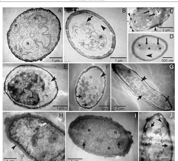
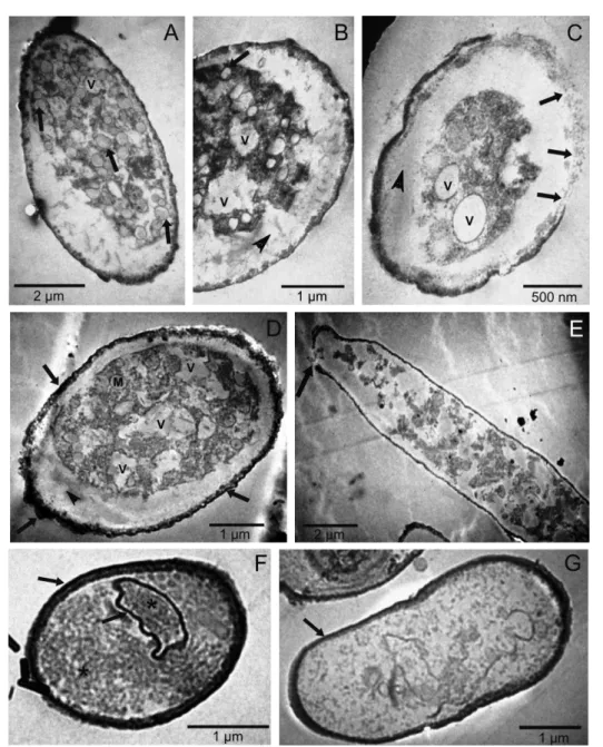
Similar alterations were observed in the fungal structures on the leaves of infected soybean plants that had been treated with higher concentrations of essential oils and with

Manuela Martins, da Universidade do Minho. 64 FERNANDES, Isabel Cristina Ferreira - Arqueologia medieval em Portugal: 25 anos de investigação. Porto, nova série, vol..