Braz. J. of Develop., Curitiba, v. 5, n. 9, p. 16620-16644 sep. 2019 ISSN 2525-8761
One-Step reverse transcriptase PCR for detection of arboviruses in serum
samples of patients assisted in Basic health Units in the State of Maranhão,
Brazil
PCR de transcriptase reversa em uma etapa para detecção de arbovírus em
amostras de soro de pacientes atendidos em Unidades Básicas de Saúde no
estado do Maranhão, Brasil
DOI:10.34117/bjdv5n9-203Recebimento dos originais: 20/08/2019 Aceitação para publicação: 27/09/2019
Jadna Patricia Pinheiro Nunes
Mestre
Programa de Mestrado em Biologia Parasitária - Universidade CEUMA
Endereço: Rua Josué Montello, 01, Renascença II, São Luis-MA, Brazil. CEP: 65075-120 E-mail: jadnapatricia7@hotmail.com
Bruna de Oliveira de Melo Graduada em Biomedicina
Programa de Mestrado em Biologia Parasitária - Universidade CEUMA
Endereço: Rua Josué Montello, 01, Renascença II, São Luis-MA, Brazil. CEP: 65075-120 E-mail: brunaoliv96@gmail.com
Silvio Gomes Monteiro Doutor
Programa de Mestrado em Biologia Microbiana da Universidade CEUMA
Endereço: Rua Josué Montello, 01, Renascença II, São Luis-MA, Brazil. CEP: 65075-120 E-mail: silvio_gm@yahoo.com.br
Viviane da Silva Sousa Almeida Mestranda
Universidade Ceuma, Programa de Mestrado em Biologia Microbiana, São Luis -MA Endereço: Rua Josué Montello, 01, Renascença II, São Luis-MA, Brazil. CEP: 65075-12
E-mail: viviane.sousa@hotmail.com Andrea de Souza Monteiro
Doutora
Programa de Mestrado em Biologia Microbiana da Universidade CEUMA
Endereço: Rua Josué Montello, 01, Renascença II, São Luis-MA, Brazil. CEP: 65075-120 E-mail: andreasmont@gmail.com
Lécia Maria Sousa Santos Cosme Especialista em Saúde Pública
Laboratório Central de Saúde Pública do Estado do Maranhão-MA Endereço: Rua Osvaldo Cruz, S/N. São Luis-MA. CEP: 65015-280
Braz. J. of Develop., Curitiba, v. 5, n. 9, p. 16620-16644 sep. 2019 ISSN 2525-8761 Conceição de Maria Fernandes da Silva Pinto
Especialista em Saúde Pública
Laboratório Central de Saúde Pública do Estado do Maranhão-MA Endereço: Rua Osvaldo Cruz, S/N. São Luis-MA. CEP: 65015-280
E-mail: cmfpinto@yahoo.com.br Luena Maria Souza Silva Especialista em Saúde Pública
Laboratório Central de Saúde Pública do Estado do Maranhão-MA Endereço: Rua Osvaldo Cruz, S/N. São Luis-MA. CEP: 65015-280
E-mail: luenamsilva@yahoo.com.br Rosimary de Jesus Gomes Turri
Doutora
Programa de Graduação em Farmácia da Universidade Federal do Maranhão (UFMA) Endereço: Av. dos Portugueses, 1966 - Vila Bacanga, São Luís - MA, 65080-805
E-mail: rositurri@gmail.com Maria de Fátima Castro Mendes
Doutoranda
Doutorado em Biodiversidade e Biotecnologia da Amazônia Leal (BIONORTE). Endereço: Rua Josué Montello, 01, Renascença II, São Luis-MA, Brazil. CEP: 65075-120
E-mail: fcmendes02@hotmail.com
Maria Rosa Quaresma Bomfim Doutora
Programa de Mestrado em Biologia Microbiana da Universidade CEUMA / Doutorado em Biodiversidade e Biotecnologia da Amazônia Leal (BIONORTE). Rua Josué Montello, 01,
Endereço: Renascença II, São Luis-MA, Brazil. CEP: 65075-120 E-mail: mrqbomfim@gmail.com
ABSTRACT
Background: Brazil has a high prevalence of infections caused by different arboviruses. The standard method used for diagnosis is an Enzyme-linked immunosorbent assay for IgM capture (MAC-ELISA). This study aimed to optimize and evaluate a one-step reverse transcription-polymerase chain reaction to detect acute infections caused by dengue, zika, chikungunya, and mayaro virus in clinical samples. Methods: We evaluated 620 sera samples collected from March 2016 to March 2018 and provided by the Central Health Laboratory of Maranhão (LACEN-MA). Total RNA was isolated from clinical specimens and used as the template for one-step RT-PCR assays with specific-primers designed for this study. Results: Of the 620 sera evaluated, 386 (62.2%) were positive, among them 330 (85.5%) amplified a specific fragment for chikungunya, 55 (14.2%) showed a fragment compatible with dengue serotype 4, and 1 (0.3%) exhibited profile for mayaro virus. Conclusions: The results obtained here were more sensitive than IgM-ELISA because the viral RNA was detected in serum samples from patients, not only from 1 to 6 days but also from 7 to 10 days after the beginning of clinical signs (convalescent period). Besides, the mayaro virus was detected in one serum sample that was IgM-ELISA negative for dengue, zika, and chikungunya.
Braz. J. of Develop., Curitiba, v. 5, n. 9, p. 16620-16644 sep. 2019 ISSN 2525-8761 RESUMO
Antecedentes: O Brasil tem uma alta prevalência de infecções causadas por diferentes arbovírus. O método padrão usado para o diagnóstico é um ensaio imunossorvente ligado à enzima para captura de IgM (MAC-ELISA). Este estudo teve como objetivo otimizar e avaliar uma reação em cadeia da polimerase com transcrição reversa em uma etapa para detectar infecções agudas causadas pelo vírus da dengue, zika, chikungunya e mayaro em amostras clínicas. Métodos: Avaliamos 620 amostras de soro coletadas de março de 2016 a março de 2018 e fornecidas pelo Laboratório Central de Saúde do Maranhão (LACEN-MA). O RNA total foi isolado a partir de amostras clínicas e utilizado como modelo para ensaios de RT-PCR em uma etapa com iniciadores específicos projetados para este estudo. Resultados: Dos 620 soros avaliados, 386 (62,2%) foram positivos, dentre eles 330 (85,5%) amplificaram um fragmento específico para chikungunya, 55 (14,2%) apresentaram fragmento compatível com o sorotipo 4 da dengue e 1 (0,3%) exibiu perfil para o vírus mayaro. Conclusões: Os resultados aqui obtidos foram mais sensíveis que o IgM-ELISA, pois o RNA viral foi detectado em amostras de soro de pacientes, não apenas de 1 a 6 dias, mas também de 7 a 10 dias após o início dos sinais clínicos (período de convalescença). Além disso, o vírus mayaro foi detectado em uma amostra de soro negativa para IgM-ELISA para dengue, zika e chikungunya.
Palavras-chave: vírus Chikungunya; Vírus zika; Vírus Mayaro. 1. INTRODUCTION
The arboviruses are a heterogeneous group of viruses that are transmitted to humans via the bite of arthropod vectors, especially mosquitoes [1,2]. The female of the Aedes aegypti is the primary vector of the dengue virus (DENV), Zika (ZIKV), and chikungunya (CHIKV) [3,4,5]. These mosquitoes have a diurnal activity and peridomestic habits; it's depositing their eggs preferentially in containers of water [6]. These eggs are resistant to dissection, and this property allows them to withstand unfavorable environmental conditions [7].
In Brazil, several arboviruses have been detected in the last years, among which DENV, CHIKV, ZIKV, Yellow Fever virus (YFV), and mayaro virus (MAYV) [8]. DENV, ZIKV, and YFV belong to the Flaviviridae family and genus Flavivirus, whereas CHIKV and MAYV are members of the Togaviridae family and genus Alphavirus [6,9]. The clinical signs and symptoms caused by these arboviruses are very similar, making it difficult to diagnose these infections [10,11] accurately.
Dengue human infections are caused by four genetically and antigenically distinct serotypes, DENV-1 to DENV-4, each generating a unique host immune response to infection [12,13]. The occurrence of DENV cases in the American continent in recent years has exceeded 2 million cases per year. The increase in the number of DENV infections, including severe cases and deaths, has been growing significantly and is becoming alarming [14]. In Brazil,
Braz. J. of Develop., Curitiba, v. 5, n. 9, p. 16620-16644 sep. 2019 ISSN 2525-8761 3,659,093 cases of DENV were reported, with 2,089 deaths occurring from 2015 to 2018 [14,15,16,17,18].
Historically, the first case of CHIKV infection occurred in Uganda in 1947; since then, the number of diseases has been growing and expanding worldwide since 2004 [19]. However, the first outbreak was only confirmed in 2007 in Micronesia. There been an increase in the transmission area since then, expanding to islands in the Pacific Ocean and Polynesia [20,21]. At the same time, alterations in the envelope protein (E1) of the African viral lineage and adaptation processes gave A. albopictus a higher capacity of infection; this increased the number of cases. These mutations increased the epidemic risk relative to other tropical, subtropical, and temperate regions [22].
In Brazil, the first autochthonous case of CHIKV infection was reported in September 2014 [23], and since then, it has been responsible for periodic outbreaks in all Brazilian states. Epidemiological reports of CHIKV cases issued by the Ministry of Health from 2015 to 2018 presented a total of 583,901 cases and 383 deaths [14,15,16,17,18].
The entry of ZIKV to Brazil occurred in 2015 with confirmed cases in the state of Bahia [24]. It subsequently spread to other regions, like Rio de Janeiro, São Paulo, and Pernambuco, where patients experienced exanthematic fever [25]. Since the introduction of ZIKV in Brazil in 2015, the Ministry of Health recorded a total of 241,191 confirmed cases and more than 300 deaths [14,15,16,17,18].
Mayaro virus (MAYV) is another arbovirus in an emergency in Brazil. It is transmitted by Haemagogus spp., mosquitoes, especially the species Haemagogus janthinomys, which is the primary known vector [26,27]. This vector maintains a wild cycle, and it can be found in the treetops, the primates that move from tree to tree are stung by these mosquitoes, becoming natural reservoirs of this virus [28, 29, 30].
The first report of MAYV isolation occurred in 1954 in Trinidad and Tobago, where it was found in the blood of five rural workers who had been clinical characteristics of arbovirus infection. Since then, endemic outbreaks have been recorded in Bolivia, Brazil, French Guiana, Guyana, and Suriname [31]. MAYV is considered endemic in some Brazilian regions (North and Central West). The first isolation in the country occurred in 1955 in the state of Pará. Since then, sporadic cases of MAYV have been reported in this region [32].
The diagnosis of these arboviruses is based on three main laboratory methods: virus isolation, serological tests, including the plaque reduction neutralization test (PRNT), enzyme-linked immunosorbent assay (ELISA), and immunofluorescence test (IFT) [33]. Besides, the
Braz. J. of Develop., Curitiba, v. 5, n. 9, p. 16620-16644 sep. 2019 ISSN 2525-8761 reverse transcription reaction followed by polymerase chain reaction (RT-PCR) technique has been widely used [33]. Viral isolation is time-consuming, and it is limited by the high cost and training requirements [34]. PRNT uses paired samples and presents cross-reactivity among members of the flavivirus group, and this method can present a cross-reaction with other serotypes in the case of a secondary DENV infection [35].
The serological diagnosis is confirmed by direct detection of IgM antibodies or by determination of a four-fold increase in specific antibodies titers in acute and convalescent samples using ELISA, IFT, or PRNT tests [33]. PCR-based assays are suitable for clinical diagnosis because of the closed-tube assay format and high sensitivity and specificity [33].
The arboviruses DENV, CHIKV, and ZIKV [36], and MAYV are currently co-circulating in Brazil. The increase in the number of people infected by arboviruses and the increased morbimortality rate is related to the lack of an effective vaccine, specific treatments, lack of planning to control and prevent the vectors responsible for transmitting the viruses [16]. This study aimed to evaluate molecular assays to diagnose arbovirus infections in serum samples from patients with clinical signs suggesting acute infection.
2. MATERIALS AND METHODS
2.1 CLINICAL SERA SAMPLES AND SOCIODEMOGRAPHIC DATA
For this study, 620 serum samples were randomly selected from the Central Laboratory of Public Health of the State of Maranhão (LACEN-MA) serum bank. These samples were from patients who sought public health services from March 2016 to March 2018. Of the 620 serum samples, 351 (56.6%) were from patients from 1 to 6 days after onset of signs and symptoms suggesting arbovirus infection, and 269 (43.4%) sera were 7 to 10 days after infection.
All these samples had been previously screened by LACEN technicians to detect IgM antibodies against DENV, CHIKV, and ZIKV. The following diagnostic kits were used to detect antibodies against these arboviruses: DENV ELISA IgM kit (PANBIO, Brisbane, Australia), anti-CHIKV (IgM) ELISA Kit (EUROIMMUN, LÜBECK, GERMANY), and ZIKV anti-ZIKV ELISA kit IgM and IgG (EUROIMMUN, LÜBECK, GERMANY). According to the data collected in the Laboratory Management System (GAL) of LACEN-MA, 536 (86.5%) sera were IgM-ELISA positive, and 84 (13.5%) were negative.
Braz. J. of Develop., Curitiba, v. 5, n. 9, p. 16620-16644 sep. 2019 ISSN 2525-8761 The sociodemographic data of each patient were collected in the GAL system, but in this database no clinical data on the patients were found, only age, sex, ethnicity and period of infection, including the date of the beginning of the clinical signs and symptoms until the day the patient sought the Basic Health Unit (UBS).
2.2 ARBOVIRAL LINEAGES USED AS A POSITIVE CONTROL
Aliquots of the culture medium of the dengue reference strains (DENV-1, Hawaii, DENV-2, ThNH7/93, DENV-3, PhMH-J1-97, and DENV- 4, SLMC 318), CHIKV (S27-African), ZIKV (BeH819966), and MAYV (BeAr20290) were kindly provided by the Department of Arboviroses of the University of São Paulo (USP)/School of the Medicine of Ribeirão Preto. As a negative control, we used 30 aliquots of sera from individuals who had never had any of the arboviruses screened. These samples are part of our private sera collection.
2.3 ISOLATION OF MAYARO VIRUS
For the isolation of MAYV, we used the Vero cell line (ATCC CCL-81) originating from African green monkey kidneys in Dulbecco's modified Eagle's medium (DMEM) supplemented with Earle's salts, L-glutamine (SIGMA-ALDRICH PRODUCTS, USA), and supplemented with 10% fetal bovine serum (FBS), 1 % antibiotic/antimycotic solution (10,000 units/mL penicillin, 10,000 μg/mL streptomycin, and 25 μg/mL amphotericin B) (Gibco-BioSciences, Ireland). Incubation of the culture in an atmosphere containing 5% CO2 at 37 °C was monitored until 80% confluency. An aliquot of 10 ul of the patient's serum was inoculated in the culture bottle with 1 mL of DMEM medium with 2% FBS plus antibiotics, then was incubated for one hour until adsorption of the viral particle. The medium was removed, and 4 ml of the same medium was added; the culture bottle was incubated under the same conditions. The cytopathic effect was monitored until total lysis of the cell monolayer. Aliquots of up to 5 passages were stored at -80 °C in cryotubes containing SFB plus 2% of dimethylsulfoxide (DMSO; MERCK, GERMANY).
2.4 RNA EXTRACTION AND MOLECULAR ASSAYS
To obtain the total RNA for the molecular assays, we used the QIAMP RNA VIRAL Kit (QIAGEN, HILDEN, GERMANY) according to the manufacturer's instructions. The total viral RNA was quantified using NanoDrop (THERMO SCIENTIFIC, WALTHAM, MASSACHUSETTS, USA) and stored at -80 °C until use.
Braz. J. of Develop., Curitiba, v. 5, n. 9, p. 16620-16644 sep. 2019 ISSN 2525-8761 The primers used to amplify the genetic material of DENV, CHIKV, and MAYV were designed from the non-structural protein NSP1, whereas the ZIKV primers started at the end of the envelope glycoprotein until at the end of the dimerization domain II. To optimize the one-step RT-PCR conditions for each pair of primers was used as the template RNA from the reference strains DENV (1 to 4), CHIKV, ZIKV, and MAY. The sequences of all primers used and conditions of the amplification for each arbovirus are described in Table 1.
The initial trials of one-step RT-PCR were made to amplify the four DENV serotypes individually. Each reaction had a final volume of 50 µl, containing 10 picomoles of each pair, plus OneStep RT-PCR Enzyme Mix (1×), 5x QIAGEN OneStep RT-PCR Buffer (1×), dNTP Mix (10 mM each), 5× Q-Solution (1×), RNase-Free Water (1×) (QIAGEN, HILDEN, GERMANY), and 100 nanograms of RNA from the reference serotype as template. The amplification occurred under the following conditions: one initial cycle of 50 ºC for 30 minutes, denaturation at 94 ºC for 15 minutes, 35 cycles of 94 ºC for 1 minute, annealing at 56 ºC for 1 minute, extension at 72 °C for 2 minutes and final extension at 72 °C for 10 minutes.
After the optimization with each pair of primers, the one-step RT-PCR was used to detect the DENV serotypes in clinical samples. We used the same conditions of amplification to amplify RNA from reference strains CHIKV, ZIKV, and MAYV. However, the temperature of the primers that were specific for each of these arboviruses (Table 1).
Finally, after the optimization process, the same one-step RT-PCR conditions were used to detect the presence of CHIKV, ZIKV, and MAYV viral RNA in the serum samples of patients with clinical suspicion of infection.
2.4.1 One-step rt-pcr sensitivity and pecificity
The sensitivity of the one-step RT-PCR assays was verified with a serial dilution of 100 nanograms of the total CHIKV reference lineage RNA. This RNA was previously quantified in the NanoDrop Thermo Scientific NanoDrop 2000 spectrophotometer (Thermo Fisher Scientific, Waltham, MA, USA). Serial dilution was started from 100 ng RNA as follows 1: 2 (50 ng); 1: 4 (25 ng); 1: 8 (12.5 ng); 1:16 (6.25 ng); 1: 32 (3.12 ng) and 1:64 (1.56 ng). For the reaction, a final volume of 50 μL consisting of specific reagents from the QIAGEN OneStep RT-PCR kit was used as described above with 10 picomoles of each pair of primers. The reaction tubes had a decreasing template input amount: 50 ng, 25 ng, 12.5 ng, 6.25 ng, 3.12 ng, and 1.56 ng. Amplification conditions were the same as those described above, but with a specific melting temperature of the primers for CHIKV (Table 1).
Braz. J. of Develop., Curitiba, v. 5, n. 9, p. 16620-16644 sep. 2019 ISSN 2525-8761 The specificity of the primers designed exclusively for this study was verified with 100 ng of the total RNA extracted from the yellow fever virus (YFV), and Saint Louis Encephalitis virus (SLEV). Amplification reactions were performed using the same conditions described above, with the specific temperature of each primer pair (Table 1).
2.4.2 Electrophoretic analysis of one-step rt-pcr products
All results obtained by the molecular method were verified on a 2% agarose gel electrophoresis; thus it was used 8 µL of each amplified product, plus 2 µL of sample buffer (Blue Orange 6× Loading Dye-PROMEGA, USA), and electrophoresis was performed as previously described [37]. The run occurred in 0.5× TBE buffer (Tris-borate-EDTA) (100 mM Tris-base; 2.0 mM EDTA solution (pH 8.0) and 50 mM boric acid) (PROMEGA, USA) at 85 V for 1 hour and 30 minutes. After the electrophoresis, the gels were stained in ethidium bromide solution (0.5 μg / mL) for 15 minutes, and observed using an ultraviolet transilluminator, and photographed to document the results.
2.4.3 One-step rt-pcr product purification, sequencing, and sequence analysis
To sequence the product of one-step RT-PCR it was used the Wizard® SV Gel and PCR Clean-Up kit, according to the manufacturer's guidelines (PROMEGA, USA). The sequencing was perforrmed with the DyEnamic ™ kit ET Dye Terminator in a MegaBaceTM 1000 automatic sequencer (Amersham Biosciences), according to a previously described method [38].
The ChromasPro program (Technelysium Pty. Ltd -
http://www.technelysium.com.au/chromas.html) was used to analyze the electropherograms provided by MegaBace during the sequencing process. To check the similarity of the obtained sequences, we used the program BLASTn in the package BLAST 2.0 (Basic Alignment Search Tool - http://www.ncbi.nlm.nih.gov/BLAST/) [39]. The MEGA software version 6 [40] was used to align the sequences obtained with MAYV sequencing in Genbank.
2.5 STATISTICAL ANALYSIS
The sociodemographic data collected in the GAL system of each patient selected were tabulated in an Excel spreadsheet and analyzed by statistical program IBM SPSS Statistics 25.0 [41] and R Commander [42]. The descriptive statistic analysis (minimum, maximum, mean, and standard deviation) was applied to analyze numerical variables (age and days) concerning
Braz. J. of Develop., Curitiba, v. 5, n. 9, p. 16620-16644 sep. 2019 ISSN 2525-8761 gender. The evaluation of the association of the sociodemographic variables and the day of onset of signs and symptoms about the results of ELISA-IgM and one step-RT-PCR assays were performed using the parametric Chi-square independence test. The level of significance of the tests was 5% or a p-value of <0.05 and a 95% confidence interval.
2.6 ETHICS STATEMENT
The present study was submitted to the Research Ethics Committee of the Universidade Ceuma, and it was approved under a consubstantiated opinion number: 1,570,391/2016, emitted on 12 May 2016.
3. RESULTS
3.1 OPTIMIZATION OF ONE-STEP RT-PCR ASSAY WITH RNA FROM REFERENCE STRAINS
In this study, we report the evaluation of a one-step RT-PCR assay for the diagnosis or detection of arbovirus infections. We used 620 sera samples that had been previously screened by ELISA for the detection of IgM antibodies against DENV, ZIKV, and CHIKV. The results of IgM ELISA assays showed that 536 (86.5%) sera were positive, and 84 (13.5%) were negative. Of these, 498 (49.1%) were positive for CHIKV, and 38 were positive for DENV, and none of the samples evaluated by ELISA were reactive to ZIKV.
Initially, it was optimized and evaluated the one-step RT-PCR assays with RNA from reference strains. Specific fragments with the expected molecular size, for each of the arboviruses screened, were viewed on agarose gels (Figure 1), being 1A: profiles of CHIKV; 1B: ZIKV; 1C: MAYV and DENV serotypes (1-4).
3.1.1 Evaluation of one-step rt-pcr assays with rna obtained from clinical samples
Of the 620 samples evaluated by one-step RT-PCR assays, a total of 386 (62.2%) were positive for arboviruses, CHIKV, DENV, and MAYV. Of these, 78.0% (301) were serologically positive, and 22.0% (85) were IgM ELISA negative by anti-CHIKV (IgM) ELISA Kit (EUROIMMUN). Of the positive IgMs, 89.7% (270) were from patients who were in the acute phase of infection, and 31 (10.3%) were in the convalescent period. Regarding the serum IgM negative samples, 81 (95.3%) were in the acute phase, and 4 (4.7%) in the convalescent phase (Table 2).
Braz. J. of Develop., Curitiba, v. 5, n. 9, p. 16620-16644 sep. 2019 ISSN 2525-8761 Of the 386 positive samples by one-step RT-PCR, 330 (85.5%) were positive for CHIKV based on the amplification of the specific 623 bp fragment, as shown in Figure 2. It is interesting to note that, out the 330 serum samples, 67 (20.3%) were negative in the IgM ELISA. These sera were from patients who sought health services within a period of 1 to 8 days after the onset of signs and symptoms. The remaining 263 sera were from patients who went to hospitals from the first to the tenth day after the beginning of clinical signs and symptoms. These sera samples were positive by the IgM ELISA anti-CHIKV (IgM) ELISA Kit (EUROIMMUN) test (Table 2).
The one-step RT-PCR assay detected 55 (14.2%) serum as DENV positive with the profile for serotype 4 based on the presence of the specific of 434 bp fragment, as shown in Figure 3. Of the DENV positive samples, 17 (30,9%) were serologically negative using the DENV ELISA IgM kit (PANBIO). These serum samples were from patients from 1 to 3 days after the onset of clinical signs and symptoms suggesting DENV infection. The other 38 sera samples were from 1 to 7 days after onset of the disease were ELISA positive.
It is important to note that a serum sample that had been tested with different IgM ELISA kits for the detection of antibodies against dengue, zika, and chikungunya arboviruses was always negative. This RNA was submitted for one-step RT-PCR to detect DENV, CHIKV, and ZIKV, but no amplification was observed. However, when specific primers for MAYV were used in one-step RT-PCR, the specific 370 bp fragment was amplified (Figure 4). This result triggered the isolation of MAYV from the patient's serum sample. The one-step RT-PCR product was purified and sequenced, and its sequence was deposited in GenBank with the accession number MK956954.
The presence of the ZIKV genetic material was not detected in any of the clinical samples evaluated in this study. However, the amplification of the RNA from reference viral strain occurred in all one-step RT-PCR assays (Figure 5).
3.1.2 Sensitivity and pecificity of the primers and one-step RT-PCR assays
The sensitivity of the one-step RT-PCR was performed using a serial dilutions of the RNA of CHIKV reference lineage showed amplification for the following dilutions: 1:2 (50 ng), 1:4 (25 ng), 1:8 (12.5 ng), 1:16 (6.25 ng), and 1:32 (3.12 ng). Thus, we verified a detection limit of 3.12 ng of viral RNA (Figure 6). The same limit of detection was observed for DENV, ZIKV, and MAYV (data no shown). Moreover, the primers evaluated here were specific because it did not amplify YFV and SLEV RNA.
Braz. J. of Develop., Curitiba, v. 5, n. 9, p. 16620-16644 sep. 2019 ISSN 2525-8761 4. DISCUSSION
Brazil has been considered as a hyperendemic area for the occurrence of arboviral infections [43]. Regarding the high number of cases of arbovirus infections that have been diagnosed by the ELISA method, the current rates are not known. This is because, in endemic areas, as is here, the vast majority of the population has secondary antibodies that hinder the serological diagnosis [44]. Besides, the correct diagnosis of an arbovirus infection is not restricted to the use of only one technique, but the differential diagnosis is essential for effective patient management, clinical care, and epidemiologic surveillance [45].
Molecular methods have emerged as an alternative tool for the use of serological tests commonly used for the diagnosis of infections caused by arboviruses. Currently, numerous PCR-based assays have been evaluated, and the results have been promising [46,47].
The one-step RT-PCR evaluated in the present study detected arboviruses in both acute and convalescent-phase (IgM positive) and negative sera by IgM ELISA from both phases. These findings are significant, as positive cases are no reported to the epidemiological surveillance services. This result is in agreement with the findings of Fortuna et al. [48] who detected the presence of viral RNA in both IgM ELISA positive and negative IgM sera using specific probes with qPCR, additionally revealing that is possible to have IgM antibodies in the acute-phase due to a previous infection.
The results of the detection and amplification of viral RNA obtained from the clinical samples showed a high prevalence of arbovirus infections, in the state of Maranhao. CHIKV infections were detected in 85.5% of the positive samples by one-step RT-PCR assays. The state of Maranhão has a hot and humid tropical climate, which favors the proliferation of mosquitoes A. aegypti, with an index of infestation in at least 73.3% of its municipalities [16, 49]. It is interesting to note that the successive outbreaks of CHIKV occurring in various parts of the world and may cause specific adaptive genetic changes that allow the Mosquito A. aegypti to contract the infection and transmit it to humans [50-52].
The high index of CHIKV infection observed here, showed the imminent risk of an expansion of new outbreaks. This result agrees with that observed by Wahid et al. [53] as, after the introduction of CHIKV into a new geographical site, high incidence rates occur because of the rapid spread. Moreover, Costa-da-Silva et al. [54] detected for the first time, a natural A. aegypti infection by the CHIKV genotype East-Central South African (ECSA) in the Northeast region, and they considered this species a vector capable of transmitting this arbovirus in urban areas in the region.
Braz. J. of Develop., Curitiba, v. 5, n. 9, p. 16620-16644 sep. 2019 ISSN 2525-8761 Regarding infections caused by DENV, only the serotype 4 was detected, and no other Dengue serotype was found. This fact is important since DENV-4 was introduced in the state of Maranhao at the end of 2011 [18]. As a result, when a new variant is introduced at a specific site, and the prevalence of this lineage overlaps with the circulating variants, demonstrating its potential for dispersion [55]. The theory of sequential infection by DENV is characterized by pre-existing antibody-dependent immuno-amplification and has been widely accepted [56].
Sanchez-Carbonel et al. [57] and Fortuna et al. [48] evaluated a one-step RT real-time PCR (qPCR) assays using a specific Taqman probe for DENV detection. This technique differs from our methodology because the construction of probes and the application of qPCR are expensive and require skilled labor. The methodology evaluated here costs less, and it is faster and does not demand specialized personnel.
The detection of MAYV by one-step RT-PCR in a clinical sample is important because by serology were not detected antibodies against DENV, CHIKV, and ZIKV. Regarding the occurrence of an autochthonous case of MAYV fever caused by the infection probably occurred through a bite of A. aegypti, and an experimental study showed that the transmission of MAYV by this vector is possible [58].
The presence of MAYV raises an alert for the public health services from Brazil, especially of the state of Maranhão, as there are no reports of its presence in any state of the Northeast region. However, it has sporadically circulated in the North and Central West states [18]. Zuchi et al. [59] detected by nested-PCR the MAYV circulating in Mato Grosso during an outbreak of DENV. These results show the need for molecular screening of this arbovirus in patients with suspected dengue infection, where this virus may silently circulate.
Most of the primers evaluated here were designed from the NS1 protein because there are found specific markers for the diagnosis of the infections by DENV, CHIKV, and MAYV [60]. However, for the specific primers for ZIKV was used a preserved region of the glycoprotein E (env), this region is responsible for the neutralization of human antibodies by the occurrence of viral infection [61].
The results of the sensitivity and specificity obtained by one-step RT-PCR assays were significant due to the use of these specific-primers sets. The literature does not report any direct sensitivity technique for DENV infections. While that, previous results refer to sensitivity using other methods such as nested-PCR with dot blot [62]; real-time PCR with the use of specific probes [63]; and the hybridization [64].
Braz. J. of Develop., Curitiba, v. 5, n. 9, p. 16620-16644 sep. 2019 ISSN 2525-8761 The frequency of the distribution of the sociodemographic variables from the 386 positive patients evaluated here, for arbovirus infections in the molecular test, showed that 296 (76.7%) were female. A total of 35.7% were 16 to 30 years old (Table 3). Studies from Anker et al. [65], Mishra et al. [66], and Wangdi et al. [67] have demonstrated that these arboviruses affect younger and female people. While Kalayanarooj et al. [68] reported that this is due to the peridomicilliary habit of the vector, and its proliferation is often related to the accumulation of water in containers placed inside or around the house. Then, as women and children commonly stay at home longer, they to become more susceptible to mosquito bites.
5. CONCLUSIONS
In this study, we have described the first detection and molecular identification of MAYV in the Northeast of Brazil, especially in the state of Maranhão. Besides, we found a high prevalence of CHIKV infection and a decrease in dengue cases during the study period. The one-step RT-PCR assay evaluated herein detected the RNA from different arboviruses in IgM-positive and IgM-negative ELISA sera samples. It was sensitive, specific, and could diagnose the presence of the viral RNA in sera samples from patients, both in the acute and convalescent phases of infection. Moreover, the optimized and evaluated method was reproducible in all trials and demonstrated that CHIKV, DENV, and MAYV are co-circulating in patients from different municipalities of the state of Maranhão. Therefore, the simultaneous circulation of these arboviruses and the increased incidence of infected individuals demonstrated the need for more effective preventive measures and faster and more sensitive diagnostic tests that could expedite clinical care for the patient.
Author Contributions: JPPN, BOM and MFCM extracted the RNA from the serum samples and did molecular biology assays. SGM and VSSA performed the statistical analyzes of the results. LMSSC and CMFSP randomly selected the records of patients with clinical suspicion of arbovirus infection in the LACEN-MA database. LMSS separated the serum samples in the freezers -80 ºC stored in the LACEN-MA. RJGT and ASM prepared excel spreadsheets for the analysis of the results. MRQB designed and conducted the study, analyzed the data and drafted the manuscript. In addition to the activities already mentioned, all authors participated in the analyzes of the results and the writing of the manuscript.
Braz. J. of Develop., Curitiba, v. 5, n. 9, p. 16620-16644 sep. 2019 ISSN 2525-8761 Funding: The authors thank the Fundação de Amparo à Pesquisa e Desenvolvimento Científico do Maranhão (FAPEMA) for the financial support of the research from project number: PPSUS-05970/2016.
Acknowledgments: The authors thank Dr. Eurico de Arruda Neto and Maria Lucia Silva for supplying the reference strains of the researched arboviruses used as positive control for molecular assays. We thank technical staff from LACEN-MA who performed the ELISA in the serum samples. We would like to thank Editage (www.editage.com) for English language editing.
Conflicts of Interest: The authors declare no conflict of interest. The funders had no role in the design of the study; in the collection, analyses, or interpretation of data; in the writing of the manuscript, or in the decision to publish the results.
Figure 1. Electrophoretic profiles obtained in the optimization of one-step RT-PCR for CHIKV, ZIKV, MAYV,
Braz. J. of Develop., Curitiba, v. 5, n. 9, p. 16620-16644 sep. 2019 ISSN 2525-8761 Caption: 2% agarose gel stained with ethidium bromide showing the profiles obtained with total RNA from
reference arbovirus A: CHIKV 623 pb; B: ZIKV 911 pb; C: MAYV 370 bp and D: DENV-1 518 bp; DENV-2 595 bp; DENV-3 372 bp; DENV-4 434bp; MM - molecular marker (100 bp DNA ladder, Promega, USA) and CN negative controls. Vz – empty line.
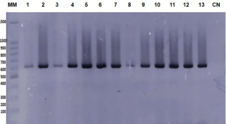
Figure 2. Specific profile for chikungunya virus obtained by one-step RT-PCR assay using as a template the RNA
extracted from some sera samples
Caption: 2% agarose gel stained with ethidium bromide. MM - molecular marker (100 bp DNA ladder, Promega,
USA). CN negative controls. Lines 1 to 12, the profile of the 623 bp fragment compatible with CHIKV, and line 13-CHIKV reference sample used as a positive control during the amplification process.
Figure 3. Electrophoretic profiles obtained by the one-step RT-PCR assays using as a template the RNA obtained
from the sera samples of patients with clinical signs and symptoms of arbovirus infection
Caption: 2% agarose gel stained with ethidium bromide. MM molecular marker (100 bp DNA ladder, Promega,
USA). Lines 1 to 7, PCR products of some clinical samples showing amplification profiles for DENV -4. Line 8, CP positive control of the serotype 4 reference (434 bp), and CN negative control.
Braz. J. of Develop., Curitiba, v. 5, n. 9, p. 16620-16644 sep. 2019 ISSN 2525-8761
Figure 4. Profiles of the one-step RT-PCR assays obtained with the RNA extracted from the MAYV reference lineage and the serum of the patient with clinical signs and symptoms suggesting arbovirus infection
Caption: 2% agarose gel stained with ethidium bromide showing the specific fragment of 370 bp obtained with the positive control, and with the sample (OSSD1) from the patient with clinical suspicion of arbovirus infection. This sample was negative by IgM-ELISA for the detection of DENV, CHIKV, and ZIKV antibodies. MM-molecular marker (100 bp DNA ladder, Promega, USA), CN, negative control.
Figure 5. Profiles of the one-step RT-PCR assays obtained with the RNA extracted from reference ZIKV lineage
and the sera sample of patients with clinical signs and symptoms suggesting arbovirus infection
Caption: 2% agarose gel stained with ethidium bromide showing the specific fragment of 911 bp obtained with the ZIKV positive control reference lineage. Lines 1 to 14: Profiles of the sera samples evaluated for detection of this arbovirus. MM. Molecular marker (100 bp DNA ladder, Promega, USA); CN, Negative control of the reagents used in the reaction.
Braz. J. of Develop., Curitiba, v. 5, n. 9, p. 16620-16644 sep. 2019 ISSN 2525-8761
Table 1. Primers used in the present study
Arboviruses Primers sequence (5’ – 3’) Size in base pairs (pb) Melting temperature * Dengue 1 FW: CACCGGCTAATGTCAGCTGC RV: CCTGCAGAGACCATTGACTTA 518 56 Dengue 2 FW:AGGAATCATGCAGGCAGGAA RV:TCCACAGTCCTCAGTCACCAC 595 Dengue 3 FW:CATAATAGACGGACCAAACACAC RV:TGTGGTTGTGTTGCGAGATAG 372 Dengue 4 FW:GACCAAAGGCAAGAGAGCACT RV:GCATCTGGCTTTCCAGCACT 434 Chikungunya FW: ACCGAATGACCATGCYAATGCTAGAG RV: ACAGCACACGGTCGCACGGT 623 62 Zika FW: ATGATACTGTTGATTGCCCC RV:TTCAGGCGACATTTCAAGTG 911 53
Mayaro Mayv1FW: GCATTCTCGCATCTGGCTAC Mayv2RV: GGAAGTACAAAGAAGTCGGTGC
370 56
Caption: FW: primer sense. RV: primer antisense. * temperature in degrees Celsius.
Table 2. Distribution of the sociodemographic variables from patients positive by the one-step RT-PCR assays
Number of days after the onset of clinical signs and symptoms
Molecular method ELISA IgM
p value Positive Negative Positive Negative
Chikungunya detection
1 to 3 days 185 0 140 45 p<0.0001
4 to 6 days 113 0 94 19 p<0.0001
7 to10 days 32 234 263 3 p<0.0001
Braz. J. of Develop., Curitiba, v. 5, n. 9, p. 16620-16644 sep. 2019 ISSN 2525-8761 1 to 3 days 28 0 15 13 p<0.0001 4 to 6 days 24 0 21 3 p<0.0001 7 to 10 days 3 0 2 1 p=0.117 Mayaro detection 1 to 6 days 1 0 0 1 p=0.317
Caption: From 1 to 6 days: the acute phase, and from 7 to 10 days convalescent period. Table 3. Comparison of the results of the one-step RT-PCR concerning IgM-ELISA assays for acute and convalescent phases of the disease
REFERENCES
Sigfrid, L.; Reusken, C.; Eckerle, I.; Nussenblatt, V.; Messina, J.; Kraemer, M.; Ergonul, O.; Papa, A.; Koopmans, M.; Horby P. Preparing clinicians for (re-) emerging arbovirus
Variables Positive by one-step RT-PCR Percentage (%) Sex Female Male 296 90 76,68 23,32 Age (years) ≤ 1 - 15 16 - 30 31 - 45 46 - 60 61 - 75 76 - 90 41 138 114 47 30 16 10,62 35,75 29,53 12,18 7,77 4,15 Race Brown White Black 217 141 28 217 141 28 Area Urban Rural 330 56 85,49 14,51 Total 386 100
Braz. J. of Develop., Curitiba, v. 5, n. 9, p. 16620-16644 sep. 2019 ISSN 2525-8761 infectious diseases in Europe. Clin. Microbiol. Infect. 2018, 3, 229-239, doi: 10.1016/j.cmi.2017.05.029.
Braack, L.; Gouveia de Almeida, A.P.; Cornel, A.J.; Swanepoel, R.; de Jager, C. Mosquito-borne arboviruses of African origin: review of key viruses and vectors. Parasit. Vectors. 2018, 1, 29, doi: 10.1186/s13071-017-2559-9.
Patterson, J.; Sammon, M.; Garg, M. Dengue, Zika and Chikungunya: Emerging Arboviruses
in the New World. Western. J. Emerg. Med. 2016, 6, 671-679,
doi: 10.5811/westjem.2016.9.30904.
Boyer, S.; Calvez, E.; Chouin-Carneiro, T.; Diallo, D.; Failloux, A.B. An overview of mosquito vectors of Zika virus. Microbes Infect. 2018, 11-12, 646-660, doi: 10.1016/j.micinf.2018.01.006.
Mourya, D.T.; Gokhale, M.D.; Majumdar, T.D.; Yadav, P.D.; Kumar, V.; Mavale M.S. Experimental Zika virus infection in Aedes aegypti: Susceptibility, transmission & co-infection with dengue & chikungunya viruses. Indian. J. Med. Res. 2018, 1, 88-96, doi: 10.4103/ijmr.IJMR_1142_17.
Day, J.F. Mosquito Oviposition Behavior and Vector Control. Tabachnick WJ. Insects. 2016, 4, 65, doi:10.3390/insects7040065.
Almeida, M.C.; Assunção, R.M.; Proietti, F.A.; Caiaffa, W.T. Intra-urban dynamics of dengue epidemics in Belo Horizonte, Minas Gerais State, Brazil, 1996–2002. Cad. Saúde. Publ. 2008, 24, 2385–2395.
Donalisio, M.R.; Freitas, A.R.R.; Zuben, A.P.B.V. Arboviruses emerging in Brazil: challenges for clinic and implications for public health. Rev Saude Publica. 2017, 10, 51:30, doi.org/10.1590/S0102-311X2008001000019.
Paixão, E.S.; Teixeira, M.G.; Rodrigues, L.C. Zika, chikungunya and dengue: the causes and threats of new and re-emerging arboviral diseases. BMJ Glob Health. 2017, 3, e000530, doi: 10.1136/bmjgh-2017-00053.
Rodriguez-Morales, A.J. No era suficiente con dengue y chikungunya: llegó también Zika. Arch. Med. 2014, 2-3, 1-4, doi: 10.3823/1245.
Paniz-Mondolfi, A.E.; Rodriguez-Morales, A.J.; Blohm, G.; Marquez, M.; Villamil-Gomez, W.E. ChikDenMaZika Syndrome: the challenge of diagnosing arboviral infections in the midst of concurrent epidemics. Ann. Clin. Microbiol. Antimicrob. 2016, 15, 42, doi: 10.1186/s12941-016-0157-x.
Braz. J. of Develop., Curitiba, v. 5, n. 9, p. 16620-16644 sep. 2019 ISSN 2525-8761 Mustafa, M.S.; Rasotgi, V.; Jain, S.; Gupta, V. Discovery of fifth serotype of dengue virus (DENV-5): A new public health dilemma in dengue control. Med. J. Armed. Forces. India. 2014, 1, 67-70, doi: 10.1016/j.mjafi.2014.09.011.
Uno, N.; Ross, T.M. Dengue virus and the host innate immune response. Emerg Microbes Infect. 2018, 1,167. doi: 10.1038/s41426-018-0168-0.
Brasil. Ministério da Saúde. Secretaria de Vigilância em Saúde Monitoramento dos casos de dengue, febre de chikungunya e febre pelo vírus Zika até a Semana Epidemiológica 45. Boletim Epidemiológico. 2015, 46, 36.
Brasil. Ministério da Saúde. Secretaria de Vigilância em Saúde. Departamento de Vigilância das Doenças Transmissíveis. Dengue: diagnóstico e manejo clínico: adultos e crianças [recurso eletrônico] / Ministério da Saúde, Secretaria de Vigilância em Saúde, Departamento de Vigilância das Doenças Transmissíveis. – 5. ed. – Brasília: Ministério da Saúde. 2016, 58 p. Brasil. Boletim Epidemiológico Monitoramento dos casos de dengue, febre de chikungunya e febre pelo vírus Zika. Brasília: Ministério da Saúde, Secretaria de Vigilância em Saúde. 2017, 48, 29.
Brasil. Boletim Epidemiológico. Monitoramento dos casos de dengue, febre de chikungunya e febre pelo vírus Zika. Brasília: Ministério da Saúde, Secretaria de Vigilância em Saúde. Ministério da Saúde. 2019, 50, 4.
Brasil. Ministério da Saúde. Secretaria de Vigilância em Saúde. Departamento de Vigilância das Doenças Transmissíveis. Nota Informativa. 2015, 6. CIEVS/ DEVIT/SVS/MS. Brasília: Ministério da Saúde. 2015.
Sergon, K.; Yahaya, A.A.; Brown, J.; Bedja, S.A.; Mlindasse, M.; Agata, N.; Allaranger, Y.; Ball, M.D.; Powers, A.M.; Onyango, C; et al. Seroprevalence of Chikungunya virus infection on Grande Comore Island, union of the Comoros, 2005. Am. J.Trop. Med. Hyg. 2007, 6, 1189-93.
Brito, C.A.; Cordeiro, M.T. One year after the Zika virus outbreak in Brazil: from hypotheses to evidence. Rev. Soc. Bras. Med. Trop. 2016, 5, 537–43, doi.org/10.1590/0037-8682-0328-2016.
Plourde, A.R.; Bloch, E.M. A Literature Review of Zika Virus. Emerg. Infect. Dis. 2016, 7, 1185-92, doi: 10.3201/eid2207.151990.
Coffey, L.L.; Forrester, N.; Tsetsarkin, K.; Vasilakis, N.; Weaver. S.C. Factors shaping the adaptive landscape for arboviruses: implications for the emergence of disease. Future Microbiol. 2013, 2, 155-76, doi:10.2217/fmb.12.139.
Braz. J. of Develop., Curitiba, v. 5, n. 9, p. 16620-16644 sep. 2019 ISSN 2525-8761 Teixeira, M.G.; Andrade, A.M.S.; Costa, M.C.N.; Castro, J.M.; Oliveira, F.L.; Goes, C.S.; Maia, M.; Santana, E.B.; Nunes, B.T.; Vasconcelos, P.F. East/Central/ South African genotype Chikungunya virus, Brazil, 2014. Emerg. Infect. Dis. 2015, 5, 906-7, doi: 10.3201/eid2105.141727.
Campos, G.S.; Bandeira, A.C.; Sardi, S.I. Zika Virus Outbreak, Bahia, Brazil. Emerg. Infect. Dis. 2015, 10, 1885-6, doi: 10.3201/eid2110.150847.
Freire, C.C.M.; Lamarino, A.; Lima, N.D.F.; Sall, A.A.; Zanotto, P.M.A. Spread of the pandemic Zika virus lineage is associated with NS1 codon usage adaptation in humans. BioRxiv. 2015, doi: https://doi.org/10.1101/032839.
Muñoz, M.; Navarro, J.C. Virus Mayaro: un arbovirus reemergente en Venezuela y Latinoamérica. Biomed. 2012, 32, 288–302, doi.org/10.7705/biomedica.v32i2.647
Acosta-Ampudia, Y.; Monsalve, D.M.; Rodríguez, Y.; Pacheco, Y.; Anaya, J.M.; Ramírez-Santana, C. Mayaro: an emerging viral threat?. Emerg. Microb. Infect. 2018, 1, 163, doi: 10.1038/s41426-018-0163-5.
Hoch, A.L.; Peterson, N.E.; Leduc, J.W.; Pinheiro, F.P. An outbreak of Mayaro virus disease in Belterra, Brazil. III. Entomological and ecological studies. Am. J. Trop. Med. Hyg. 1981, 30, 689–698, doi: 10.4269/ajtmh.1981.30.67.
Figueiredo, L.T. Emergent arboviruses in Brazil. Rev. Soc. Bras. Med. Trop. 2007, 40, 224– 229, doi.org/10.1590/S0037-86822007000200016.
Batista, P.M.; Andreotti, R.; Almeida, P.S.; Marques, A.C.; Rodrigues, S.G.; Chiang, J.O.; Vasconcelos, P. F. da C. Detection of arboviruses of public health interest in free-living New World primates (Sapajus spp.; Alouatta caraya) captured in Mato Grosso do Sul, Brazil. Rev. Soc. Bras. Med. Trop. 2013, 6, 684-690, doi.org/10.1590/0037-8682-0181-2013.
Lednicky, J.; Rochars, V.; Elbadry, M.; Loeb, J.; Telisma, T.; Chavannes, S.; Gina, A.; Eleonora, C.; Massinno, C.; Bernard, O; et al. Mayaro Virus in Child with Acute Febrile Illness, Haiti, 2015. Emerg Infect Dis. 2016, 11, 2000-2002, doi.org/10.3201/eid2211.161015. Napoleão-Pego, P.; Gomes, L.P.; Provance-Jr, D.W.; Simone, S.G. Mayaro Virus Disease. J. Hum. Virol. Retrovirol. 2014, 3:0018, doi: 10.15406/jhvrv.2014.01.00018.
Patel. P.; El Wahed, A.; Faye, O.; Prüger. P.; Kaiser, M.; Thaloengsok, S.; Sukathida, U.; Sakuntabha, A.; Leparc-Goffart, I.; Hufert, F.T; et al. A Field-Deployable Reverse Transcription Recombinase Polymerase Amplification Assay for Rapid Detection of the
Chikungunya Virus. PLoS. Negl.Trop. Dis. 2016, 10, e0004953,
Braz. J. of Develop., Curitiba, v. 5, n. 9, p. 16620-16644 sep. 2019 ISSN 2525-8761 Xu, Q.; Liu, H.; Yuan, P.; Zhang, X.; Chen, Q.; Jiang, X.; Zhou, Y. Development of a simplified RT-PCR without RNA isolation for rapid detection of RNA viruses in a single small brown planthopper (Laodelphax striatellus Fallén). Virol. J. 2017, 1, 90. doi: 10.1186/s12985-017-0732-6.
Roehring JT, Hombach J, Barrett ADT. Guidelines for plaque-reduction neutralization testing of human antibodies to dengue viruses. Viral. Immunol. 2008, 21, 123-32, doi: 10.1089/vim.2008.0007.
Lima-Camara, T.N. Emerging arboviruses and public health challenges in Brazil. Rev, Saude. Publica, 2016, 50, 36, doi.org/10.1590/S1518-8787.2016050006791.
Sambrook, J.; Fritsch, E.F.; Maniatis, T. Molecular Cloning: A Laboratory Manual, C.S.H. Laboratory, 2nd edition, Cold Spring Harbor, NY, USA, 1989.
Sanger, F.; Nickelen, S.; Coulson, A.R. DNA sequencing with chain-terminating inhibitors. Proc. Natl. Acad. Sci. USA. 1977, 74, 5463-5467, doi:10.1073/pnas.74.12.5463.
Altschul, S.F.; Madden, T.L.; Schäffer, A.A.; Zhang, J.; Zhang, Z.; Miller, W.; Lipman, D.J. Gapped BLAST and PSI-BLAST: a new generation of protein database search programs. Nucleic. Acids. Res. 1997, 25, 3389-3402.doi:10.1093/nar/25.17.3389.
Tamura, K.; Stecher, G.; Peterson, D.; Filipski, A.; Kumar, S. MEGA6: Molecular Evolutionary Genetics Analysis Version 6.0. Molecular Biology and Evolution. 2013, 30, 2725–2729, doi: 10.1093/molbev/mst197.
IBM Corp. Released 2017. IBM SPSS Statistics for Windows, Version 25.0. Armonk, NY: IBM Corp.
Fox, J.; Bouchet-Valat, M. Rcmdr: R Commander. R package version. 2.5-1. 2018
Terzian, A.C.B.; Zini, N.; Sacchetto, L.; Rocha, R.F.; Parra, M.C.P.; Del Sarto, J.L.; Dias, A.C.F.; Coutinho, F.; Rayra, J.; Silva, R. A, et al. Evidence of natural Zika virus infection in neotropical non-human primates in Brazil, 2018, Scientific. Reports, 8, 16034.
Changal, K.H.; Raina, A.H.; Raina, A.; Raina, M.; Bashir, R.; Latief, M.; Mir, T. Changal, Q. H. Differentiating secondary from primary dengue using IgG to IgM ratio in early dengue: an observational hospital based clinic-serological study from North India. BMC. Infect. Dis, 2016, 1, 715.
Leon, F.; Meyer, A.; Reynier, R.; Blanc, E.; Bruyère-Ostells, L.; Brès, J.C.; Simonin, Y.; Salinas, S.; Gallian, P., et al. An Innovative Multiplexed And Flexible Molecular Approach For The Differential Detection Of Arboviruses, J. Mol. Diagn. 2018, 1, 81-88.
Braz. J. of Develop., Curitiba, v. 5, n. 9, p. 16620-16644 sep. 2019 ISSN 2525-8761 Jacobsen, S.; Patel, P.; Schmidt-Chanasit, J.; Leparc-Goffart, I.; Teichmann, A.; Zeller, H.; Niedrig, M. External quality assessment studies for laboratory performance of molecular and serological diagnosis of Chikungunya virus infection. J. Clin. Virol. 2016, 76, 55-65, doi: 10.1016/j.jcv.2016.01.008.
Ramírez, A.L.; van den Hurk, A.F.; Meyer, D.B.; Ritchie, S.A. Searching for the proverbial needle in a haystack: advances in mosquito-borne arbovirus surveillance. Parasit. Vectors. 2018, 1, 320, doi: 10.1186/s13071-018-2901-x.
Fortuna, C.; Remoli, M.E.; Rizzo, C.; Benedetti, E.; Fiorentini, C.; Bella, A.; Argentini, C.; Farchi, F.; Castilletti, C.; Capobianchi, M.R. Arbovirus Working Group, Venturi G. Imported arboviral infections in Italy, July 2014-October 2015: a National Reference Laboratory report. BMC. Infect. Dis. 2017, 1, 216, doi: 10.1186/s12879-017-2320-1.
Matheus, S.; Boukhari, R.; Labeau, B.; Ernault, V.; Bremand, L.; Kazanji, M.; Rousset, D. Specificity of Dengue NS1 Antigen in Differential Diagnosis of Dengue and Zika Virus Infection. Emerg. Infect. Dis. 2016, 9, 1691-3, doi: 10.3201/eid2209.160725.
Schuffenecker, I.; Iteman, I.; Michault, A.; Murri, S.; Frangeul, L.; Vaney, M.C.; Lavenir, R.; Pardigon, N.; Reynes, J.M.; Pettinelli, F; et al. Genome microevolution of chikungunya viruses causing the Indian Ocean outbreak. PLoS. Med. 2006, 7:e263, doi: 10.1371/journal.pmed.0030263.
Vazeille, M.; Moutailler, S.; Coudrier, D.; Rousseaux, C.; Khun, H.; Huerre, M.; Thiria, J.; Dehecq, J.S.; Fontenille, D.; Schuffenecker, I. Two chikungunya isolates from the outbreak of la Reunion (Indian Ocean) exhibit different patterns of infection in the mosquito, Aedes albopictus. PLoS One. 2007, 2, e1168. doi.org/10.1371/journal.pone.0001168.
Tsetsarkin, K.A.; Vanlandingham, D.L.; McGee, CE.; Higgs, S. A single mutation in chikungunya virus affects vector specificity and epidemic potential. PLoS Pathog. 2007, 3:e201, doi: 10.1371/journal.ppat.0030201
Wahid, B.; Ali, A.; Rafique, S.; Idrees, M. Global expansion of chikungunya virus: mapping the 64-year history. Int. J. Infect. Dis. 2017, 58, 69-76.doi: 10.1016/j.ijid.2017.03.006.
Costa-da-Silva, A.L.; Ioshino, R.S.; Petersen, V.; Lima, A.F.; Cunha, M.P.; Wiley, M.R.; Ladner, J.T.; Prieto, K.; Palacios, G.; Costa, D.D; et al. First report of naturally infected Aedes aegypti with chikungunya virus genotype ECSA in the Americas. PLoS. Negl. Trop. Dis. 2017, 11:6:e0005630.
Koo, C.; Tien, W.P.; Xu, H.; Ong, J.; Rajarethinam, J.; Lai, Y.L.; Lee-Ching, N.G.; Hapuarachchi, H.C. Highly Selective Transmission Success of Dengue Virus Type 1 Lineages
Braz. J. of Develop., Curitiba, v. 5, n. 9, p. 16620-16644 sep. 2019 ISSN 2525-8761 in a Dynamic Virus Population: An Evolutionary and Fitness Perspective. iScience. 2018, 6, 38-51, doi: 10.1016/j.isci.2018.07.008.
de Alwis, R.; Williams, K.L.; Schmid, M.A.; Lai, C.Y.; Patel, B.; Smith, S.A.; Crowe, J.E.; Wang, W.K.; Harris, E.; de Silva, A.M. Dengue viruses are enhanced by distinct populations of serotype cross-reactive antibodies in human immune sera. PLoS Pathog. 2014, 10:e1004386. doi: 10.1371/journal.ppat.1004386.
Sánchez-Carbonel, J.; Tantaléan-Yépez, D.; Aguilar-Luis, M.A.; Silva-Caso, W.; Weilg, P.; Vásquez-Achaya, F.; Costa, L.; Martins-Luna, J.; Sandoval, I.; Del Valle-Mendoza, J. Identification of infection by Chikungunya, Zika, and Dengue in an area of the Peruvian coast. Molecular diagnosis and clinical characteristics. BMC Res Notes. 2018, 1, 175. doi: 10.1186/s13104-018-3290-0.
Long, K.C.; Ziegler, S.A.; Thangamani, S.; Hausser, N.L.; Kochel, T.J.; Higgs, S.; Tesh, R.B Experimental transmission of Mayaro virus by Aedes aegypti. Am J Trop Med Hyg. 2011, 85, 750–757. doi: 10.4269/ajtmh.2011.11-0359.
Zuchi, N.; Heinen, L.B.; Santos, M.A.; Pereira, F.C.; Slhessarenko, R.D. Molecular detection of Mayaro virus during a dengue outbreak in the state of Mato Grosso, Central-West Brazil. Mem Inst Oswaldo Cruz. 2014, 6, 820-823. doi.org/10.1590/0074-0276140108.
Rastogi, M.; Sharma, N.; Singh, S.K. Flavivirus NS1: a multifaceted enigmatic viral protein. Virol J. 2016, 13, 131. doi: 10.1186/s12985-016-0590-7.
Chunpeng, Y. C.; Gong, R.; De Val, N. Development of Neutralizing Antibodies against Zika Virus Based on Its Envelope Protein Structure. Virolog Sin. 2019, 34, 168. doi: 10.1007/s12250-019-00093-5.
Lanciotti, R.S.; Calisher, C.H.; Gubler, D.J.; Chang, G.J.; Vorndam, A.V. Rapid detection and typing of dengue viruses from clinical samples by using reverse transcriptase-polymerase chain reaction. J Clin Microbiol. 1992, 33, 545, PMID: 1372617.
Esposito, D.L.A.; Fonseca, B.A.L.D. Sensitivity and detection of chikungunya viral genetic material using several PCR-based approaches. Rev. Soc. Bras. Med. Trop. 2017, 50, 4. doi.org/10.1590/0037-8682-0403-2016
Dong, D.; Fu, S.H.; Wang, L.H.; Zi, LV.; Li, T.Y.; Liang, G.D. Simultaneous detection of three arboviruses using a triplex RT-PCR: enzyme hybridization assay. Virol Sin. 2012, 3, 179-86, doi: 10.1007/s12250-012-3246-9.
Braz. J. of Develop., Curitiba, v. 5, n. 9, p. 16620-16644 sep. 2019 ISSN 2525-8761 Anker, M.; Arima, Y. Male-female differences in the number of reported incident dengue fever cases in six Asian countries. Western Pac Surveill Response J. 2011, 2, 17-23, doi: 10.5365/WPSAR.2011.2.1.002
Mishra, S.; Ramanathan, R.; Agarwalla, S.K. Clinical profile of dengue fever in children: a
study from southern Odisha, India. Scientifica. 2016, 2016,
6391594.doi.org/10.1155/2016/6391594.
Wangdi, K.; Clements, A.C.A.; DU, T.; Nery, S.V. Spatial and temporal patterns of dengue infections in Timor-Leste, 2005–2013. Parasi Vectors. 2018, 1:9, doi: 10.1186/s13071-017-2588-4.
Kalayanarooj, S.; Rimal, H.S.; Andjaparidze, A.; Vatcharasaevee, V.; Nisalak, A.; Jarman, R.G; et al. Clinical intervention and molecular characteristics of a dengue hemorrhagic fever outbreak in Timor Leste, 2005. Am J Trop Med Hyg. 2007, 3, 534-7. PMID.17827374.