0
Clínica Universitária de Oftalmologia
Corneal properties and Glaucoma – a review of
the literature and meta-analysis
Ricardo Gaspar
1
Junho
’2017
Clínica Universitária de Oftalmologia
Corneal properties and Glaucoma – a review of
the literature and meta-analysis
Ricardo Gaspar
Orientado por:
2 RESUMO
Objetivo: A literatura publicada sugere que as propriedades biomecânicas da
córnea, nomeadamente a espessura central da córnea (ECC) e a histerese corneana (HC), influenciam a medição da pressão intraocular (PIO). Este estudo teve como objetivo investigar a associação entre a ECC e a HC e o desenvolvimento de glaucoma.
Métodos: Revisão da literatura e meta-análise. Foram incluídos estudos
observacionais, publicados entre 2006 e 2016, que integrassem um grupo-controlo e um grupo de pacientes com glaucoma em que estes dois grupos apresentassem, igualmente, a ECC e a HC como outcomes. Dezanove estudos foram considerados elegíveis e a diferença média (MD) daqueles parâmetros nos dois grupos foi utilizada para análise estatística.
Resultados: Estudaram-se um total de 1213 olhos com glaucoma e 1055 olhos
saudáveis. A análise quantitativa revelou que a HC é significativamente mais baixa no grupo de doentes com glaucoma quando comparada com o grupo-controlo (𝑀𝐷 = -1.54 µm, intervalo de confiança de 95% [-1.68, -1.41], P <0.00001). A ECC foi, também, significativamente mais baixa no grupo glaucoma quando comparada com os indivíduos saudáveis (𝑀𝐷 = -8.49 µm, intervalo de confiança de 95% [-11.36, -5.62], P <0.001).
Conclusão: Os pacientes com glaucoma parecem possuir propriedades corneanas
diferentes das que apresentam os indivíduos saudáveis. Os nossos resultados enfatizam a importância das propriedades biomecânicas da córnea na interpretação da PIO e devem contribuir para novos estudos sobre a influência da HC e da ECC no rastreio e diagnóstico do glaucoma.
Palavras-chave: Glaucoma · Espessura central da córnea · Histerese corneana ·
Meta-análise
3 ABSTRACT
Purpose: There is evidence suggesting that corneal biomechanical properties
influence intraocular pressure (IOP) measurement, namely corneal central thickness (CCT) and corneal hysteresis (CH). This study aimed to investigate the association between CH and CCT with glaucoma development.
Methods: Review of the literature and meta-analysis of observational studies
(2006-2016) including both adult glaucoma patients and a control group, reporting CCT and CH as outcomes. Nineteen studies were considered eligible and the mean difference (MD) between groups (patient - control) of both variables was used for statistical analyses.
Results: A total of 1213 glaucoma and 1055 healthy eyes were studied.
Quantitative analysis suggested that CH was significantly lower in the glaucoma group compared to the control group (MD = -1.54 µm, 95% CI [-1.68, -1.41], P < 0.00001). The CCT was also significantly lower in the glaucoma group compared to healthy controls (MD = -8.49 µm, 95% CI [-11.36, -5.62], P < 0.001).
Conclusion: Glaucoma patients seem to have different corneal properties than
healthy controls. Our results emphasize the importance of corneal biomechanical properties in IOP interpretation, and should trigger further studies on the influence of CH and CCT in glaucoma screening and diagnosis.
4 ÍNDICE
INTRODUCTION ... 5
METHODS ... 7
Eligibility criteria ... 7
Information sources and Search ... 7
Study selection: ... 8
Data collection process and statistical analysis ... 8
RESULTS ... 9
Synthesis of results ... 9
Summary of evidence, limitations and conclusions ... 10
REFERENCES ... 12
FIGURES ... 14
5 INTRODUCTION
Glaucoma is the leading cause of irreversible blindness worldwide [1, 2]. This disease consists in a chronic and progressive optic neuropathy [1, 3] characterised by loss of retinal ganglion cells [3], which leads to visual field deterioration [1, 3, 4]. Moreover, glaucoma is associated with vehicle accidents, restricted mobility and falls, thus, affecting quality of life [1]. An important risk factor in glaucoma is intraocular pressure (IOP), and its decrease is the mainstay of treatment [3].
Measurement of IOP has been a matter of debate for years. In 1950, Goldmann introduced a way of measuring IOP that is currently the gold standard - applanation tonometry [3–5]. However, this device is related to the elasticity of the cornea, which means that it is dependent on its thickness and hysteresis [4]. Goldmann assumed that the average central corneal thickness (CCT) would be approximately 500 µm [4–7], meaning that excessively thin or thick corneas would generate underestimations or overestimations of the IOP, respectively [4, 7, 8]. With the advent of more sophisticated devices capable of measuring CCT, it became clear that it is much more variable than Goldmann predicted [5–7]. More recently, some studies like the Ocular Hypertension Treatment Study (OHTS) stated CCT as an important confounder of Goldmann applanation tonometer (GAT) measurements [5, 6, 8]. Beyond this, factors like astigmatism, the examiner’s competence, direction of gaze, tear thickness, corneal hydration, connective tissue composition, bioelasticity, corneal curvature and other corneal biomechanical properties are also important sources of error in GAT [2–4, 8]. Currently, there is not yet an accepted formula to correct IOP [4, 6, 7].
The Ocular Response Analyzer (ORA) was introduced in 2005, classified as a non-contact tonometer [2, 3, 5, 9]. This tonometer allows the measurement and evaluation of corneal biomechanical properties, namely the corneal hysteresis (CH), corneal resistance factor (CRF), corneal compensated intraocular pressure (IOPcc) [3, 5] and also CCT and Goldmann correlated intraocular pressure (IOPg) [3, 5]. Briefly, the ORA produces a rapid air pulse that deforms the cornea curvature [2, 3, 5, 9] and records the corneal deformation [2, 9]. When the cornea is moving inwards, it reaches a first applanation state (P1) [2, 3, 9]. After a slightly concave state [2, 3, 9], the air pulse pressure decreases and the cornea moves outwardly, passing through a second applanation state (P2) [2, 3, 9]. The average of P1 and P2 is IOPg - analogous to the IOP
6
measured by GAT [2, 5, 9] - being the difference between these two values the value of CH [2, 3, 5].
The OHTS revealed that CCT is an important and independent risk factor for the development of glaucoma [4–6, 10]. These results were validated in the European Glaucoma Prevention Study (EGPS) [4, 5]. In fact, it was found a two-fold increased risk for the progression to glaucoma over 5 years for each 40 mm thinning of the central cornea [4], meaning that a patient with a thinner cornea has more risk of glaucoma progression [4, 6]. However, this was not true in other studies. For instance, in the Early Manifest Glaucoma Trial (EMGT), with 5 years of follow up, CCT was not a significant predictive factor for glaucoma progression [4]. The value of CCT as significant predictive factor for the progression of glaucoma was only true for those patients with higher baseline IOP and not for those with lower baseline IOP after 11 years of follow up [4]. Furthermore, other studies, such as the Barbados Eye Study, the ones by Chauhan et al and Congdon et al, did not find any association between CCT and glaucoma [2, 4].
Interestingly, Nathan Congdon and colleagues showed that CH is associated with glaucoma progression risk [2, 5, 9]. This evidence suggests that low CH is associated with glaucomatous visual field damage and optic nerve defects [2, 9]. In fact, CH may be more strongly associated with glaucoma diagnosis, risk of progression and effectiveness of glaucoma treatments than CCT itself [2, 9].
All in all, the biological link between the biomechanical properties of the eye and glaucoma development and progression [4–6] remains to be understood.
Our review aimed to investigate the association between CH and CCT with glaucoma development.
7 METHODS
Our study is the first review of the literature and meta-analysis collecting CCT and CH data from adults with glaucoma and heathy controls in order to discuss differences in those two outcomes for both groups. This study started on July 2016.
Eligibility criteria
In this study, we only considered observational studies including adult patients (with a diagnosis of open angle glaucoma) and a control group reporting CCT and CH as outcomes.
Other studies with any other ophthalmologic diagnosis that could affect IOP, studies not written in English, with an interventional design, with a non-healthy control group, paediatric patients and volunteers (age < 18 years) and which did not provide outcome values for each group separately, were excluded.
We used this selective criteria to obtain a homogenous glaucoma and glaucoma related population with a healthy control group. We excluded any other diagnosis as a cause of the IOP and all interventional studies in order to reduce the possible bias associated with a heterogeneous group of diagnosis and the possible bias of procedures and medications performed during the studies.
Information sources and Search
MEDLINE was used as an information source and the search terms used were “hysteresis”, “glaucoma” and “corneal thickness”, from 2006 throughout July-2016. Since CH and CCT were our primary outcomes, we used “glaucoma”, “hysteresis” and “corneal thickness” as search terms in order to get access to a non-restrictive group of studies on this topic for further consideration.
8 Study selection:
A total of 124 articles were found with this search criteria. The abstract from each article was used for screening, one of them was found to be duplicated. After screening, we found n = 45 studies, from which 2 were written in French, 2 written in German, 1 was written in Czech, 3 included paediatric populations, 1 had no outcome information, 2 studies had a case group including more than just glaucoma diagnosis, 2 provided the data from control and case groups together, 6 had a non-healthy control group and 7 were interventional studies.
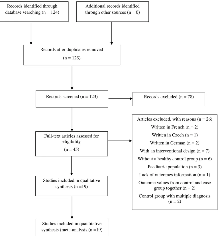
For comparative and quantitative purposes, 19 studies, from 2008 throughout 2016, were considered for analysis. This information is presented in Figure 1, according to Preferred Reporting Items for Systematic Reviews and Meta-Analyses (PRISMA) guidelines [11].
Data collection process and statistical analysis
The selected full texts were collected and assessed for demographic data and reported outcomes. For the statistical analysis, we used the mean difference (MD) between groups (patient - control) of CH and CCT, respectively.
9 RESULTS
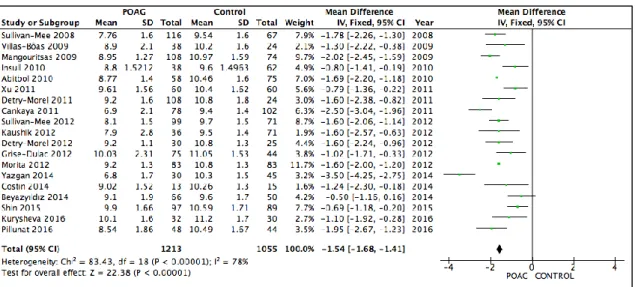
From a total of 124 studies screened, only 19 of them complied with our eligibility criteria, as shown in Figure 1. Table 1 and Figures 2-3 summarize the mean and standard deviation (SD) of both CH and CCT for the control and case groups of each study.
Synthesis of results
A total of 1213 glaucoma eyes, arising from 1159 glaucoma patients and 1055 healthy eyes from 1021 healthy subjects, were considered in our study. Table 1 shows the baseline characteristics of these participants and their eye-related parameters.
A quantitative analysis showed us that CH was significantly lower in the glaucoma group when compared to the control group (MD = -1.54 µm, 95% CI [-1.68, -1.41], P < 0.00001) as shown in Figure 2. As we can observe in Figure 3, CCT was also significantly lower in the glaucoma group when compared to healthy controls (MD = -8.49 µm, 95% CI [-11.36, -5.62], P < 0.001).
10 DISCUSSION
Summary of evidence, limitations and conclusions
The latest evidence is still unclear regarding the true value of CCT as a risk factor for glaucoma. While some studies stand up for CCT as an important risk factor for the development of glaucoma [4–6, 10], others like EMGT, Barbados Eye Study, Chauhan and Congdon, did not find such a simple and linear relationship between those two parameters [2, 4]. According to our study, there is a significantly lower CCT value among glaucoma patients (mean difference 8.49 µm, range [-11.36, -5.62], 95% CI; P = 0.0005) compared to the control group. However, it is hard to draw simple conclusions about the meaning of this difference for the two groups, since it is different to applanate a thinner or a thicker surface, becoming easier or harder to applanate the cornea, respectively [9]. So, in other words, CCT may result in a confounding factor for IOP measured by GAT rather than an independent risk factor for the disease.
The ORA device give us several biomechanical properties that are assumed to be less influenced by CCT when compared to GAT, namely CH, which is a biomechanical property related to the viscoelasticity of the cornea. According to our results, there is a significantly lower CH among glaucoma patients, compared to healthy controls (mean difference 1.54 mmHg, range [-1.68, -1.41], 95% CI; P < 0.00001), which is in agreement with previous results from other studies [2, 9]. Since ORA is a non-contact tonometer [2, 3, 5, 9], parameters measured by this device may be more reliable than GAT [2, 3].
From these study results, a relevant question that rises is about the applicability of CH as an instrument in clinical practice and its reliability. Standard CCT measurements have been widely used and may help interpret IOP findings, but up to this day it remains undetermined whether this variable per se is useful for assessing a patient’s risk factor on developing the disease. In this sense, by providing further information about the corneal biomechanics, CH may be different. However, there is still not a consolidated evidence that allow us to replace the use of CCT for other marker such as CH, in the management of glaucoma patients. The fact that CH is not theoretically influenced by CCT [3, 5, 8, 9], which shows a large variability in the overall population [5–7], is very important, so it can become a valuable tool, for example, on the assessment of the stratification risk for glaucoma patients or even for prognosis. Yet, ORA is an instrument that is not commonly found in ophthalmology clinics worldwide, which limits the knowledge of CH in glaucoma.
11
The results of our study show a strong evidence on the subject. Furthermore, it should be pointed out that this is the first study review of the literature and meta-analysis about this topic involving corneal hysteresis in glaucoma. However, we recognize that our study has some limitations. We highlighted the fact that we include several glaucoma diagnosis [POAG, normal tension glaucoma (NTG), pre-perimetric POAG, pseudoexfoliative glaucoma (PEXG) and exfoliative glaucoma (EXG)], which can also bias the results, once the different physiopathology of each type of glaucoma may have a different impact on the cornea. Additionally, our study only considered articles from 2008 through 2016, based on a strict criteria. Furthermore, the ORA device was only introduced in 2005. So, these two factors resulted in a relatively short period under review in our study. Finally, we also recognise some other limitations, namely, the fact that it is not a systematic review, that it has not a risk of bias evaluation and that we have only included observational studies with the purpose to eliminate the risk of bias from interventions in the groups.
Concluding, our study reveals a significant difference on CH and CCT between glaucoma patients and healthy controls. These results show that maybe the true assessment is beyond CCT measurement alone. So, it is important to keep searching for new and more sophisticated tools to measure corneal properties, as CH, to deepen our knowledge in this subject.
12 REFERENCES
1. Garway-Heath DF, Crabb DP, Bunce C, Lascaratos G, Amalfitano F, Anand N, et al. Latanoprost for open-angle glaucoma (UKGTS): A randomised, multicentre, placebo-controlled trial. The Lancet. 2015;385(9975):1295–304.
2. Deol M, Taylor DA, Radcliffe NM. Corneal hysteresis and its relevance to glaucoma. Current Opinion in Ophthalmology. 2015;26(2):96–102.
3. Yaoeda K, Fukushima A, Shirakashi M, Fukuchi T. Comparison of intraocular pressure adjusted by central corneal thickness or corneal biomechanical properties as measured in glaucomatous eyes using noncontact tonometers and the Goldmann applanation tonometer. Clinical Ophthalmology. 2016;10:829–34.
4. Iester M, Mete M, Figus M, Frezzotti P. Incorporating corneal pachymetry into the management of glaucoma. Journal of Cataract and Refractive Surgery. 2009;35(9):1623–28.
5. Brandt JD. Central corneal thickness, tonometry, and glaucoma risk—a guide for the perplexed. Canadian Journal of Ophthalmology. 2007;42(4): 562–6.
6. European Glaucoma Prevention Study Group. Central Corneal Thickness in the European Glaucoma Prevention Study. Ophthalmology. 2007;114(3):454–9. 7. Brandt JD, Beiser JA, Kass MA, Gordon MO; Ocular Hypertension Treatment
Study Group. Central corneal thickness in the Ocular Hypertension Treatment Study (OHTS). Ophthalmology. 2001;108(10):1779–88.
8. Hong Y, Shoji N, Morita T, Hirasawa K, Matsumura K, Kasahara M, et al. Comparison of corneal biomechanical properties in normal tension glaucoma patients with different visual field progression speed. International Journal of Ophthalmology. 2016;9(7):973–78.
9. Garcia-Porta N, Fernandes P, Queiros A, Salgado-Borges J, Parafita-Mato M, González-Méijome JM. Corneal Biomechanical Properties in Different Ocular Conditions and New Measurement Techniques. ISRN Ophthalmology. 2014;2014:1–19.
10. Brandt JD, Gordon MO, Beiser JA, Lin SC, Alexander MY, Kass MA; Ocular Hypertension Treatment Study Group. Changes in Central Corneal Thickness over Time. The Ocular Hypertension Treatment Study. Ophthalmology. 2008;115(9):1550–6.
13
11. Moher D, Liberati A, Tetzlaff J, Altman DG; The PRISMA Group. Preferred Reporting Items for Systematic Reviews and Meta-Analyses: The PRISMA Statement. Physical Therapy. 2009;89(9):873–80.
14 FIGURES
Full-text articles assessed for eligibility
(n = 45)
Records screened (n = 123) Records after duplicates removed
(n = 123)
Studies included in qualitative synthesis (n =19) Records identified through
database searching (n = 124)
Additional records identified through other sources (n = 0)
Articles excluded, with reasons (n = 26) Written in French (n = 2)
Written in Czech (n = 1) Written in German (n = 2) With an interventional design (n = 7) Without a healthy control group (n = 6)
Paediatric population (n = 3) Lack of outcomes information (n = 1) Outcome values from control and case
group together (n = 2)
Control group with multiple diagnosis (n = 2)
Records excluded (n = 78)
Studies included in quantitative synthesis (meta-analysis (n =19)
15
Figure 2: Corneal Hysteresis - Forest plot
CI = confidence interval. SD = Standard deviation.
Figure 3: Central Corneal Thickness - Forest plot
16 TABLES
Article Diagnosis No. of
patients No. of eyes
CH CCT Mean ± SD (mmHg) Mean ± SD (µm) Kurysheva, 2016 Glaucoma 32 32 10,1 ± 1,6 548,1 ± 31,3 Control 30 30 11,2 ± 1,7 549,3 ± 30,8 Pillunat, 2016 Glaucoma 48 48 8,54 ± 1,86 530,6 ± 38,4 Control 44 44 10,49 ± 1,67 556,2 ± 37 SHIN, 2015 Glaucoma 97 97 9,9 ± 1,66 548,3 ± 34,82 Control 89 89 10,59 ± 1,71 558,77 ± 31,19 Beyazyıldız, 2014 Glaucoma 66 66 9,1 ± 1,9 550,4 ± 36,3 Control 50 50 9,6 ± 1,7 537,3 ± 38,5 Yazgan, 2014 Glaucoma 30 30 6,8 ± 1,7 509 ± 36 Control 45 45 10,3 ± 1,5 546,3 ± 28 Costin, 2014 Glaucoma 13 13 9,02 ± 1,52 546,7 ± 35 Control 15 15 10,26 ± 1,3 546,1 ± 35,5 Insull, 2010 Glaucoma 38 38 8,8 ± 1,5212 532 ± 33,466 Control 62 62 9,6 ± 1,4963 550 ± 35,4397 Sullivan-Mee, 2012 Glaucoma 116 116 7,76 ± 1,6 541 ± 36 Control 67 67 9,54 ± 1,6 552 ± 35 Kaushik, 2012 Glaucoma 36 36 7,9 ± 2,8 523,5 ± 35,5 Control 71 71 9,5 ± 1,4 530,7 ± 33,4 Detry-Morel, 2012 Glaucoma 30 30 9,2 ± 1,1 544 ± 37 Control 25 25 10,8 ± 1,6 554 ± 19 Morita, 2012 Glaucoma 83 83 9,2 ± 1,3 535,4 ± 24,9 Control 83 83 10,8 ± 1,3 541,4 ± 26,8 Cankaya, 2011 Glaucoma 78 78 6,9 ± 2,1 537,9 ± 35,2 Control 102 102 9,4 ± 1,4 539,8 ± 25,9 Grise-Dulac, 2012 Glaucoma 38 75 10,03 ± 2,31 551,5 ± 38,9 Control 22 44 11,05 ± 1,53 550,7 ± 29,3 Detry-Morel, 2011 Glaucoma 108 108 9,2 ± 1,6 536 ± 61 Control 24 24 10,8 ± 1,8 550 ± 36 Xu, 2011 Glaucoma 60 60 9,61 ± 1,56 541,4 ± 37,46 Control 60 60 10,4 ± 1,62 541,75 ± 26,07 Abitbol, 2010 Glaucoma 58 58 8,77 ± 1,4 535,34 ± 42,7 Control 75 75 10,46 ± 1,6 560,2 ± 36,3 Villas-Bôas, 2009 Glaucoma 21 38 8.90 ± 2.1 514.80 ± 41.3 Control 12 24 10.20 ± 1.6 529.00 ± 45.4 Mangouritsas, 2009 Glaucoma 108 108 8,95 ± 1,27 526,77 ± 35,73 Control 74 74 10,97 ± 1,59 537,84 ± 41,93 Sullivan-Mee, 2008 Glaucoma 99 99 8,1 ± 1,5 541 ± 41 Control 71 71 9,7 ± 1,5 546 ± 33
Table 1: Baseline characteristics
CCT = central corneal thickness. CH = corneal hysteresis. No = number. SD = standard deviation