Arq Bras Oftalmol. 2010;73(4): 346-9 3 4 6
A
R T I G O SO
R I G I N A IS|
OR I G I N A L AR T I C L E SWork carried out at Glaucoma Service, Hospital São Geraldo, Universidade Federal de Minas Gerais - UFMG - Belo Horizonte (MG), Brazil.
1Physician, Ophthalmology Department, Faculdade de Medicina, Universidade Federal de
Minas Gerais - UFMG - Belo Horizonte (MG), Brazil.
2Undergraduate student, Faculdade de Medicina, Universidade Federal de Minas Gerais
-UFMG - Belo Horizonte (MG), Brazil; Scholar of Scientific Initiation (PIBIC - CNPq). None of the authors had proprietary interest in the development and marketing of any products mentioned in the article.
Correspondence address: Sebastião Cronemberger. Rua Martim de Carvalho, 410/ 501 - Belo Horizonte (MG) - CEP 30190-090 - E-mail: [email protected]. Recebido para publicação em 05.07.2009
Última versão recebida em 18.04.2010 Aprovação em 29.06.2010
Importance of intraocular pressure measurement at 6:00 a.m. in bed
and in darkness in suspected and glaucomatous patients
A importância da medida da pressão intraocular às 6 horas no leito
e no escuro em suspeitos e portadores de glaucoma
SEBASTIÃO CRONEMBERGER1,ANDRÉA CRISTIANE LOPESDA SILVA2,NASSIM CALIXTO1
INTRODUCTION
T
he current consensus states that the elevated intraocular pres-sure (IOP) is the main risk factor for the development and the progression of primary open-angle glaucoma (POAG)(1-4).Recently, it was demonstrated in the Advanced Glaucoma Inter-vention Study (AGIS) that long-term IOP fluctuation is associated with a progression of visual field loss in patients with low mean IOP but not in patients with high mean IOP(5).This fact has been well-ABSTRACT
Purpose: To assess the importance of intraocular pressure measurement obtai-ned at 6:00 a.m. in bed and darkness for the diagnosis and intraocular pressure control of primary open-angle glaucoma.
Methods: Retrospective analysis of the daily curve of intraocular pressure of suspects and glaucomatous patients under treatment. Suspects were classified as intraocular pressure values ranging from 19 to 24 mmHg in isolated measu-rements and/or cup/disc ratio ≥ 0.7 in one or both eyes and/or asymmetry of cup/disc ratio ≥ 0.3 and a normal visual field. Each daily curve of intraocular pressure comprised five to seven IOP measurements with Goldmann applana-tion tonometer at 9:00 a.m., 12:00 p.m., 3:00 and/or 6:00 and 10:00 p.m. and/or 12:00 a.m. and in the following day morning at 6:00 a.m. in supine position in bed and in darkness with Perkins tonometer before the patient had stood up. Only the daily curves of intraocular pressure that presented an intraocular pressure peak (difference between the higher and the lesser intraocular pressure value) >6 mmHg were analyzed. In these daily curves, the average intraocular pressure and the standard deviation were calculated. The average intraocular pressure and standard deviation values were compared with the normal superior limits: average + two standard deviation of average intraocular pressure and standard deviation of intraocular pressure daily curve from normal patients of the same age group. Daily curves were considered abnormal when their average intrao-cular pressure and standard deviation values were above the normal superior limits. Secondary and congenital glaucoma were excluded.
Results: Daily curves of intraocular pressure of 565 eyes were analyzed; 361 suspected eyes and 204 eyes with primary open-angle glaucoma. In suspects, 64.3% presented an intraocular pressure peak at 6:00 a.m. in bed. In primary open-angle glaucoma, 68.6% presented an intraocular pressure peak at 6:00 a.m. in bed. In 5.3% of the suspects and in 5.9% of primary open-angle glaucoma patients, the daily curve of intraocular pressure profile was inverted (lesser intrao-cular pressure at 6:00 a.m.).
Conclusion: Intraocular pressure peaks at 6:00 a.m. were responsible for the diagnosis of preperimetric glaucoma in 64.3% of suspects and revealed inadequate intraocular pressure control in 68.6% of eyes with primary open-angle glaucoma. The daily curve of intraocular pressure including the intraocular pressure measurement at 6:00 a.m. in bed and in darkness is of vital importan-ce in doubtful cases in order to confirm the diagnosis of preperimetric glaucoma in suspects as well as for the adequate intraocular pressure evaluation of antiglau-comatous treatment.
Keywords: Intraocular pressure/physiology;Ocular hypertension;Glaucoma, open-angle/diagnosis; Circadian rhythm/physiology; Tonometry, ocular; Moni-toring, physiologic
RESUMO
Objetivo: Avaliar a importância da medida da pressão intraocular (Po) às 6 h no leito e no escuro para o diagnóstico de glaucoma pré-perimétrico e o controle do tratamento do glaucoma primário de ângulo aberto (GPAA).
Métodos: Análise retrospectiva da curva diária de pressão intraocular (CDPo) de suspeitos e glaucomatosos com perda de campo visual em tratamento. Suspeitos: Po de 19 a 24 mmHg e/ou relação escavação/disco (E/D) ≥ 0,7 num olho ou nos dois olhos e/ou assimetria de E/D ≥ 0,3 e campo visual normal. Cada curva diária de pressão intraocular: cinco a sete medidas (tonômetro de Goldmann) feitas às 9:00, 12:00, 15:00 e/ou 18:00 e 22:00 e/ou 24:00 h e na manhã seguinte às 6:00 h (tonômetro de Perkins) com o paciente em decúbito dorsal e no escuro, antes de levantar-se. Analisadas apenas as curvas diária de pressão intraocular que apresentavam pico de Po (diferença entre o maior e o menor valor de Po) >6 mmHg. Nessas curvas diária de pressão intraocular, calculamos a pressão média (Pm) e a variabilidade (V) e as comparamos com os limites superiores da normalidade: média + dois desvios-padrões da Pm e da V obtidos no Serviço de pacientes normais do mesmo grupo etário. As curvas diária de pressão intraocular com Pm e/ou V acima dos limites superiores da normalidade foram consideradas anormais. Excluídas: CDPos de glaucomas secundário e congênito.
Resultados: Analisadas curvas diária de pressão intraocular de 565 olhos: 361 olhos de suspeitos e 204 de glaucomatosos. Picos de Po às 6:00 h foram encontrados em 64,3% dos suspeitos e em 68,6% dos glaucomatosos. Em 5,3% dos suspeitos e em 5,9% dos glaucomatosos, o perfil da CDPo foi invertido (menor valor da Po às 6:00 h).
Conclusão: Picos de Po às 6:00 h foram responsáveis pelo diagnóstico de glau-coma pré-perimétrico em 64,3% dos suspeitos e revelaram inadequado con-trole da Po em 68,6% dos olhos glaucomatosos. Em casos duvidosos, a medida da Po às 6:00 h no leito e no escuro com um tonômetro de aplanação é indis-pensável para confirmar o diagnóstico de glaucoma pré-perimétrico e para a adequada avaliação do tratamento antiglaucomatoso.
CRONEMBERGER S, SILVA ACL, ET AL.
Arq Bras Oftalmol. 2010;73(4): 346-9 3 4 7
known since Maklakov’s research at the beginning of the 20th
century using a Maklakov applanation tonometer(6).
It is widely recognized that the IOP varies throughout the twenty-four hours. Also, IOP reduction is the only goal in the treatment of glaucoma(1-5). Despite this, only a few
ophthalmo-logists carry out a rigorous investigation of IOP using the daily curve of intraocular pressure (DCPo) by taking the IOP measu-rement at 6:00 a.m. with the patient in a supine position in bed and in darkness before the patient had stood up(7-9).
For some authors, the DCPo with the IOP measurement taken with an applanation tonometer at 6:00 a.m. in a supine position in bed and darkness has great importance to esta-blish the diagnosis of suspected patients and the assessment of IOP control of glaucomatous patients(10-14).
However, other authors have studied the diurnal IOP varia-tions by diurnal curves (DC) with 4-6 IOP readings generally taken between 8 a.m. and 6:30 p.m. of the same day(13).
Thiel (1925) was the first to report, without the benefit of the Goldmann applanation tonometer (GAT), that the IOP is more elevated in the morning between 5:00 and 7:00 a.m. before the patient stands up(14). He reported that the nocturnal IOP
elevation is caused by the supine position that leads to cerebral and ocular venous stasis that reduces the aqueous outflow(14).
Some authors did the DCPo with the IOP measurement taken at 6:00 a.m. in a supine position but they used the TonoPen or the pneumotonometer to measure the IOP(10,15).
This paper evaluates the importance of measuring the IOP at 6:00 a.m. in supine position in bed and in darkness using an applanation tonometer before the patient has stood up in the management of suspected and glaucomatous patients.
METHODS
A retrospective analysis of the DCPo of suspects and glauco-matous patients was performed. Suspects presented an IOP, without medication, ranging from 19 to 24 mmHg in an isolated measurement and/or cup/disc ratio (C/D) equal to or higher than 0.7 in one or both eyes and/or an asymmetry of C/D equal to or higher than 0.3 and a normal visual field. In suspects, the DCPos were carried out for diagnostic purposes. In glaucomatous pa-tients, who presented a C/D ratio equal to or higher than 0.7 and a typical visual field loss, the DCPos were performed to assess the effectiveness of antiglaucomatous treatment in reducing the IOP. Glaucoma progression was not evaluated. All glau-comatous patients were using antiglauglau-comatous medications, such as prostaglandin analogs, β-blockers, α2-agonists or topic carbonic anhydrase inhibitors when admitted. Each DCPo con-sisted from five to seven IOP measurements performed with GAT at 9:00 a.m. and 12:00 p.m. and at 3:00 and/or 6:00 and 10:00 p.m. and/or 12:00 a.m. and in the morning of the follo-wing day at 6:00 a.m. with Perkins tonometer with the patient in a supine position in bed and in darkness and before he or she had stood up. IOP measurements were performed by two authors (SC and NC). Only the DCPos that presented an IOP peak (difference between the higher and the lesser IOP value)
superior to 6 mmHg (abnormal IOP variation(16)) were
consi-dered. In these DCPos, the average IOP (Pm) and the stan-dard deviation (variability - V) were calculated. Pm and V values were compared with the normal superior limits: average + two standard deviation of Pm and average + two standard deviation of V of the DCPo from normal patients of the same age group available in our service(12).The DCPos were
con-sidered abnormal when their values of Pm and/or V were above the normal superior limits (Table 1). The DCPos of non-POAG were excluded.
RESULTS
Three hundred and thirty-one patients were enrolled (21.2%), representing the DCPos of 565 eyes equaling 206 (62.2%) suspects (361 eyes) and 125 (37.8%) POAG cases (204 eyes). We excluded 1229 (78.8%) out of the 1560 DCPos analyzed because they did not fill the inclusion criteria. Among the suspects, 64.3% of the eyes presented abnormal Pm and/or V when compared to those of the DCPo from normal patients of the same age group(12).
This percentage of abnormal Pm and/or V value(s) was due to an abnormal IOP peak at 6:00 a.m. in supine position in bed and in darkness (Table 2). Therefore, based on the abnormal values of Pm and/or V and the normal result of the visual field, we definitively confirmed the diagnosis of preperimetric glaucoma in almost 2/3 of the suspects by measuring the IOP at 6:00 a.m. In glaucomatous patients, 68.6% of the eyes presented an abnormal Pm and/or V in the DCPo due to an abnormal IOP peak at 6:00 a.m. (Table 2). These data certainty revealed that these glaucomatous patients presented an inadequate IOP control. Moreover, in 5.3% of the suspects and in 5.9% of the glaucomatous, the DCPo profile revealed inverted values (lesser IOP value at 6:00 a.m.).
DISCUSSION
POAG is a challenging disease in which the elevated IOP is the primary risk factor. Taking into account that up to now the only treatment for that optic neuropathy is IOP reduction, it is clear that a rigorous IOP investigation should be mandatory.
Papers about DCPo have been published for many years(8-12).
However, the majority of authors have studied the variations of the DC with 4-6 IOP readings generally taken between 8 a.m. and 6:30 p.m. of the same day(13).Some authors have done the
DCPo including the IOP measurement at 6:00 a.m. in supine
position, but using the TonoPen or pneumotonometer(15).
To-noPen is an electronic tonometer and the pneumotonometer is an indentation tonometer and, therefore, neither are gold-standard instruments for IOP assessment because their reliabi-lity is variable(17-18).The OBF-pneumotonometer and the
Tono-Pen can register false values because they are more influenced by corneal thickness than the GAT(17-18). The pneumotonometer is
affected by ocular rigidity and the TonoPen tends to underes-timate Goldmann’s IOPs in patients with IOP equal to or greater than 30 mmHg.
Some authors, using the pneumotonometer, found that the nocturnal supine IOP (11:00 p.m. - 7:00 a.m.) was a little lower than the diurnal upright IOP (7:00 a.m. - 11:00 p.m.), resulting in the diagnosis of 24 untreated early glaucomatous patients(15).
In the present paper, in which only applanation tonometers (GAT and Perkins) were used, opposite results were obtained. Other authors analyzed 690 DC of the right eye out of a total of 2272 DC obtained from 1178 patients. They reported
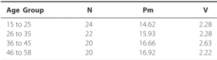
Table 1. Normal superior limits (average + two standard deviations) of the average IOP (Pm) and of the V of the DCPo in normal patients according to their age(12)
Age Group N Pm V
15 to 25 24 14.62 2.28
26 to 35 22 15.93 2.28
36 to 45 20 16.66 2.63
46 to 58 20 16.92 2.22
DCPo= diurnal curve of intraocular pressure; Pm= average intraocular pressure; V= standard deviation; N= number of eyes
Arq Bras Oftalmol. 2010;73(4): 346-9 3 4 8
IM P O R T A N C E O F I N T R A O C U L A R P R E S S U R E M E A S U R E M E N T A T 6:00 A.M. I N B E D A N D I N D A R K N E S S I N S U S P E C T E D A N D G L A U C O M A T O U S P A T I E N T S
that in 40% of the cases, the highest IOP was found at the earliest morning measurement with 65% of peaks occurring before noon(13). Again, our data were completely different.
There are more differences between the findings of the present paper and those of the works above(13,15).First, to the
best of our knowledge, this paper analyzed the greatest sam-ple of DCPos in which the IOP of suspected and glaucomatous eyes was measured at 6:00 a.m. with an applanation tonome-ter (Perkins) in bed and in darkness before the patient had stood up. Second, besides the important finding that the highest percentage of IOP peaks occurs at 6:00 a.m. in suspected (64.3%) and glaucomatous patients under inadequate treatment (65.8%), two other findings must be emphasized. Firstly, the relatively low percentage (23%) of IOP peaks at 9:00 a.m. and secondly, the even lower percentage of IOP peaks at the end of the morning (0.01% at 11:00 a.m.) and during normal office hours from 12:00 to 6:00 p.m. (8.8%). This is, precisely, the most common time period when most ophthalmologists perform the IOP measurements.
Undetected IOP peaks at 6:00 a.m. in bed and darkness can be responsible for the failure to substantiate an early diagnosis of preperimetric glaucoma. Also, some authors reported that IOP peaks can be responsible for glaucoma progression in some patients(8,19-20). In this paper, the glaucoma progression was not
evaluated.
To assure early glaucoma diagnosis and adequate IOP control in glaucoma patients, we always perform a DCPo with at least five IOP measurements including the IOP taken at 6:00 a.m. (at the hospital or home’s patient[H1]) using the Perkins tono-meter before the patient has stood up.
In literature, a few papers show similar results to those of this paper(8-9,14,19).In the past, one author reported that in 140
untreated glaucomatous eyes, 60% of the IOP peaks occurred at 6:00 a.m. in spite of the fact that the IOP measurement had been recorded with a certified Schiötz tonometer and a 5.5 gm. weight(8).It is not clear in that paper if the IOP at 6:00 a.m. was
measured in bed in a supine position. A Brazilian paper made a comparison between the DCPo and DC performed by seven residents about IOP peak detection and the verification of the influence of postural variation on 6:00 a.m. measurement(11). In
DC, they considered the IOP measurements at 9:00 a.m. - noon - 3:00 p.m. - 6:00 p.m. At 6:00 a.m., IOP measurements were performed in the dark, with patients in supine position, using Perkins tonometer. Then, another measurement was taken with GAT and these same patients were seated. Mean IOP and IOP
peaks (IOP ≥ 21 mmHg) in the DCPo and DC were compared,
as well as the time when the peaks occurred. The comparison showed that mean IOP was higher at 6:00 a.m. when taken in bed. The DC was unable to detect respectively 60.42% and 88.24% of IOP peaks in 68 glaucomatous and 57 suspected
eyes(11). Another paper also demonstrated that the 24-hour
IOP peak was higher than the peak noted during previous office visits in 40 (62%) out of 64 eyes(20). The IOP peak was
recorded outside of office hours in at least 1 eye in 22 (69%) out of 32 patients(20).Those authors suggested that in glaucoma
pa-tients with advanced disease or with progression that is dispro-portionate to known IOP measurements, 24-hour IOP monitoring can reveal higher peaks and wider fluctuation of IOP than those found during typical office hours, measured either in multiple office visits or repeatedly during a single day(20).
In one study of 16 patients with normal pressure glauco-ma suspicion, we found an abnorglauco-mal DCPo in 14 (87.5%) of them. Only two patients had normal DCPo. However, one patient presented severe circulation alterations of both inter-nal carotid arteries and the other presented pathological tono-graphic coefficients(21).
As demonstrated in one paper dating from 1925(14),along
with others(11,21), the data of this paper emphasize that is
impos-sible to establish the diagnosis of normal-tension glaucoma without a complete and adequate IOP evaluation with its mea-surement taken with an applanation tonometer (Perkins) at 6:00 a.m. in bed and in darkness before the patient stood up. A substantial variation in the IOP is common in these patients, truly characterizing an abnormality which was not registered(19).
Li-kewise, all epidemiological studies for evaluation of the diag-nosis and treatment of this disease that do not take into account the IOP variations are incomplete and, therefore, unable to demonstrate the true role of the IOP in suspected and glauco-matous patients.
In the most recent paper, the authors analyzed retrospec-tively the 24-hour IOP curves of 29 healthy subjects (10 young adults, 19 elderly) and 30 patients with untreated glaucoma(22).
IOP measurements were taken at 9 a.m.; 12, 3, 6, and9 p.m.;
and 12, 3, and 6 a.m., both in supine and sitting (GAT) posi-tions. During the night, IOP measurements were made with an electronic tonometer (Tonopen XL) when the patient was in supine position and with a GAT when the patient was seated in front of a slit lamp. Peak, mean, and fluctuation of 24-hour IOP curves were compared with office-hour measurements
obtainedin subjects in the sitting position alone and with
combinedpressures obtained in the sitting and supine positions (fourmeasurements in each body position from 9 a.m. to 6 p.m.).
The percentageof subjects with estimates of all IOP
parame-ters within a cutoffof ± 1 (peak and mean) and ± 2 mmHg
(fluctuation)was calculated.They reported that office-hour
sitting measurements correctly identifiedpeak, mean, and IOP
fluctuation in 10% of the young adults,32% of the elderly
control subjects, and 20% of the patientswith glaucoma,
whe-reas the combination of supine and sittingmeasurements
cor-rectly identified them in 30%, 85%, and 46%of the cases,
respectively. It is noteworthy that office-hour measurements did not characterize any 24-hour parameter in 20% of patients
with glaucoma.The authors recommended that supine and
sitting IOP data be collected during office hours, at least in patients with abnormal tonometric functional behavior (i.e., unexplained progression) to reduce their need for 24-hour monitoring. We are in total disagreement with these authors Table 2. IOP Peak time (∆∆∆∆∆OP>6 mmHg) of 331 DCPos (565 eyes)
Diagnosis IOP Peak 6:00 a.m. (bed) 9:00 a.m. 12:00 p.m. 3:00 p.m. 6:00 p.m. 10:00 p.m. 2:00 a.m. Total
N 232 99 16 4 5 1 4 361
Suspects 64.3% 27.4% 4.5% 1.1% 1.4% 0.3% 1.1% 100%
N 140 31 17 3 7 2 4 204
POAG 68.6% 15.2% 8.3% 1.5% 3.4% 1.0% 2.0% 100%
N 372 130 33 7 12 3 8 565
Total 65.8% 23.0% 5.9% 1.2% 2.1% 0.5% 1.4% 100%
CRONEMBERGER S, SILVA ACL, ET AL.
Arq Bras Oftalmol. 2010;73(4): 346-9 3 4 9 because: 1. The IOP evaluation in supine and sitting position
during office hours is less reliable and more time-consuming than the DCPo we have done including the IOP measurement taken at 6:00 a.m.; 2.Tonopen, used in that paper, can register false values because it is more influenced by corneal thickness than the GAT(18); 3. The change of patient’s position from supine
to sitting alters the behavior of the IOP in POAG and normal tension glaucoma patients, as well as in normal eyes. However, the raise in IOP neither is significant when compared among the 3 groups nor is consistent with visual field defects(23).
Unfortunately, most ophthalmologists manage their glaucomatous patients on the basis of sporadic IOP measure-ments that are taken during regular office hours, generally in the afternoon.
We think that is meaningless to follow suspected or glau-comatous patients under treatment performing the IOP mea-surements only in the afternoon.
CONCLUSION
On average, (65.8%) of the IOP peaks in suspects and glaucomatous patients with inadequate treatment were dis-covered by measuring the IOP at 6:00 a.m. in supine position in bed and in darkness before the patient stood up with an applanation tonometer. Also, at 6:00 a.m., on average, 5.6% of the eyes of suspects and glaucomatous patients with inade-quate IOP control presented the lesser IOP value.
In dubious cases, in order to correctly diagnose preperime-tric glaucoma in suspects as well as for the adequate evaluation of clinical treatment of POAG, the DCPo correctly performed is of vital importance.
REFERENCES
1. The Advanced Glaucoma Intervention Study (AGIS). 7. The relationship between control of intraocular pressure and visual field deterioration. The AGIS Investigators. Am J Ophthalmol. 2000;130(4):129-40. Comment in: Am J Ophthalmol. 2000; 130(4):490-1. 2. Kass MA, Heuer DK, Higginbotham EJ, Johnson CA, Keltner JL, Miller JP, et al. The
Ocular Hypertension Treatment Study: a randomized trial determines that topical ocular hypotensive medications delays or prevents the onset of primary open-angle glaucoma. Arch Ophthalmol. 2002;120(6):701-13; discussion 829-30. Comment in: Arch Ophthalmol. 2004;122(7):1088-9; author reply 1089. Arch Ophthlamol. 2003; 121(7):1070; author reply 1070.
3. Heijl A, Leske MC, Bengtsson B, Hyman L, Bengtsson B, Hussein M; Early Manifest Glaucoma Trial Group. Reduction of intraocular pressure and glaucoma progres-sion: results from the Early Manifest Glaucoma Trial. Arch Ophthalmol. 2002;120(10): 1268-79. Comment in: Arch Ophthalmol. 2002;120(10):1371-2. JAMA. 2002;288(20): 2607-8. Optom Vis Sci. 2002;79(12):741-2.
4. Bergea B, Bodin L, Svedbergh B. Impact of intraocular pressure regulation on visual fields in open-angle glaucoma. Ophthalmology. 1999;106(5):997-1005; discussion 1004-5. 5. Caprioli J, Coleman AL. Intraocular pressure fluctuation a risk factor for visual field
progression at low intraocular pressures in the advanced glaucoma intervention study. Ophthalmology. 2008;115(7):1123-9. Comment in: Ophthalmology. 2009; 116(4):817. 6. Maklakov A. L’ophtalmotonometrie. Arch Ophtalmol. 1885;5:159.
7. Musch DC, Lichter PR, Guire KE, Standardi CL. The Collaborative Initial Glaucoma Treatment Study: study design, methods, and baseline characteristics of enrolled patients. Ophthalmology. 1999;106(4):653-62.
8. Drance SM. The significance of the diurnal tension variations in the normal and glaucomatous eyes. Arch Ophthalmol. 1960;64:494-501.
9. Sampaolesi R, Calixto N, De Carvalho CA, Reca R. Diurnal variation of intraocular pressure in healthy, suspected, and glaucomatous eyes. Med Problem Ophthal-mol. 1968;74:1-23.
10. Hughes E, Spry P, Diamond J. 24-hour monitoring of intraocular pressure in glauco-ma glauco-management; a retrospective review. J Glaucoglauco-ma. 2003;12(3):232-6. 11. Rodrigues LD, Silva MR, Schellini AS, Jorge EN. Picos de pressão intraocular:
comparação entre curva tensional diária, minicurva e medida da pressão intraocu-lar às 6 horas. Arq Bras Oftalmol. 2004;67(1):127-31.
12. Calixto NS. Pressão intraocular, curva diária de pressão intraocular, rigidez parietal e coeficientes tonográficos (Médias de normalidade em diferentes grupos etários) [tese]. Belo Horizonte; Universidade Federal de Minas Gerais; 1967. p.51. 13 David R, Zangwill L, Briscoe D, Dagan M, Yagev R, Yassur Y. Diurnal intraocular
variations: an analysis of 690 diurnal curves. Br J Ophthalmol. 1992;76(5):280-3. 14 Thiel R. Die physiologischen und experimental erzeutgen Schankungen des
in-traocularen Druckes im gesundem und glaukomatosen. Auge Arch Augenheilk. 1925;96:331-54.
15. Liu JH, Zhang X, Kripke DF, Weinreb RN. Twenty-four-hour intraocular pattern associated with early glaucomatous changes. Invest Ophthalmol Vis Sci. 2003; 44(4):1586-90.
16. Duke-Elder S. The phasic variations in the ocular tension in primary glaucoma. Am J Ophthalmol. 1952;35(1):1-21.
17. Gupta V, Sony P, Agarwal HC, Sihota R, Sharma A. Inter-instrument agreement and influence of central corneal thickness on measurements with Goldmann, pneumo-tonometer and noncontact pneumo-tonometer in glaucomatous eyes. Indian J Ophthalmol. 2006;54(4):261-5.
18. Amaral WO, Teixeira RM, Alencar LM, Cronemberger S, Calixto N. [Central and peripheral corneal thickness: influence on the iop measurement by Tonopen]. Arq Bras Oftalmol. 2006;69(1):41-5. Portuguese.
19. Asrani S, Zeimer R, Wilensky J, Gieser D, Vitale S, Lindenmuth K. Large diurnal fluctuations in intraocular pressure are an independent risk factor in patients with glaucoma. J Glaucoma. 2000;9(2):134-42. Comment in: J Glaucoma. 9(6):487-8. 20. Barkana Y, Anis S, Liebmann J, Tello C, Ritch R. Clinical utility of intraocular pressure
monitoring outside of normal office hours in patients with glaucoma. Arch Ophthal-mol. 2006;124(6):793-7.
21. Calixto N, Meira DM, Cronemberger S. Estudo de pacientes com suspeita diagnós-tica de glaucoma de pressão normal. Rev Bras Oftalmol. 1997;56(11):823-35. 22. Fogagnolo P, Orzalest N, Ferreras A, Rossetti L. The circadian curve of intraocular
pressure: can we estimate its characteristics during office hours? Invest Ophthalmol Vis Sci. 2009;50(5):2209-15.