ANDREA RIPOL MALO
PROSPECTION OF BIOACTIVITIES, BIOACCESSIBILITY, AND BIOCHEMICAL CHARACTERIZATION OF GREEN SEAWEEDS GROWN IN INTEGRATED
MULTI-TROPHIC AQUACULTURE ENVIRONMENTS
A THESIS For the Degree of Master of Science in Marine Biology
Under the supervision of: Dr. João Varela, Centre of Marine Sciences (CCMAR), Marine Biotechnology research
group, University of Algarve Dr. Narcisa Bandarra, Division of Aquaculture and Upgrading (DivAV), Portuguese Institute for the Sea and Atmosphere (IPMA, IP)
ii Prospection of Bioactivities, Bioaccessibility, and Biochemical Characterization of Green Seaweeds Grown in Integrated Multi-Trophic Aquaculture Environments
Declaro ser a autora deste trabalho, que é original e inédito. Autores e trabalhos consultados estão devidamente citados no texto e constam da listagem de referências incluída.
(Andrea Ripol Malo)
Copyright , todos os direitos são reservados à autora Andrea Ripol Malo
A Universidade do Algarve tem o direito, perpétuo e sem limites geográficos, de arquivar e publicitar este trabalho através de exemplares impressos reproduzidos em papel ou de forma digital, ou por qualquer outro meio conhecido ou que venha a ser inventado, de o divulgar através de repositórios científicos e de admitir a sua cópia e distribuição com objetivos educacionais ou de investigação, não comerciais, desde que seja dado crédito ao autor e editor.
iii
Acknowledgments
Foremost, I would like to express my sincere gratitude to my advisor Dr. Carlos Cardoso for the continuous support, for his patience, motivation, enthusiasm, and immense knowledge. His guidance helped me in all the time of research and writing of this thesis, making of it an enjoyable journey; I could not have imagined having a better advisor and mentor for my Master Thesis. You are a very special person!
Besides my advisor, I would of course like to thank my two supervisors: Dr. Narcisa Bandarra and Prof. João Varela for the opportunity and trust they put on me. Their encouragement, insightful comments, and experience have made this work truly meaningful.
In addition, I would like to thank all the colleagues that contributed to the work: Claudia Afonso, Hugo Quental-Ferreira, Pedro Pousão-Ferreira and the colleagues of National Health Institute Doutor Ricardo Jorge.
I would also like to thank my parents for their wise counsel and constant support. You are always there for me.
Finally, there are my friends and boyfriend. We were not only able to support each other by deliberating over our problems and findings, but also by happily talking about things other than just our papers and work.
iv
I. Abstract
The nutritional composition of five species of green seaweeds (Rhizoclonium
riparium, Ulva lactuca, Ulva prolifera, Chaetomorpha linum, Ulva intestinalis) grown
in multi-trophic aquaculture systems were studied. Firstly, fucose and total polyphenols, as relevant bioactive constituents, were analyzed and antioxidant and anti-inflammatory activities were measured. The effects of bioaccessibility on these aspects were also assessed. Though lipid content was very low (less than 3 g/100 g dry weight), there were qualitative differences between lipid fractions, since fatty acid profiles varied considerably between the five seaweed species. The fucose content also depended on the particular species. Total polyphenol content and antioxidant activity presented a significant correlation. U. prolifera had the highest total polyphenol content and antioxidant activity, whereas no polyphenol or antioxidant activity was found in the bioaccessible fraction. The anti-inflammatory activity was highest in U. prolifera and C.
linum extracts with high COX-2 inhibition (ranging between 18 and 27 %) at a
concentration of 100 µg/mL. Despite the compounds causing this anti-inflammatory activity were not rendered bioaccessible, U. prolifera seems to be a potential source of bioactive substances, provided that adequate methods for their extraction are used or tisanes are developed that are able to enhance their bioaccessibility. Secondly, the lipid composition of the five species of green seaweeds was studied. In particular, the overall fatty acid (FA) profile and the FA profile of each main lipid class found in these seaweed species were thoroughly analysed. It was found that every seaweed had a specific FA profile, whose specificities were rendered more obvious with the study of the FA profile per lipid class. However, between U. lactuca and U. intestinalis, there were only minor differences. Nonetheless, it was possible to identify significant differences between the palmitic acid content in the PL class of each seaweed. A clear distinction between the FA profiles of R. riparium and C. linum (Cladophorales) and those of Ulva (Ulvales) was also determined. Moreover, there were also differences among lipid classes, yielding large contrasts between PL and TAG as well as between MAG and FFA. This study also found evidence supporting the location of particular FA in specific TAG positions. Finally, the mineral composition was studied. The elemental bioaccessibility in these species was also investigated through the application of an innovative in vitro digestive model. It was observed that R. riparium had the highest levels of Mn, Sr, Cd, Sn, and I and that U. lactuca had the highest Ni and Cu concentrations. The daily amounts of dried green seaweed required for achieving specific dietary intakes were calculated: 7 g of dried U. lactuca (for meeting Cu Recommended Daily Allowance, RDA); 173 g of dried U. lactuca (Zn RDA); 78 g of dried C. linum (Se RDA); 41 g of dried C. linum (Mo RDA); and 0.5 g of dried R.
riparium (I Dietary Reference Intake, DRI). Mn and Cu had the highest values of
elemental bioaccessibility, always above 50 %, whereas I was always poorly bioaccessible, in the range of 14-31 %. The bioaccessibility range of R. riparium (31-100 %) was higher than the ranges for other species, particularly that of C. linum (≤ 56 %). The bioaccessibility results entailed higher quantities of dried seaweed for reaching dietary intakes: 10 g of dried U. lactuca (Cu RDA); 290 g of dried R. riparium (Zn RDA); and 2 g of dried R. riparium (I DRI). Accordingly, R. riparium is a very rich I source. This study showed the importance of taking into account bioaccessibility results in estimating dietary intakes.
Keywords: Green seaweed; IMTA; antioxidant activity; anti-inflammatory activity;
v
II. Resumo
A composição nutricional de cinco espécies de algas verdes (Rhizoclonium
riparium, Ulva lactuca, Ulva prolifera, Chaetomorpha linum, Ulva intestinalis) em
sistemas de aquicultura multitróficos foi estudada. Em primeiro lugar, a fucose e os polifenóis totais, como constituintes bioativos relevantes, foram analisados e as atividades antioxidantes e anti-inflamatórias foram medidas. Os efeitos da bioacessibilidade nesses aspectos também foram avaliados. Embora o teor de lipídios tenha sido muito baixo (menos de 3 g / 100 g de peso seco), houve diferenças qualitativas entre as frações lipídicas, uma vez que os perfis de ácidos graxos variaram consideravelmente entre as cinco espécies de algas marinhas. O teor de fucose também dependeu das espécies específicas. O conteúdo total de polifenóis e a atividade antioxidante apresentaram correlação significativa. U. prolifera apresentou o maior teor total de polifenóis e atividade antioxidante, enquanto que nenhuma atividade de polifenol ou antioxidante foi encontrada na fração bioaccessível. A atividade antiinflamatória foi maior nos extratos U. prolifera e C. linum com alta inibição de COX-2 (variando entre 18 e 27%) a uma concentração de 100 μg / mL. Apesar dos compostos que causam essa atividade antiinflamatória não serem tornados bioaccessíveis, a U. prolifera parece ser uma fonte potencial de substâncias bioativas, desde que sejam utilizados métodos adequados para sua extração ou desenvolvidos desenvolvendo tisanas que possam aumentar sua bioacessibilidade. Em segundo lugar, estudou-se a composição lipídica das cinco espécies de algas verdes. Em particular, o perfil geral de ácidos graxos (FA) e o perfil FA de cada classe principal de lipídeos encontrados nestas espécies de algas marinhas foram cuidadosamente analisados. Verificou-se que todas as algas tinham um perfil FA específico, cujas especificidades foram mais evidentes com o estudo do perfil FA por classe de lipídios. No entanto, entre
U. lactuca e U. intestinalis, houve apenas pequenas diferenças. No entanto, foi possível
identificar diferenças significativas entre o teor de ácido palmítico na classe PL de cada alga. Uma clara distinção entre os perfis de R. riparium e C. linum (Cladophorales) e os de Ulva (Ulvales) também foi determinada. Além disso, houve diferenças entre as classes de lipídios, produzindo grandes contrastes entre PLs e TAG, bem como entre MAGs e FFA. Este estudo também encontrou evidências que suportam a localização de FA específicas em posições TAG específicas. Finalmente, a composição mineral foi estudada. A bioacessibilidade elementar nessas espécies também foi investigada através da aplicação de um modelo digestivo in vitro inovador. Observou-se que R. riparium apresentou os níveis mais altos de Mn, Sr, Cd, Sn e I e que a U. lactuca apresentou as maiores concentrações de Ni e Cu. As quantidades diárias de algas verdes secas necessárias para a obtenção de ingestão dietética específica foram calculadas: 7 g de U.
lactuca secas (para a reunião Cu recomendado diariamente, RDA); 173 g de U. lactuca
seca (Zn RDA); 78 g de C. linum seca (Se RDA); 41 g de C. linum seca (Mo RDA); e 0,5 g de R. riparium seca (I Dietary Reference Intake, DRI). Mn e Cu tiveram os valores mais elevados de bioacessibilidade elementar, sempre acima de 50%, enquanto que sempre fui praticamente bioacessível, na faixa de 14-31%. A faixa de bioacessibilidade de R. riparium (31-100%) foi maior do que as faixas para outras espécies, particularmente a de C. linum (≤ 56%). Os resultados de bioacessibilidade implicaram maiores quantidades de algas secas para atingir a ingestão dietética: 10 g de U. lactuca
vi seca (Cu RDA); 290 g de R. riparium seca (Zn RDA); e 2 g de R. riparium seca (I DRI). Consequentemente, R. riparium é uma fonte muito rica. Este estudo mostrou a importância de levar em consideração resultados de bioacessibilidade na estimativa de ingestão dietética.
Palavras-chave: Algas verdes; IMTA; atividade antioxidante; atividade
anti-inflamatória; classes de lipídios; composição de ácidos graxos; composição mineral; bioacessibilidade.
vii
III. Table of contents
1. Introduction ... 1
1.1 Integrated multi-trophic aquaculture (IMTA) ... 1
1.2 Seaweeds properties ... 3
1.2.1 Antioxidants ... 3
1.2.2 Lipids and fatty acids ... 4
1.2.3 Polysaccharides ... 5 1.2.4 Essential elements ... 6 1.2.5 Toxic elements ... 7 1.3 Chaetomorpha linum ... 8 1.4 Rhizoclonium riparium ... 9 1.5 Ulva intestinalis ... 10 1.6 Ulva lactuca ... 12 1.7 Ulva prolifera ... 12 1.8 Digestive process ... 14 1.8.1 Human digestion ... 14
1.8.2 Bioaccessibility and bioavailability of nutrients and contaminants ... 15
1.8.3 Evaluation of the bioaccessibility in vitro digestion model ... 16
2. Objective ... 17
3. Materials and Methods ... 17
3.1 Cultivation conditions ... 17
3.2 Samples ... 18
3.3 Proximate composition ... 18
3.4 Lipid extraction ... 18
3.5 Lipid class analysis ... 19
3.6 Lipid classes separation for fatty acid analysis ... 20
3.7 Fatty acid profile ... 21
3.8 Fucose content ... 22
3.9 Total polyphenol content ... 23
3.10 Antioxidant activity ... 24
3.11 Anti-inflammatory activity ... 25
3.11.1 Extract preparation for in vitro anti-inflammatory activity ... 25
3.11.2 Cyclooxygenase (COX-2) inhibition assay ... 25
viii
3.12.1 Calculation of bioaccessibility... 27
3.13 Mineral composition analysis ... 28
3.14 Statistical analysis ... 28
4. Composition, Biological Activity, and Bioaccessibility of Green Seaweeds from an Integrated Multi-Trophic Aquaculture System ... 30
4.1 Introduction ... 31
4.2 Results and Discussion ... 32
4.2.1 Seaweed proximate composition ... 32
4.2.2 Seaweed fucose content ... 33
4.2.3 Seaweed total polyphenol content and antioxidant activity ... 34
4.2.4 Seaweed anti-inflammatory activity ... 36
4.3 Conclusions ... 38
5. Fatty Acid Profiles of the Main Lipid Classes of Green Seaweeds from Fish Pond Aquaculture ... 39
5.1 Introduction ... 40
5.2 Results and Discussion ... 41
5.2.1 Seaweed fatty acid profile ... 41
5.2.2 Seaweed lipid class distribution ... 43
5.2.3 Fatty acid profile of main lipid classes ... 44
5.3 Conclusions ... 48
6. Mineral Composition and Bioaccessibility of Green Seaweeds from Fish Pond Aquaculture ... 49
6.1 Introduction ... 50
6.2 Results and Discussion ... 51
6.2.1 Elemental composition ... 51 6.2.2 Elemental bioaccessibility ... 55 6.3 Conclusions ... 59 7. General conclusion ... 60 8. Future perspectives ... 61 9. References ... 62 10. Annexes ... 77
ix
IV. List of Figures
Figure 1: Conceptual diagram of an Integrated Multi-Trophic Aquaculture (IMTA) (Chopin, 2013). ... 2 Figure 2: Chaetomorpha linum in Mar Menor, Murcia, Spain. 11 Jul 2011. Isabel Rubio Perez (Guiry & Guiry 2017). ... 9 Figure 3: Sample from a floating mat of Rhizoclonium riparium from Aquaculture Research Station, Olhão (EPPO), Portuguese Institute for the Sea and Atmosphere (IPMA, IP). ... 10 Figure 4: Detail of Rhizoclonium riparium from EPPO (Olhão) in 2015. ... 10 Figure 5: Studied sample of Ulva intestinalis from Aquaculture Research Station, Olhão (EPPO), Portuguese Institute for the Sea and Atmosphere (IPMA, IP). ... 11 Figure 6: Ulva intestinalis floating before sampling at EPPO (Olhão). Under the surface we can discern large quantities of Chaetomorpha linum. ... 11 Figure 7: Ulva lactuca in Zeeland delta, Netherlands. 26 Mar 2011. Mat Vestjens & Anne Frijsinger (Guiry & Guiry 2017)... 12 Figure 8: Ulva prolifera grown on a rock from the Aquaculture Research Station in Olhão. ... 13 Figure 9: Studied sample of Ulva prolifera from the Aquaculture Research Station, Olhão (EPPO), Portuguese Institute for the Sea and Atmosphere (IPMA, IP). ... 13 Figure 10: Sample after centrifugation. Three phases can be differentiated: water, seaweed fibers and organic phase containing the lipids. ... 19 Figure 11: Organic phase containing lipids after filtration... 19 Figure 12: Distribution of the lipid classes through analytical TLC for the five seaweeds: a) before spraying with 10 % phosphomolybdic acid in ethanol and b) when imaged at a digital scanner. ... 20 Figure 13: Preparative TLC for Chaetomorpha linum (a) after hexane:diethyl ether:acetic acid solution (b) in UV chamber after dichlorofluorescein (c) with lipid fractions identified using sigma standards (TAG - glyceryltrioleate, DAG - glyceryl
1,3-x dipalmitate, MAG - Dmonoolein, FFA - oleic acid, and PL -
L--phosphatidylcholine). ... 21
Figure 14: Differentiation of phases during acid-catalyzed transesterification ... 22
Figure 15: Final result of the polyphenol extraction ... 23
Figure 16: Sample solutions (a) before and (b) after adding DPPH. ... 24
Figure 17: Spectrophotometer with plate for prostanoid reading ... 26
Figure Figure 18: Comparison between in vivo and in vitro digestion model applied .. 26
Figure 19 – Bioaccessibility (%) of elements in the five studied green seaweed species (Rr - Rhizoclonium riparium, Ul - Ulva lactuca, Up - Ulva prolifera, Chl - Chaetomorpha linum, Ui - Ulva intestinalis). Different letters within a series regarding a specific element correspond to statistical differences (p<0.05). ... 56
xi
V. List of Tables
Table 1 - Proximate crude composition (g/100 g wet weight and for ash, protein, fat, and carbohydrate also g/100 g dry weight) in the five studied green seaweed species .. 33 Table 2 – Fucose content (mg/g dry weight) in the five studied green seaweed species before (initial) and after digestion (bioaccessible) and fucose bioaccessibility (%). ... 34 Table 3 – Total polyphenol content (mg GAE/g dry weight) and antioxidant activity (% inhibition) in the five studied green seaweed species before (initial) and after digestion (bioaccessible). ... 34 Table 4– Anti-inflammatory activity (% inhibition of COX-2) in the five studied green seaweed species before (initial) and after digestion (bioaccessible). ... 36 Table 5 – Overall fatty acid profile (%) in the five studied green seaweed species. ... 42 Table 6 – Lipid class distribution (%) as determined by TLC of the five studied green seaweed species. ... 43 Table 7 – Phospholipid+glycolipid and triacylglycerol fatty acid profile (% of total fatty acids) in the five studied green seaweed species. ... 77 Table 8– Monoacylglycerol and free fatty acid profile (% of total fatty acids) in the five studied green seaweed species. ... 78 Table 9 - Elemental composition (mg/kg dry weight) of the five studied green seaweed species. ... 52 Table 10 – Bioaccessible elemental contents (mg/kg; calculated taking into account the mass of sample input in the in vitro digestion and subtracting blank interference) of the five studied green seaweed species. ... 55
1
1. Introduction
1.1 Integrated multi-trophic aquaculture (IMTA)
The aquaculture production has grown dramatically worldwide. Over the past three decades, aquaculture production has increased from 6.2 million t in 1983 to 70.2 million t in 2013 (FAO, 2015). In 2013, aquaculture surpassed the supplies from the capture fisheries and contributed nearly 51% to the global fish production. This growth in marine aquaculture industry has introduced many concerns about the environmental impacts of aquaculture waste on the ecosystems (Mente et al., 2006; Reid et al., 2008; Tacon & Forster, 2014). Intensive fish farming can release significant quantities of nutrients to the vicinity of the farm site in the form of uneaten feed, feces and other excretory products. These metabolic wastes from farm effluents, mostly ammonia, may contribute to an increment of nutrients and, consequently, eutrophication in the farm. One of the major challenges for the sustainable development of aquaculture industry is to minimize environmental degradation concurrently with its expansion.
In many monoculture farming systems, the fed aquaculture species (e.g. carnivorous fish, shrimps) and the organic/inorganic extractive aquaculture species (e.g. bivalves, herbivorous fishes and aquatic plants) are independently farmed in different geographical locations, resulting in a pronounced shift in the environmental processes (Sasikumar & Viji, 2006). Integrated multi-trophic aquaculture (IMTA) is a practice in which the by-products (wastes) from one species are recycled by becoming inputs (fertilizers, food and energy) for another species (Fig. 1). IMTA involves cultivating in the appropriate proportions, fed aquaculture species (e.g. finfish/shrimps) with organic extractive aquaculture species (e.g. suspension feeders/deposit feeders/herbivorous fish) and inorganic extractive aquaculture species (e.g. seaweeds) for a balanced ecosystem management approach that takes into consideration site specificity, operational limits, and food safety guidelines and regulations. The goals are to achieve environmental sustainability through biomitigation, economic stability through product diversification and risk reduction, and social acceptability through better management practices (Barrington et al., 2009).
2 Figure 1: Conceptual diagram of an Integrated Multi-Trophic Aquaculture (IMTA) where the organic extractive aquaculture species (e.g., shellfish) take advantage of the enrichment in small particulate organic matter (POM) coming from the excretion products of fed aquaculture (e.g., finfish); inorganic extractive aquaculture species (e.g., seaweeds) take advantage of the enrichment in dissolved inorganic nutrients (DIN). Deposit organic extractive aquaculture species (e.g., echinoids, holothuroids, and polychaetes), take advantage of the enrichment in large particulate organic matter (POM) and feces and pseudo-feces (F&PF) from suspension-feeding organisms. The bioturbation on the bottom also regenerates some DIN, which becomes available to the seaweeds (Chopin, 2013).
The appropriate selection and proportions of the different species will provide different ecosystem functions and it is crucial for the correct balance of the biological and chemical processes in an IMTA. However, the co-cultured species should be more than just biofilters; they should also be harvestable crops of commercial value (Barrington et al., 2009). Seaweed aquaculture is a rapidly growing component of marine aquaculture, with about 0.17% of all named marine seaweed having been cultured to date and a growth rate of global marine seaweed production at 7.5% per year. In parallel, the range of sectors demanding products of seaweed farming has widened, from an initial focus to direct food supply to humans, to include bio-energy, cosmetics, biomedical applications, and formulation of feeds for aquaculture animals (Mazarrasa et al., 2014). Marine macroalgae are considered to be important reservoirs of several bioactive compounds that display important biological activities, which may be relevant for the improvement of human health, including antioxidant, anti-inflammatory and antitumor activities (Custódio et al., 2016). Several authors have referred to the nutritional high value of several seaweeds and suggest that they may be
3 included as a food ingredient. For example, Phaeophyta seaweeds can be a good source of biological active compounds, such as antioxidants, fatty acids, polysaccharides, vitamins (A, B12, C, D and E) and minerals (iron, Fe, magnesium, Mg, iodine, I, calcium, Ca) among others (Cian et al., 2014a).
In the Far East and Pacific, there has been a long tradition of consuming seaweeds as sea vegetables, while in Western countries, commercial applications of seaweed products have been restricted to the manufacture of gelling agents, stabilizers and food additives, pharmaceuticals and fertilizers (Burtin, 2003). However, with the increased awareness for the need of a healthy diet, the interest in seaweeds for food consumption is on the rise in Western countries. Depending on the concentration of metals in the environment and the bioavailability ratio from the seaweed, macroalgae can accumulate metals at levels several thousand times higher than those found in the surrounding seawater. Although seaweeds are a source of essential minerals for humans, they also might present a risk for human health because they are accumulators of non-essential elements, some of them widely recognized for their high toxicity, such as arsenic (Kim, 2015). Bioaccesibility will be nevertheless the final determining factor affecting bioavailability and human health. In order to exert their beneficial or harmful effects, nutrient content in seaweeds need firstly to be released from the food matrix and secondly to be absorbed at intestinal level (Moreda-Piñeiro et al., 2012).
1.2 Seaweeds properties
1.2.1 Antioxidants
Antioxidant molecules are important to the living organisms since they act against reactive oxygen species (ROS) like hydrogen peroxide (H2O2), nitric oxide
(NO), superoxide anion (O2-) and hydroxyl radical (OH.). These ROS are produced
during the cellular metabolism with harmful effects, since they are highly reactive and tend to initiate chain reactions that promote irreversible damage to proteins, lipids, and DNA (Balboa et al., 2013; Hamed et al., 2015).
Several seaweeds species can be found belonging to three different groups on the basis of their color: Chlorophyta (green), Rhodophyta (red) and Phaeophyta (brown). The color in green seaweeds is due to the presence of chlorophyll a and b in the same proportions as in ‘higher’ plants. Phycoerythrin and phycocyanin are responsible for the color of red seaweeds by masking the pigments such as chlorophyll
4
a. The brown coloration is due to the presence of xanthophylls such as fucoxanthin that
will mask, like in red algae, the chlorophylls present in the seaweed (Gupta and Abu-Ghannam, 2011).
Brown and red seaweeds are considered rich sources of antioxidants, including pigments such as fucoxanthin and polyphenols such as phlorotannins (Chakraborty et al., 2013). However, in a study with Sargassum siliquastrum, a brown seaweed, the total phenolic content did not correlate with antioxidant activity. These inconsistent results imply that not only the total phenolic content, but also other constituents, such as chlorophyll and carotenoids, may affect the antioxidant activity of extracts from marine algae. Despite extensive research on the antioxidant potential of extracts from various types of marine algae, few studies have been performed on the antioxidant compounds originated from green seaweeds, which are ubiquitous, easily cultivated and important natural resources (Cho et al., 2011). The antioxidants levels present in algae can be affected by a number of parameters such as location, salinity, sun exposure, season, and seaweed age (Holdt and Kraan, 2011).
1.2.2 Lipids and fatty acids
The total lipid (TL) contents in seaweeds is, in general, in the range of 1-6% dry weight (dw), varying in green seaweeds according to its phylogeny (Fleurence and Levine, 2016). The major lipids found in seaweeds are glycoglycerolipids, representing more than half of the total lipid content in some species, followed by phospholipids, triacylglycerols, sterols and pigments (Holdt and Kraan, 2011; Miyashita et al., 2013).
Glycoglycerolipids are very important to seaweeds and high plants since they collaborate in the photosynthesis and serve as markers for cellular recognition because of their association with cell membranes (Holdt and Kraan, 2011; Miyashita et al., 2013). Glycoglycerolipids are abundant in chloroplasts whose composition is rarely rich in highly unsaturated fatty acids. On the contrary, chloroplasts are rich in polyunsaturated fatty acids (PUFA). Polyunsaturated fatty acids (PUFA) are essential nutrients which cannot, or only to a limited extent, be synthesized by mammals. Therefore, they must be ingested via dietary sources. The two main PUFA groups are omega-3 (n-3) and omega-6 (n-6). The n-3 PUFA are provided by fish and microalgae, whereas the n-6 PUFA are ingested mainly via vegetable oil.
5 It is important to maintain an appropriate balance of n-3 and n-6 in the diet as these Fatty Acids (FA) work together to promote health: some n-3 PUFA, especially DHA, are major components of brain cells and crucial for proper development and functioning of the brain and the nervous system. Besides this, n-3 FA, particularly EPA, have been recognized to exhibit anti-inflammatory and antioxidant activity. Until now the major source of n-3 long-chain PUFAs, EPA and DHA, is fish oil. However, it is noteworthy that the original source of these long-chain PUFA is not the fish itself, but marine algae and phytoplankton which form their major dietary source (van Ginneken et al., 2011). The most common PUFA found in green seaweeds is alpha linolenic acid (ω3 C18:3), followed by EPA and DHA (Burtin, 2003).
1.2.3 Polysaccharides
Seaweeds contain large amounts of cell wall structural polysaccharides and storage polysaccharides (Davis, Volesky and Mucci, 2003; Holdt and Kraan, 2011). Polysaccharides are polymers of simple sugars (monosaccharides) linked together by glycosidic bonds, and have numerous commercial applications as food/feed additives (Holdt and Kraan, 2011). The total polysaccharide concentrations in the seaweed species range from 4% to 76% of dry weight, which makes them of major interest to the industry (Holdt and Kraan, 2011).
Polysaccharides are classified according to their biological function in two groups: energy storage and structural polysaccharides (Stadnik & Freitas, 2014). The principal cell wall polysaccharides in green seaweeds are ulvans, representing 8 to 29% of the algal dry weight (Vera at al., 2011). Because of its texturizing properties (gelling, thickening) and chemical specificities (presence of sulphate groups and rare sugars), ulvan offers numerous opportunities for applications in different industrial sectors: agri-food, cosmetics, pharmaceuticals, agriculture, chemicals industry, etc (Lahaye & Robic, 2007; Yanagisawa, Kawai, & Murata, 2013).
Sulfated fucans are one of the most well-studied sulfated polysaccharide and contain the monomer fucose, being present in all brown algae. Nevertheless, these polysaccharides may occur in minor amounts in green algae (Chlorophyta), red algae (Rhodophyta), and golden algae (Xanthophyta) (Mao, Zang, Li, & Zhang, 2006; Pomin & Mourão, 2008). One of the reasons why fucoidan has been so intensively studied is the numerous biological properties with potential human health applications that have
6 been shown such as: antitumor, anticoagulant, antioxidant activities, antiviral, antithrombotic, in addition to the impact on the inflammatory and immune systems (Davis et al., 2003; Holdt and Kraan, 2011; Hamed et al., 2015).
1.2.4 Essential elements
The mineral content of seaweeds is usually high enough (8–40% w/dw) to fulfill the recommended daily intakes of essential macroelements and trace elements for human nutrition (Kumar et al., 2011). This elemental content includes both macro- and trace elements such as magnesium, calcium, iron, copper, sodium, potassium, zinc, manganese, cobalt and especially iodine that are usually present in higher concentrations in seaweeds than in higher plants (Mabeau and Fleurence, 1993; Mohamed at al., 2012). Seaweeds are high in minerals due to their marine habitat, and the diversity of the minerals they absorb is wide. Important minerals, such as Ca, accumulate in seaweeds at much higher levels than in terrestrial foodstuffs. This is illustrated in an 8 g portion of Ulva lactuca, which provides 260 mg of Ca, equaling approximately 37% of the RDA of Ca for an adult male (Macartain et al., 2007). Nevertheless, mineral composition deeply varies according to the phylum, season, environment, geography, and physiology (El-Said & El-Sikaily, 2013; Kumar et al., 2011; Moreda-Piñeiro et al., 2012).
Having a diet rich in minerals is really important for human health, since they can interfere and/or be a part of key pathways necessary for the well-functioning of the human body. For example, Mg that is present in high quantities in seaweeds, works as co-factor for DNA and protein synthesis, oxidative phosphorylation, neuro-muscular excitability, enzyme activity and regulation of parathyroid hormone secretion (Saris et al., 2000; Romani, 2011). Mg deficiency or hypomagnesaemia is fairly common, with an estimated prevalence in the general population ranging from 2.5 to 15%, with higher rating, up to 65%, in patients in intensive care settings (Ayuk and Gittoes, 2014).
Iodine is also present in high quantities in seaweeds, and it essential for the production of the thyroid hormones thyroxine and triiodothyronine, which regulate many important physiological processes in humans. An iodine deficiency can cause several problems including an effect on growth and development due to insufficient formation of the thyroid hormones leading to spontaneous abortion, stillbirth, cretinism, goiter, and mental defects (Andersson at al., 2012; Hamed et al., 2015).
7 Trace elements, such as Zn, are present in seaweeds and some of these, such as arsenic, have negative health effects (Macartain et al., 2007). Severe Zn deficiency causes thymic atrophy, severe depression of immunity and acrodermatitis. Growth failure, delayed puberty, pregnancy complications, teratology, poor healing and decreased immunity occur in less severe Zn deficiency (Sahdstead & Smith, 1996). In the case of As, further analysis of speciation indicates that the type of As is important in assessing toxicity; since many types of As are not metabolized, these do not pose a risk to health. For the vast majority of seaweeds, the levels of heavy metals are below food safety limits naturally (Macartain et al., 2007).
Copper is an essential micronutrient that forms part of several proteins involved in a variety of biological processes indispensable to sustain life (Olivares et al., 2011). Minerals such as Cu and Fe are present in seaweeds at higher levels than in many well-known terrestrial sources of minerals, like meats and spinach. Therefore, regular seaweed consumption can help regulate Cu and other minerals dietary requirements (Macartain et al., 2007).
1.2.5 Toxic elements
Because of increasing levels of pollution in the oceans, seaweeds also tend to bioaccumulate contaminants like lead, cadmium, mercury, and arsenic, which makes them good environmental biomonitors (Reis et al., 2014). The toxicity of these elements is related to reactions that end up in electron transfer, formation of oxygen free radicals that can damage DNA, leading to increased mutagenicity, teratogenicity, genotoxicity, and carcinogenicity (Afonso, 2009).
Cadmium pollution may result from mining operations, metallurgical industries, corrosion of galvanized zinc batteries and the use of fertilizers (Afonso, 2009). In the human body, Cd competes with Zn, Cu and Fe which might lead to inhibiting their absorption. Cadmium can also replace Ca leading to bone changes that can provoke bone pain, osteoporosis, and osteomalacia (Del Ramo at al., 1993; Goyer, 1997).
Contamination of the environment by Pb is due to industrialization and the use of petrol (leaded) as fuel, as well as, the use of lead in cooking utensils, in pipes, paints, in pottery glazed with lead or industrial emissions, among others (Goyer and Clarkson, 1996). Nevertheless, the replacement of leaded petrol by unleaded fuel resulted in a decrease of the Pb contamination levels (Afonso, 2009). In individuals subjected to
8 occupational exposure, the most common effects are observed on the hematopoietic system and central nervous system which lead to encephalopathies. In extreme cases, Pb intoxication can lead to coma and death (Del Ramo at al., 1993; Goyer and Clarkson, 2001).
Arsenic is a ubiquitous element in the environment and can be derived from natural or anthropogenic sources. Anthropogenic causes comprise the release of As from primary Cu, Zn, and lead smelters, glass manufacturers that add As to the raw materials and chemical manufactures (Goyer and Clarkson, 2001). Arsenic is most toxic in its inorganic form as As(III) and As(V) but their methylated metabolites, monomethylarsonic acid and dimethylarsinic acid, are less toxic, while other major organoarsenicals found in seafood, arsenobetaine, trimethylarsine oxide, and arsenocholine have low or negligible toxicity (Moreda-Piñeiro et al., 2011). Chronic exposure to inorganic As compounds may lead to neurotoxicity of both the peripheral and central nervous systems, liver injuries, cardiovascular diseases, increased risk of diabetes mellitus, skin and lung cancer (Goyer and Clarkson, 2001).
Precisely, the wide range of constituents found in seaweeds has stimulated research into their potential as a source of bioactives and their ability to yield practical applications in animal and human food, cosmetics, and other biotechnological fields. This has, in turn, fostered the interest in producing seaweeds in larger quantities and under controlled conditions, thus leading to the study of aquaculture systems with a seaweed production component. In particular, green seaweeds are frequently part of aquaculture systems mainly to their spontaneous growth. Particularly, Ulva sp. is usually chosen to integrate aquaculture environments due to it fast growth and high uptake rates (Winberg et al., 2009). This study is going to focus on five different species of green seaweeds that spontaneously grow in the ponds of an Integrated Multitrophic Aquaculture system.
1.3 Chaetomorpha linum
Chaetomorpha linum, also known as spaghetti algae, is a green seaweed that
grows as a loosely entangled filamentous mass (Fig. 2). Usually free-floating, it may also be attached to rocks and shells. The filaments themselves are unbranched and usually between 5 and 30 cm in length. The unattached filaments are wiry, stiff and curled in appearance. It is bright light to dark green in color. Spaghetti algae, though not
9 palatable to many herbivorous species, is popular in reef aquariums for its ability to remove nitrates, assist in buffering pH, uptake carbon dioxide producing oxygen, and assist in balancing trace elements. It also provides hiding spaces for small creatures.
Chaetomorpha linum is an intertidal and supralittoral species that can be found in
groups of hundreds or thousands of individuals in sandy areas, on rocks or around tide pools (Barnes, 2008).
Figure 2: Chaetomorpha linum in Mar Menor, Murcia, Spain. 11 Jul 2011. Isabel Rubio Perez (Guiry & Guiry 2017).
1.4 Rhizoclonium riparium
Rhizoclonium riparium is a cosmopolitan filamentous alga, which occurs in a
variety of habitats including semienclosed intertidal zones, marshy areas of estuaries and abandoned aquaculture ponds (Fig. 3). It prefers brackish water, such as the intertidal zone, to full marine conditions and is especially abundant in standing water (Chao et al., 2005). It consists of mats or loose fine entangled threads 50-100 mm long that can be attached to other substrates by rhizoids or occur as floating mats (Fig. 4). It
10 is a common species and has a broad geographical distribution (Guiry & Guiry, 2017;
Z, Huan, L, & H, 2016).
Figure 3: Sample from a floating mat of Rhizoclonium riparium from Aquaculture Research Station, Olhão (EPPO), Portuguese Institute for the Sea and Atmosphere (IPMA, IP).
Figure 4: Detail of Rhizoclonium riparium from EPPO (Olhão) in 2015.
1.5 Ulva intestinalis
Ulva intestinalis is a green macroalgal species, frequently found in the coastal
zone of seas and oceans. It is found also in sweetened out habitats connected with estuary waters (Messyasz & Rybak, 2008). U. intestinalis is a well-known bright grass-green seaweed, consisting of irregularly constricted tubular fronds that grow from a small discoid base. Fronds are typically unbranched with the tips usually rounded (Fig. 5). Fronds may be 10-30 cm or more in length and 6-18 mm in diameter. Like other members of the genus, Ulva intestinalis is a summer annual, decaying and forming masses of bleached white fronds towards the end of the season (Fig. 6). It occurs in a
11 wide range of habitats on all levels of the shore. Where suitable support is available, it will grow on rocks, mud, sand and in rock pools. The seaweed may become detached from the substratum and rise to the surface, where it continues to grow in floating masses (Budd & Pizzola 2008).
Figure 5: Studied sample of Ulva intestinalis from Aquaculture Research Station, Olhão (EPPO), Portuguese Institute for the Sea and Atmosphere (IPMA, IP).
Figure 6: Ulva intestinalis floating before sampling at EPPO (Olhão). Under the surface we can discern large quantities of Chaetomorpha linum.
12
1.6 Ulva lactuca
Ulva lactuca, also known as sea lettuce, is a small green alga (up to 30 cm
across) with a broad, crumpled frond that is tough, translucent and membranous (Fig. 7). The sea lettuce is found at all levels of the intertidal, although in more northerly latitudes and in brackish habitats it is found in the shallow sublittoral. Usually it is attached to rocks but in very sheltered conditions. Plants that have become detached from the substrate can continue to grow, forming extensive floating communities. The plant tolerates brackish conditions and can be found on suitable substrata in estuaries. It presents green to dark green in color.
Figure 7: Ulva lactuca inZeeland delta, Netherlands. 26 Mar 2011. Mat Vestjens & Anne Frijsinger (Guiry & Guiry 2017).
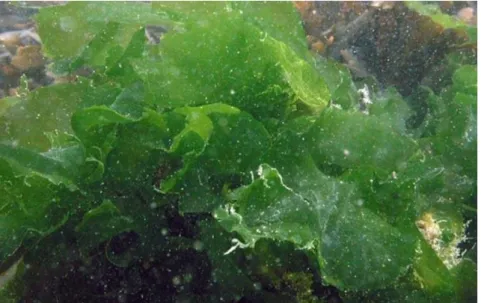
1.7 Ulva prolifera
Ulva prolifera is a common green alga which grows near the top of the shore, on
rocks (Fig. 8) or other algae. It can be found on open coasts or in estuaries and harbors, where it may grow mixed with U. intestinalis or other species of the genus. The fronds are tubular, tough and often more or less flattened (Brodie et al, 2007) (Fig. 9).
Free-floating Ulva prolifera is one of the causative species of green tides that occur along the shoreline in many countries which not only seriously affects the inshore environment, but also threatens the offshore environment and the ecological balance of the marine community. The very high growth rate in addition to the rapid proliferation
13 of produced spores causes the rapid biomass accumulation characteristic of green tides (Huan et al 2016).
Figure 8: Ulva prolifera grown on a rock from the Aquaculture Research Station in Olhão.
Figure 9: Studied sample of Ulva prolifera from the Aquaculture Research Station, Olhão (EPPO), Portuguese Institute for the Sea and Atmosphere (IPMA, IP).
Although green seaweeds represent a food group that is not normally ingested in Western societies, currently they are attracting increasing attention as a valuable food source. As we have seen, the potential of green seaweeds is large, with high levels of carbohydrates as well as minerals, vitamins, and trace elements such as iodine (Macartain et al., 2007). However, not only the amount of the components of a food is important, but also, they must be available for absorption after the digestive process.
14
1.8 Digestive process
The digestion is a physiologic process (through mechanic movements, chemicals and enzymes) that allows the release of nutrients and phytochemicals, among others, from the food matrix, allowing them to be later absorbed by the organism (Tagliazucchi et al., 2010; Bouayed et al., 2011).
1.8.1 Human digestion
Human digestion is considered to be extracellular and happens in what is known as “digestive tract”. In the latter, mechanical processes like mastication, swallowing and peristalsis movements occur. These are accompanied by the chemical component of digestion entailing pH variation and enzyme action. pH variation will promote the enzymatic hydrolysis of the food proteins, lipids, carbohydrates and nucleic acids. Afterwards, the monomers will be absorbed at the intestinal level to the blood stream (Diagram Group, 2005).
The digestive tract is composed of mouth, pharynx, esophagus, stomach, small intestine, large intestine, and anus. Besides these ones, other organs and glands are also associated to the digestion system such as salivary glands, liver, pancreas, and gall bladder (Lidon and Silvestre, 2010). The digestive process begins in the mouth or oral cavity, where the mechanical and chemical disaggregation of the food takes place. The initial degradation of, for instance, polysaccharides and triacylglycerols occur during mastication and salivation, with the help of teeth and tongue, which tend to facilitate the enzymatic action. Saliva is composed mainly of water and salivary amylase that initiates the digestion of carbohydrates (Diagram Group, 2005; Bouayed et al., 2011). Subsequently, the bolus is swallowed entering the esophagus through involuntary movement (rhythmic wave-like muscle contractions and relaxations) to the stomach.
In the stomach, the gastric juices, produced by glands in the stomach wall, dissolve the intercellular substances from the ingested food, helping the mechanical fragmentation initiated by the chewing process. The acid facilitates the fragmentation of various macromolecules, provides an optimum pH for protein digestion, contributes to the activation of enzymes present in the gastric juice, and exerts germicidal action. The enzymes of the gastric juice are pepsin, gastric lipase and gastric amylase. Pepsin is a proteolytic enzyme having a maximum activity at acidic pH (pH 2.0) and becoming
15 inactive at pH values above 5.0. With the action of gastric juice on food bolus it gives rise to chyme (Diagram Group, 2005; Lidon and Silvestre, 2010).
Stomach chyme passes into the small intestine stimulating duodenal mucosa to produce the hormones secretin and pancreosin, which in turn stimulate the pancreas to secrete pancreatic juice containing water, enzymes (trypsin, chymotrypsin, pancreatic amylase, pancreatic lipase, deoxyribonuclease and ribonuclease), and large amounts of sodium bicarbonate to neutralize the acidity of the chyme and thus ensure the action of pancreatic enzymes (Lidon and Silvestre, 2010). In the duodenum, the bile is also discharged from the gallbladder. Bile has no digestive enzymes but have bile salts, sodium glycocholate and taurocholate, to emulsify lipids, thereby fostering lipid digestion. In the intestine, chyme is transformed into chyle, a fluid rich in simple sugars, amino acids, fatty acids and glycerol (Lidon and Silvestre, 2010; Bouayed et al., 2011; Gião et al., 2012).
The nutrients in their simplest forms are then absorbed through the intestinal wall as water is reabsorbed. In the large intestine (which has no villi and does not secrete digestive juices) occurs the water and salts absorption, and by the action of numerous bacteria that make up the intestinal flora, proceeds to the dissolution of food remains unassimilable, thus leading to the formation of faeces. The bacterial fermentation that occurs in the large colonic intestine also plays a key role in the release of nutrients, making them available for absorption through the gut barrier (Diagram Group, 2005; Bouayed et al., 2011; Gião et al., 2012).
1.8.2 Bioaccessibility and bioavailability of nutrients and contaminants
Bioaccessibility is defined as the fraction of a compound that is released from the food matrix to the gastrointestinal tract, so it can be absorbed by the intestine. On the other hand, the bioavailability is the fraction of a bioaccessible compound that reaches the systemic circulation and becomes available to be absorbed by the various cells in any tissue of the human organism, stored and/or used in metabolic functions (Moreda-Piñeiro et al., 2011). Bioavailability and bioaccessibility of a compound can be affected by several factors, including:
• Possible interactions with other food components;
• Formation of stable compounds that are slowly metabolized;
16 • The release from the food matrix;
• The chemical state of the nutrient.
Thus, the total amount ingested of a compound may not provide an adequate guidance for the amount that is bioaccessible and bioavailable.
1.8.3 Evaluation of the bioaccessibility in vitro digestion model
The compound mobilization in the food matrix to the gastrointestinal tract is a dynamic process with constant changes in physiological conditions. In the in vitro digestion model, the digestive process is simulated in a simplified manner by applying/simulating physiological conditions that replicate the chemical composition of the digestive fluids, pH and typical residence time in each digestive step (Versantvoort and Rompelberg, 2004).
The in vitro digestion model developed by Versantvoort & Rompelberg (2004) allows to simulate the digestive process in the gastrointestinal tract (mouth, stomach and small intestine). In each compartment, the matrix is incubated at 37±2 °C. Digestion is started by adding artificial saliva to the matrix under investigation. Subsequently, gastric and duodenal juices and bile are added to simulate the digestive process in the stomach and small intestine, respectively. Subsequently, concentration of the compound of interest is determined in the bioaccessible and undigested fractions (Versantvoort and Rompelberg, 2004; C Afonso et al., 2015).
Although there are in vivo methods to estimate the bioavailability of a nutrient/contaminant, in vitro methods are preferred, even with their limitation, since are less expensive, easier to reproduce and do not raise ethical problems (García-Sartal et al., 2013).
17
2. Objective
Over the last few years, marine organisms, particularly seaweeds, have proved to be a unique source of molecules with high biotechnological interest, providing new compounds with the most diverse pharmacological and food properties.
The Aquaculture Research Station (EPPO) from the Portuguese Institute for the Sea and Atmosphere (IPMA) owns several Earth Ponds Aquaculture Systems with a Seaweed Production component where seaweeds grow spontaneously. Hence, the main objective of this work corresponded to the biochemical characterization of five seaweed species from IMTA systems and the determination of relevant bioactivities such as antioxidant and anti-inflammatory properties. This study enabled an assessment of the potential of this significant aquatic resource for future applications in the areas of human and animal nutrition, nutraceuticals, or cosmetics. Moreover, the bioaccessibility of the green seaweeds nutritional composition was evaluated, which is an area still poorly explored.
3. Materials and Methods
3.1 Cultivation conditions
At the Aquaculture Research Station, Olhão (EPPO), earth ponds with 0.2 ha and 2500 m3 in volume were used for meagre (Argyrosomus regius) experimental grow-out from 10 g to 1 kg and, in some tanks, till 2.5 kg in fish weight. All ponds had constant water renovation, with a daily average of 30 %, using pumped water from a reservoir connected directly to the Ria Formosa Lagoon. Dry feed is distributed to fish daily, starting with 2.3 (winter, cold water, low feed consumption by the fish) and increasing progressively to 44 kg/day (summer, warm water, high feed consumption by the fish), thereby reaching a total of 5,125 kg. No algicide (such as copper sulfate) was used during the grow-out and the presence of seaweed-feeders like gilthead seabream,
Sparus aurata, was low (less than 500 specimens per pond). Seaweed biomass in the
ponds was allowed to grow naturally until covering around 20 % of water surface area and was collected weekly.
18
3.2 Samples
Samples of five species of green seaweeds (Chaetomorpha linum, Rhizoclonium
riparium, Ulva intestinalis, Ulva lactuca, Ulva prolifera) were collected manually and
transported immediately in seawater to a nearby lab (< 100 m). This harvest was carried out in the summer (July). Each sample was thoroughly washed with seawater to eliminate any biofouling organisms. After washing, the frond samples were kept moist in a 20-L bucket and transported to the IPMA Lisbon Lab. Seaweeds were then finely minced. The processed biological material was frozen, freeze-dried, and stored at – 20 °C.
3.3 Proximate composition
The moisture and ash contents were determined according to AOAC methods (AOAC 2000). The protein level was quantified according to the Dumas method (Saint-Denis and Goupy 2004) and a conversion factor of nitrogen into protein of 5 was used. Crude fat was determined following the Folch extraction method (Folch et al., 1957). Carbohydrate content was determined by difference between 100 % and the sum of the moisture, protein, crude fat, and ash contents.
3.4 Lipid extraction
Bligh and Dyer (1959) method was used for extraction of total lipid content from the fresh seaweeds. Briefly, 5 mL methanol:chloroform (2:1), 1 ml of saturated NaCl solution and 2 ml of chloroform were sequentially added and homogenized with 1 g of sample. After centrifugation (2,000×g at 4 ºC for 10 min) (Fig. 10), organic phase was filtered (Fig. 11) through anhydrous sodium sulfate and evaporated in an RE 121 model rotary evaporator (Büchi, Flawil, Switzerland). Extractions were done in duplicate. Samples were stored at -20 ºC until further analyses.
19 Figure 10: Sample after centrifugation. Three phases can be differentiated: water, seaweed fibers and
organic phase containing the lipids.
Figure 11: Organic phase containing lipids after filtration
3.5 Lipid class analysis
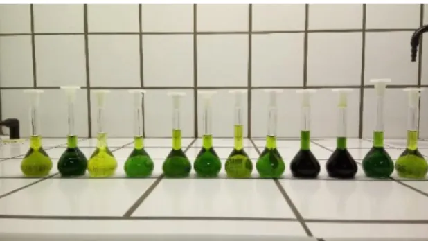
The main lipid classes were separated by analytical thin-layer chromatography (TLC) in plates coated with 0.25 mm silica gel G and developed with a mixture of hexane:diethylether:acetic acid (50:50:2 by volume), based on the method described by Bandarra et al. (1997). Extracted lipids were dissolved in chloroform (10 mg/ml concentration). A mixture of standards (Sigma Chemical Co., St. Louis, Mo) was also prepared in chloroform with the same concentration. Specifically, glyceryltrioleate (TAG), glyceryl 1,3-dipalmitate (diacylglycerol, DAG), DL--monoolein (MAG), oleic acid (FFA), L--phosphatidylcholine (PL), and monogalactosyl diacylglycerol (GL) were used. The samples and standards (10 L) were applied to the plates and each plate was immersed in 100 mL of the elution mixture inside a developing chamber. The elution front was followed visually. After elution front reached the upper limit, plates were taken out from the chamber. The developed plates were then sprayed with 10 %
20 phosphomolybdic acid in ethanol (w/v). Identification of lipid classes (polar and non polar) was done by comparison with standards. Quantification was performed using a scanner and version 4.5.2 of Quantity One 1-D Analysis software from Bio-Rad (Hercules, CA, USA) (Fig. 12). There were always two replicates.
bbb b
Figure 12: Distribution of the lipid classes through analytical TLC for the five seaweeds: a) before spraying with 10 % phosphomolybdic acid in ethanol and b) when imaged at a digital scanner.
3.6 Lipid classes separation for fatty acid analysis
The different lipid classes were fractionated using a preparative TLC. This involved applying 25 μL of a 50 mg/mL chloroform solution on several points of the
a)
21 TLC. The plate was placed in an elution vessel containing hexane:diethyl ether:acetic acid (50:50:2) and afterwards elution plates were sprayed with a 0.2 % solution of 2’,7’-dichlorofluorescein (Sigma, St. Louis, MO, USA) in ethanol. Visualization was achieved in a cabinet II model UV chamber (CAMAG, Muttenz, Switzerland). Lipid fractions were identified using sigma standards (St. Louis, MO, USA) — glyceryltrioleate (TAG), glyceryl 1,3-dipalmitate (DAG), DL--monoolein (MAG), oleic acid (FFA), L--phosphatidylcholine (phospholipid, PL), and monogalactosyl diacylglycerol (GL). There were always two replicates.
Figure 13: Preparative TLC for Chaetomorpha linum (a) after hexane:diethyl ether:acetic acid solution (b) in UV chamber after dichlorofluorescein (c) with lipid fractions identified using sigma standards (TAG - glyceryltrioleate, DAG - glyceryl 1,3-dipalmitate, MAG - D-monoolein, FFA - oleic acid, and PL - L--phosphatidylcholine).
3.7 Fatty acid profile
Fatty acid methyl esters (FAMEs) were prepared by acid-catalyzed transesterification using the methodology described by Bandarra et al. (1997). To 150 mg extracted crude fat present in a screw cap glass tube, 5 mL of a 5 % acetyl chloride methanolic solution (prepared immediately before addition) were added. These glass tubes, after vigorous agitation, were placed in a hot bath (80 C) and left there 1 hour, in accordance with the method described by Lepage and Roy (1986), modified by Cohen et al. (1988). Upon reaction completion, the solution was cooled, diluted with 1 ml water and 2 mL n-heptane and vigorously mixed, the last addition produced an organic phase (Fig. 14) that was filtered through anhydrous sodium sulfate. The resultant methyl esters were applied to a DB-WAX (Agilent Technologies, Santa Clara, USA) capillary column (film
22 thickness, 0.25 m, 30 m × 0.25 mm i.d.), integrated in a Varian Star 3800 CP gas chromatograph (Walnut Creek, CA, USA), equipped with an auto sampler with a split injector (100:1) and a flame ionization detector, both at 250 °C. The separation of the FAMEs was carried out with helium as the carrier gas and using a temperature program for the column starting at 180 ºC and increasing to 200 °C at 4 °C/min, holding for 10 min at 200 °C, heating to 210 °C at the same rate, and holding at this temperature for 14.5 min. FAMEs were identified by comparing their retention time with those of Sigma–Aldrich standards (PUFA-3, Menhaden oil, and PUFA-1, Marine source from Supelco Analytical). Analyses were always done in triplicate.
Figure 14: Differentiation of phases during acid-catalyzed transesterification
3.8 Fucose content
Free fucose was determined by the cysteine-sulfursulfuric acid method for methyl pentoses. Triplicates (50 mg) of fresh seaweed were placed into separate test tubes and mixed and homogenized at 30,000 rpm with 1 ml of Milli-Q water using a model Polytron PT 6100 homogenizer (Kinematica, Luzern, Switzerland). Afterwards, samples were subjected to an ultrasound treatment at 25 ºC for 15 min in a Sonorex Super 10 P model (Bandelin Electric, Berlin, Germany). Commercial L-fucose attained from Sigma (St. Louis, MO, USA) was used as the standard. 4.5 ml of sulfursulfuric acid (prepared by adding six volumes of concentrated sulfuric acid with one volume of water) was added into each tube (including tubes containing 1 mL of bioaccessible fraction of each seaweed) and mixed. Tubes were then put into a boiling water bath for 3 minutes. Afterwards, tubes were cooled and 0.1 mL cysteine hydrochloride solution (5
23 % cysteine hydrochloride in Milli-Q water) was added to each tube and mixed. Absorbance was read at 396 nm and 427 nm, after zeroing the spectrophotometer with a water blank treated in the same way. Fucose-specific absorbance values were calculated according to the following expression: Absorbance = A396nm – A427nm (Dische and Shettles, 1948). Interference by solutions and digestive enzymes used in the bioaccessibility method was accounted for by subtracting absorbance of the bioaccessibility blank from the absorbance measured with the bioaccessible fraction samples.
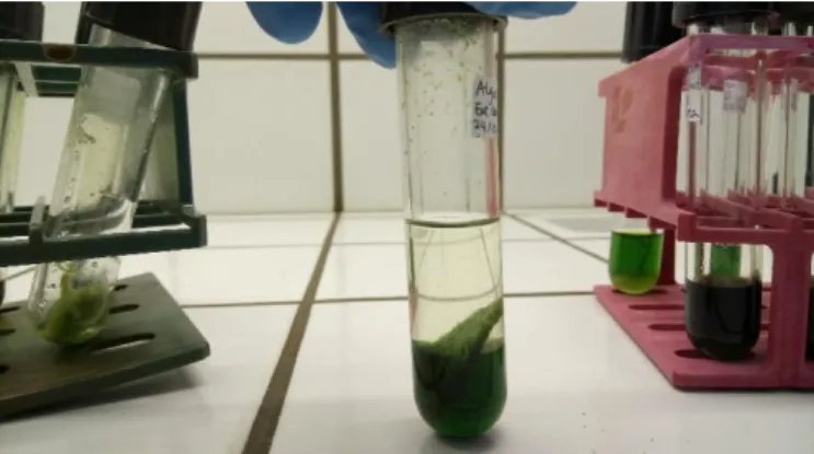
3.9 Total polyphenol content
Phenolic compounds were extracted by an appropriate solvent mixture (Siriwoharn et al., 2004) (Fig. 15) and determined by the Singleton and Rossi method using the Folin-Ciocalteu reagent (Singleton and Rossi 1965). A volume of 100 μL of each seaweed or bioaccessible extract was added to a vial. To each vial, 600 μL of MiliQ water plus 150 μL of twice-diluted Folin-Ciocalteau reagent were added and allowed to stand for 5 min at room temperature. Then, 750 μL of a 2 % w/v sodium carbonate solution were added. After 15 min reaction in the dark at room temperature, absorbance at 750 nm was measured in a Helios Alpha model (Unicam, Leeds, UK) UV-Vis spectrophotometer. Gallic acid (GA) was used as standard and phenolic content was expressed as gallic acid equivalents (mg GAE/g) through the calibration curve of gallic acid (Sigma, Steinheim, Germany). Interference by solutions and digestive enzymes used in the bioaccessibility method (see section 3.12) was taken into account by subtracting absorbance of the bioaccessibility blank from the absorbance measured with the bioaccessible fraction samples.
24
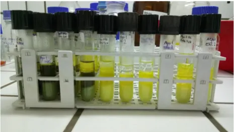
3.10 Antioxidant activity
The antioxidant activity was measured through the determination of the radical scavenging activity using DPPH (Miliauskas et al., 2004). In order to prepare the extracts, approximately 0.5 g of freeze-dried green seaweed was weighed or 5 mL of bioaccessible fraction (see section 3.12) was measured, homogenized with 25 mL of methanol 50 % v/v using a model Polytron PT 6100 homogenizer (Kinematica, Luzern, Switzerland) at a velocity of 30,000 rpm during 1 min, and agitated for 1 h on an orbital shaker. After centrifugation (5,000×g at room temperature during 10 min), the supernatant was filtered and diluted 1:5. A volume of 1 mL of the extract was prepared in triplicate for each sample and 2 ml of DPPH (Sigma, Steinheim, Germany) 0.15 mM methanolic solution was added and thoroughly mixed (Fig. 16). After 30 min of incubation at room temperature in the dark, absorbance was measured at 517 nm in a Helios Alpha model (Unicam, Leeds, UK) UV/visible light spectrophotometer. A solution containing methanol 50 % v/v was the blank.
Radical scavenging activity was calculated by the following formula:
% Inhibition = (A0 - Asample)/A0 × 100
where:
A0 – Absorbance of the blank.
Asample – Absorbance of the sample.
Figure 16: Sample solutions (a) before and (b) after adding DPPH.
25 Interference by solutions and digestive enzymes used in the bioaccessibility method was taken into account by adjusting absorbance measured with the bioaccessible fraction samples with the absorbance of the bioaccessibility blank.
3.11 Anti-inflammatory activity
3.11.1 Extract preparation for in vitro anti-inflammatory activity
For each green seaweed and each respective bioaccessible fraction (see section 3.12), an aqueous extract was prepared with the purpose of attaining a fraction with anti-inflammatory properties to be tested in vitro. Accordingly, approximately 200 mg of freeze-dried green seaweed was weighed or 5 mL bioaccessible fraction was measured and homogenized with 2 mL of Milli-Q water using a model Polytron PT 6100 homogenizer (Kinematica, Luzern, Switzerland) at a velocity of 30,000 rpm during 1 min. Afterwards, the mixture was subjected to a thermal treatment (at 80 °C for 1 h). Both the seaweed and bioaccessible extraction mixtures were centrifuged (3,000×g at 4 ºC during 10 min) and the respective supernatant was evaporated using vacuum rotary evaporator with the water bath temperature at 65 °C and inert gas (nitrogen) stream.
3.11.2 Cyclooxygenase (COX-2) inhibition assay
The cyclooxygenase (COX-2) inhibition assay is a practical and quick screening method for assessing the anti-inflammatory activity. The prepared extracts were dissolved in 100 % dimethyl sulfoxide (DMSO) to prepare a stock preparation with a concentration of 10 mg/mL. The extract was tested at 1 mg/ml and 100 μg/mL using a commercial cyclooxygenase (COX) inhibitory screening assay kit Cayman test kit-560131 (Cayman Chemical Company, Ann Arbor, MI, USA). The COX inhibitor screening assay directly measures the amount of Prostaglandin F2α generated from arachidonic acid (AA) in the cyclooxygenase reaction. A volume of 10 μL each of test extract or DMSO was used. The reaction was initiated by addition of 10 μL 10 mM AA and each reaction tube was incubated at 37 °C for 2 minutes. Reaction was terminated by addition of 50 μL 1 N HCl and saturated stannous chloride. Assays were performed using 100 units of human recombinant COX-2. An aliquot was removed and the
26 prostanoid produced was quantified spectrophotometrically (412 nm) via enzyme immunoassay (ELISA) after 18 h incubation, washing, addition of Ellman’s reagent, and further 90 min incubation (Fig. 17). Interference by solutions and digestive enzymes used in the bioaccessibility method was taken into account by subtracting COX-2 inhibition of the bioaccessibility blank from the COX-2 inhibition measured with the bioaccessible fraction samples.
Figure 17: Spectrophotometer with plate for prostanoid reading
3.12 In vitro digestion model
An in vitro digestion model was chosen for the determination of bioaccessibility in each of the five seaweed species (Fig. 18). Such model comprises three sections, which enable the simulation of digestion in three different parts of the GI tract: mouth, stomach, and small intestine (Afonso et al., 2015).
27 The composition of digestive juices (saliva, gastric, duodenal and bile) was the same described by Versantvoort et al. (2005). The chemicals KCl, NaH2PO4, Na2SO4,
NaCl, NaHCO3, HCl, CaCl2.2H2O, KH2PO4 and MgCl2 used for preparation of the
digestive fluids, were obtained from Merck (Darmstadt, Germany). NH4Cl was obtained
from Fluka (Buchs, Switzerland) and all other chemicals were obtained from Sigma (St. Louis, MO, USA). In the case of duodenal juice, trypsin and -chymotrypsin from Sigma (St. Louis, MO, USA) were also added. The quantities of these two enzymes (0.08 g trypsin and 0.87 g -chymotrypsin in 500 ml of duodenal juice) were estimated on the basis of the work by Gatellier and Santé-Lhoutellier (2009).
Briefly, approximately 1.5 g homogenized and hydrated (with up to 1.5 mL water) seaweed was weighed. For the bioaccessibility blank, 1.5 mL of Milli-Q water was used instead. Sample was mixed with 4 ml of artificial saliva at a pH 6.8 ± 0.2 for 5 min, then 8 ml of artificial gastric juice (pH 1.3 ± 0.02 at 37 ± 2 ºC) was added, and pH was lowered to 2.0 ± 0.1. The mixing lasted 2 h in a head-over-heels movement (37 rpm at 37 ± 2 ºC). Finally, 8 ml of artificial duodenal juice (pH 8.1 ± 0.2 at 37 ± 2 ºC), 4 mL of bile (pH 8.2 ± 0.2 at 37 ± 2 ºC), and 1.33 mL of HCO3- solution (1 M) was added.
The pH of the mixture was set at 6.5 ± 0.5 and agitation for 2 h was identical to gastric conditions. The mixture generated in the in vitro model was subjected to centrifugation at 2750×g for 5 min, thus yielding a non-digested portion and the bioaccessible fraction. While chemicals were of analytical grade and supplied by Merck (Darmstadt, Germany), enzymes were attained from Sigma (St. Louis, MO, USA).
3.12.1 Calculation of bioaccessibility
The percentage (%) of each seaweed constituent (C) in the bioaccessible and in the non-digested fraction was estimated as follows:
% C bioaccessible = [C]bioaccessible × 100/[S]
and
% C non-digested = [C]non-digested × 100/[S]
Being:
[C] = Concentration of constituent.