Using induced pluripotent stem cells to investigate the
mechanistic link between Gaucher disease and Parkinson
related synucleinopathies
by
Fábio Dantas Romero Monteiro Biomedical Sciences Master’s dissertation
Supervisor:
Prof. Dr. Gustavo Tiscornia
1
Using induced pluripotent stem cells to investigate the
mechanistic link between Gaucher disease and Parkinson
related synucleinopathies
by
Fábio Dantas Romero Monteiro Biomedical Sciences Master’s dissertation
Supervisor:
Prof. Dr. Gustavo Tiscornia
2
“Using induced pluripotent stem cells to investigate the mechanistic link between Gaucher disease and Parkinson related synucleinopathies”
Declaração de autoria de trabalho
Declaro ser o autor deste trabalho, que é original e inédito. Autores e trabalhos consultados estão devidamente citados no texto e constam da listagem de referências incluída.
Fábio Dantas Romero Monteiro
________________________________
Copyright, Fábio Dantas Romero Monteiro
A Universidade do Algarve tem o direito, perpétuo e sem limites geográficos, de arquivar e publicitar este trabalho através de exemplares impressos reproduzidos em papel ou de forma digital, ou por qualquer outro meio conhecido ou que venha a ser inventado, de o divulgar através de repositórios científicos e de admitir a sua cópia e distribuição com objetivos educacionais ou de investigação, não comerciais, desde que seja dado crédito ao autor e editor.
3
TABLE OF CONTENTS
ACKNOWLEDGEMENTS ... 5 ABSTRACT ... 6 RESUMO ... 7 LIST OF FIGURES ... 10 LIST OF GRAPHS ... 12 LIST OF ABBREVIATIONS ... 13 1 - INTRODUCTION ... 16 1.1 - Gaucher disease ... 161.2 - Gaucher disease and Parkinson related synucleinopathies ... 18
1.3 - Induced pluripotent stem cells and differentiation to the neuronal fate ... 20
1.4 - Objective and experimental strategy ... 22
2 - MATERIALS AND METHODS ... 23
2.1 - Tissue culture procedures ... 23
2.2 - GD FiPS 4F 21C and L-GBA rescued clones culture ... 23
2.3 - H9 hESc culture ... 24
2.4 - FiPS 3F-1 culture ... 24
2.5 - Human foreskin fibroblasts, HEK 293T and PA6 stromal cells culture ... 25
2.6 - Human foreskin fibroblasts mitotic inactivation ... 25
2.7 - hiPS differentiation to the neuronal fate ... 25
2.8 - Gelatin solution and coating preparation ... 26
2.9 - Matrigel solution and coating preparation ... 27
2.10 - Plasmid preparation (and Restriction Enzyme Digestion protocol) ... 27
2.11 – Agarose gel electrophoresis ... 27
2.12 - gDNA extraction/purification ... 28
4
2.14 - Virus preparation and titer... 28
2.15 - Infection of GD FiPS 4F 21C neural culture with lentivirus expressing β-Glucocerebrosidase ... 29
2.16 - Treatment with chaperones of GD FiPS 4F 21C embryoid bodies differentiated to the neuronal fate ... 29
2.17 - Polymerase chain reaction (PCR) ... 29
2.18 - Immunocytochemistry ... 30
2.19 - Flow cytometry ... 31
2.20 - SDS-PAGE and Western blot ... 31
2.21 - β-Glucocerebrosidase activity assay ... 32
3 – RESULTS AND DISCUSSION ... 33
3.1 – Effect of chaperone treatment on β-glucocerebrosidase and α-synuclein levels in neurons differentiated from GD iPS cells by an embryoid body/PA6 co-culture protocol ... 33
3.2 - Matrigel Based Neuronal Differentiation of iPSc ... 42
3.3 - Effect of β-glucocerebrosidase overexpression by lentiviral infection on α-synuclein levels in neurons differentiated from GD iPS cells by a Matrigel based neuronal differentiation protocol ... 47
3.4 - Rescuing of Gaucher Disease FiPS 4F 21C ... 53
3.4.1 - Introduction ... 53
3.4.2 – Virus preparation, infection and clone isolation ... 53
3.4.3 – PCR optimization ... 54
3.4.4 – Clone analysis ... 55
4 – FINAL DISCUSSION, FUTURE PERSPECTIVES AND CONCLUSION ... 59
5
ACKNOWLEDGEMENTS
I want to thank Prof. Dr. Gustavo Tiscornia and all my colleagues for the support, guidance, patience and encouragement throughout my work in the laboratory as well as my family, friends and better half for all the love and support.
6
ABSTRACT
Gaucher disease (GD) is a rare, autosomal recessive, genetic disorder caused by biallelic mutations in the β-glucocerebrosidase gene (GBA1), a lysosomal enzyme. There are numerous experimental, clinical and genetic studies linking GD patients and carriers to Parkinson disease and its related synucleinopathies, although the mechanism remains elusive. In the present study, we try to better understand this connection using neuronal populations differentiated from an induced pluripotent stem (iPS) cell line (GD FiPS 4F 21C) derived from fibroblasts of a GD patient. Neuronal cultures were treated with chaperone molecules that aid the folding of mutated β-glucocerebrosidase in the endoplasmic reticulum, increasing stability and trafficking to the lysosome, or by transduction with a lentivirus overexpressing wild-type β-glucocerebrosidase in order to rescue β-glucocerebrosidase activity and analyze α-synuclein levels. Although with variability, mutant β-glucocerebrosidase protein levels alterations due to chaperone treatment matched α-synuclein changes. Wild-type β-glucocerebrosidase overexpression by lentiviral infection did not significantly change α-synuclein levels. These results are in contrast with numerous studies that reason a loss-of-function link between mutant β-glucocerebrosidase and α-synuclein, that is, β-glucocerebrosidase deficiency leading to lysosomal disfunction and α-synuclein accumulation, but in accordance with several others that argue for toxic gain-of-function mechanistic link. It is possible that chaperone treatment aided in the stability and trafficking of mutant β-glucocerebrosidase to the lysosome, increasing its levels, where it had either a direct or indirect effect of increasing α-synuclein levels. This would also explain why when overexpressing wild-type β-glucocerebrosidase, α-synuclein levels are not changed. However, these results are preliminary and more research is needed in order to confirm our experimental data. We also established two rescued GD iPS cell lines, derived from a GD FiPS 4F 21C lentiviral infection expressing wild-type β-glucocerebrosidase and developed a novel neuronal differentiation protocol that we found to be specific to the mentioned cell line.
7
RESUMO
A doença de Gaucher é uma doença genética rara, autossómica recessiva, causada por mutações bialélicas do gene da glucocerebrosidase (GBA1), sendo a doença de depósito lisossomal mais comum. A glucocerebrosidase é uma enzima lisossomal, responsável pela degradação de glucocerebrosídeo em ceramida e glucose. Mutações homozigóticas ou heterozigóticas compostas no gene GBA1 provocam a disfunção da glucocerebrosidase, causando a acumulação de glucocerebrosídeo em diversos tipos celulares, especialmente em macrófagos visto que a maior parte de glucocerebrosídeo é derivado da fagocitose de leucócitos senescentes. Existe assim também, a acumulação de glucocerebrosídeo em diversos orgãos, especialmente no baço, fígado e medula óssea. A doença de Gaucher está divida em três subtipos, o tipo 1 ou a forma não-neuropática em que os sintomas mais comuns incluem hepatomegalia, esplenomegalia, trombocitopenia e anemia. Este é o tipo mais comum tendo uma frequência de 1 em cada 40000 pessoas sendo ainda mais comum nas populações judaicas asquenazes com uma frequência de 1 em cada 855 pessoas. Existe também o tipo 2 ou a forma neuropática aguda que é caraterizada por uma rápida neurodegeneração com envolvimento visceral e que normalmente leva à morte nos primeiros 2 anos de vida devido a problemas respiratórios. Finalmente existe o tipo 3 ou a forma neuropática crónica que embora seja varíavel, tipicamente se apresenta com manifestações neurológicas e viscerais, tal como o tipo 2, mas não de forma tão agravada. Ainda é desconhecido o mecanismo do envolvimento neurológico na doença de Gaucher tipo 2 e 3, no entanto poderá estar relacionado com defeitos da via autofágica-lisossomal, neuroinflamação e/ou a acumulação de glucocerebrosídeo, um glicolípido citotóxico, no cérebro. Relativamente ao tratamento, existem duas terapias disponíveis para tratar os sintomas viscerais da doença de Gaucher: a terapia de reposição enzimática que consiste em administrar sistemicamente glucocerebrosidase recombinante e a terapia de redução de substrato no qual há uma inibição da glucosilceramida sintase provocando a redução da síntese de glucocerebrosídeo de forma a compensar a reduzida degradação desta molécula. No entanto, estes tratamentos não são eficazes no tratamento dos sintomas neurológicos da doença de Gaucher devido à incapacidade do agente terapêutico transpor a barreira hematoencefálica. Embora existam sugestões de terapias alternativas
8
capazes de ultrapassar este obstáculo, é necessário mais investigação de forma a consolidar estas terapias.
Mutações no gene GBA1 é o mais preponderante factor de risco genético no desenvolvimento de sinucleinopatias relacionadas com Parkinson, nas quais existem um defeito no processamento da α-sinucleína e consequente acumulação, levando à formação de corpos e neurites de Lewy. Existem numerosos estudos experimentais, clínicos e genéticos associando pacientes e portadores da doença de Gaucher à doença de Parkinson e sinucleinopatias associadas a Parkinson. No entanto, o mecanismo desta associação permanece elusivo. Teorias propostas incluem: redução da atividade de glucocerebrosidase levando à ruptura do sistema autofagossomal e consequentemente à degradação reduzida de proteínas, levando assim ao aumento e acumulação de α-sinucleína ; redução de glucocerebrosidase levando à alteração da composição lipídica da membrana devido à acumulação de glucocerebrosídeo, afetando a função das jangadas lipídicas e portanto, a triagem e o transporte de proteínas relacionadas com as jangadas tais como a α-sinucleína ; redução de glucocerebrosidase levando ao sobrecarregamento da via de degradação do retículo endoplasmático e consequentemente, a sinais de stress do retículo endoplasmático e à acumulação de α-sinucleína ; entre outras.
No presente estudo tentamos compreender melhor esta relação através do uso de células pluripotentes induzidas (iPSc ; induced Pluripotent Stem cells), uma técnica relativamente recente desenvolvida pelo laboratório de Shinya Yamanaka e que consiste na reprogramação de células diferenciadas de volta a um estado pluripotente, através de certos factores de trancrição (Oct4, Sox2, Klf4 e c-Myc). Shinya Yamanaka foi galadoardo com o Prémio Nobel de Fisiologia ou Medicina de 2012 devido a este feito. Esta técnica permitiu-nos obter células pluripotentes induzidas derivadas de fibroblastos provenientes de um doente de Gaucher, a linha celular GD FiPS 4F 21C, a qual diferenciámos para o destino neuronal de forma a analisar a relação entre a doença de Gaucher e sinucleinopatias associadas a Parkinson. A diferenciação neuronal foi feita através de dois métodos: formação de corpos embrióides e co-cultura com células estromais PA6 sob meio de cultura GMEM por 21 dias ; dissociação celular através da acção de accutase e cultura em placas revestidas com Matrigel sob o meio de cultura mTeSR™1, este método tendo sido acidentalmente descoberto pelo nosso laboratório e que pesquisa subsequente mostrou ser um comportamento específico da linha celular
9
utilizada (GD FiPS 4F 21C). As culturas neuronais foram tratadas com chaperonas, moléculas que auxiliam o enovelamento de proteínas, neste caso da glucocerebrosidase mutante no retículo endoplasmático, de forma a aumentar a estabilidade, a atividade e o transporte para o lisossoma. As culturas neuronais foram também transduzidas com um vetor lentiviral que sobreexpressava glucocerebrosidase wild-type de forma a resgatar a atividade da glucocerebrosidase. Em ambos os casos o objectivo foi depois comparar os níveis de glucocerebrosidase e de α-sinucleína. No caso do tratamento com chaperonas, embora tenha havido variabilidade, as alterações dos níveis protéicos de glucocerebrosidase foram semelhantes às alterações dos níveis de α-sinucleína. No caso da sobreexpressão de glucocerebrosidase wild-type através de infecção lentiviral, não houve alterações significativas dos níveis de α-sinucleína. Estes resultados estão em contraste com numerosos estudos que argumentam um mecanismo de perda-de-função entre a glucocerebrosidase mutante e a α-sinucleína, isto é, a deficiência de glucocerebrosidase levando à disfunção lisossomal e à acumulação de α-sinucleína. No entanto, estes resultados estão de acordo com outros estudos que argumentam um mecanismo de ganho-de-função tóxico. É possível que o tratamento com chaperonas tenha ajudado na estabilização e transporte de glucocerebrosidase mutante para o lisossoma, onde teve um efeito direto ou indireto no aumento dos níveis de α-sinucleína. Esta hipótese explicaria também o porquê de não ter havido alterações nos níveis de α-sinucleína quando a glucocerebrosidase wild-type foi sobreexpressada. Estes resultados são no entanto, preliminares e mais pesquisa é necessária de forma a confirmar os nossos dados experimentais.
Durante o decurso deste estudo estabelecemos também duas linhas celulares iPS, derivadas da GD FiPS 4F 21C e resgatadas relativamente aos níveis e atividade da glucocerebrosidase. Este desenvolvimento foi realizado através da dissociação celular de colónias, infecção lentiviral sobreexpressando glucocerebrosidase wild-type e cultura em fibroblastos mitoticamente inactivados. Foram obtidos 17 clones em que através da caracterização por PCR, western blot e ensaios de atividade enzimática, restaram 2 clones que possuíam altos níveis de glucocerebrosidase com altos níveis de atividade enzimática. Estas linhas celulares serão ferramentas importantes na continuação da investigação do mecanismo de ligação entre a doença de Gaucher e sinucleinopatias associadas a Parkinson.
10
LIST OF FIGURES
Figure 3.1 – Embryoid Body Differentiation Protocol...33
Figure 3.2 - Embryoid Body Differentiation Results...34
Figure 3.3 - Characterization of Embryoid Body Differentiation Results...35
Figure 3.4 - Chaperone Treatment Protocol...36
Figure 3.5 - Protein Levels of β-glucocerebrosidase and Actin I...37
Figure 3.6 - Protein Levels of α-synuclein and β3-tubulin I...37
Figure 3.7 - Protein Levels of β-glucocerebrosidase and Actin II...39
Figure 3.8 - Protein Levels of α-synuclein and β3-tubulin II...39
Figure 3.9 - Matrigel Based Neuronal Differentiation of iPSc...43
Figure 3.10 – Matrigel Based Neuronal Differentiation of GFP Positive iPSc...43
Figure 3.11 – Early Stage Characterization of Matrigel Based Neuronal Differentiation of iPSc...44
Figure 3.12 - Late Stage Characterization of Matrigel Based Neuronal Differentiation of iPSc...44
Figure 3.13 – Mechanical Passaging of Neural Cultures...45
Figure 3.14 - Viability Assay of Neural Cultures...45
Figure 3.15 - Matrigel Based Neuronal Differentiation Protocol Applied to Other Stem Cell Lines...46
Figure 3.16 - Transduction of Neuronal Cultures Experimental Scheme...47
Figure 3.17 - Neuronal Cultures...48
Figure 3.18 – Protein Levels of β-glucocerebrosidase and Actin III...48
11
Figure 3.20 - Protein Levels of α-synuclein and β3-tubulin III...50
Figure 3.21 - Protein Levels of α-synuclein and β3-tubulin IV...51
Figure 3.22 - PCR Annealing Temperature Optimization...54
Figure 3.23 - Genotyping of Rescued GD iPSc Clones I...55
Figure 3.24 - Genotyping of Rescued GD iPSc Clones II...56
Figure 3.25 - Genotyping of Rescued GD iPSc Clones III...56
12
LIST OF GRAPHS
Graph 3.1 - Average GCase levels I (Fig. 3.5)...38
Graph 3.2 - Average α-synuclein levels I (Fig. 3.6)...38
Graph 3.3 - Average GCase levels II (Fig. 3.7)...40
Graph 3.4 - Average α-synuclein levels II (Fig. 3.8)...40
Graph 3.5 – GCase activity I...41
Graph 3.6 – Average GCase levels III (Figures 3.18 and 3.19)...49
Graph 3.7 – GCase activity II...50
Graph 3.8 – Average α-synuclein levels III (Figures 3.20 and 3.21)...51
Graph 3.9 – GCase levels (Fig. 3.26)...57
13
LIST OF ABBREVIATIONS
BSA – Bovine serum albumin; CDM – Chemically-defined medium; CO2 – Carbon dioxide;
DAPI – 4’,6-diamidino-2-phenylindole;
DMEM – Dulbecco’s modified Eagle medium; DNA – Deoxyribonucleic acid;
DPBS – Dulbecco’s phosphate-buffered saline; EDTA – Ethylenediaminetetraacetic acid; ER – Endoplasmic reticulum;
ERAD – Endoplasmic reticulum-associated degradation; FACS – Fluorescence-activated cell sorting;
FBS – Fetal bovine serum;
FiPS – Fibroblast induced pluripotent stem; GBA – β-glucocerebrosidase gene;
GCase – β-glucocerebrosidase protein; GD – Gaucher disease;
gDNA – Genomic deoxyribonucleic acid; GFP – Green fluorescent protein;
GMEM – Glasgow's minimal essential medium; HEK 293T – Human embryonic kidney 293T cell line; hES – Human embryonic stem;
14 HFF – Human foreskin fibroblasts;
hiPS – Human induced pluripotent stem; IgG – Immunoglobulin G;
iPS – Induced pluripotent stem; iPSc – Induced pluripotent stem cells; LB – Lysogeny broth;
MEM – Minimum essential medium; MOI – Multiplicity of infection; NaCl – Sodium chloride;
NCSC - Neural crest stem cells NEAA – Non-essential amino acids; NSC – Neural stem cells;
PBS – Phosphate-buffered saline; PCR – Polymerase chain reaction; PEI – Polyethylenimine;
PFA – Paraformaldehyde; PI – Propidium iodide;
PVDF – Polyvinylidene difluoride; RIPA – Radioimmunoprecipitation assay; RNA – Ribonucleic acid;
ROCK – Rho-associated protein kinase; RT – Room temperature;
15 TBS – Tris-buffered saline;
TH – Tyrosine hydroxylase; WT – Wild-type.
16
1 - INTRODUCTION
1.1 - Gaucher diseaseGaucher disease is a rare, autosomal recessive, genetic disorder caused by mutations on the β-glucocerebrosidase gene (GBA1 ; OMIM 606463), which is composed of 11 exons and 10 introns with a total of 7.6 kilobases (kb) located in chromosome 1q21.[1] β-glucocerebrosidase is a 497 amino acid long lysosomal enzyme of approximately 62 kilodaltons (kDa), responsible for the hydrolysis of the β-glucosyl linkage of glucosylceramide, breaking it into ceramide and glucose.[2] Homozygotic or compound heterozygotic mutations in GBA1 gene cause an impairment in the enzyme’s stability and function, resulting in an accumulation of glucosylceramide in several cell types and particularly in macrophages, due to the fact that glucosylceramide is mainly derived from the phagocytosis of senescent leukocytes. Utimately, an accumulation of glucosylceramide occurs in several organs, mainly spleen, liver and bone marrow. Around 300 different mutations in the GBA1 gene have been identified in Gaucher disease patients.[1] Gaucher Disease derives its name from Phillipe Gaucher, a french physician who in 1882 described a patient with hepatosplenomegaly, anemia and thrombocytopenia, bone demineralization and neurologic effects; he also described ‘Gaucher cells’, lipid laden macrophages typical of the disease.[3]
Gaucher disease is classically divided into three clinical sub-types, defined by the degree of visceral vs. neurologic involvement and speed of progression: GD Type 1 (Non-neuronopathic form), GD Type 2 (Acute Neuronopathic form) and GD Type 3 (Chronic neuronopathic form). Clinical presentation of GD Type 1 typically involves painless hepatomegaly and/or splenomegaly, thrombocytopenia or anemia. It is also possible to present hepatomegaly, chronic fatigue, skeletal disorders or thrombocytopenia-derived symptoms such as bleeding leading to bruising easily and nosebleeds. Occasionally, patients with this type of GD may have multiple myeloma, pulmonary involvement or parkinsonism. GD Type 2 is characterized by fast neurodegeneration with visceral involvement that typically leads to death in the first 2 years of life, normally caused by respiratory problems. Symptoms may include organomegaly (more commonly hepatosplenomegaly), progressive brain damage, increased tone, strabismus, eye movement disorders, seizures and swallowing abnormalities. GD Type 3 is more variable but typically also involves both visceral and
17
neurological symptoms, albeit to a lesser extent when compared to GD Type 2. It may appear during infancy or childhood and like the other types, usually presents organomegaly as well as skeletal disorders. Frequently, the only neurological symptom of this type is the slowing of horizontal saccades but patients may also present myoclonic epilepsy, dementia or learning disabilities. A rare subgroup of this type exists in the Norrbottnian region of Sweden, consequence of a homozygotic L444P mutation. In this subgroup, symptoms like visceral, skeletal and oculomotor disorder usually appears during early childhood. They may present as well symptoms like cognitive problems, dementia and seizures.[2, 4] The mechanism underlying the neurological pathology in GD Type 2 and Type 3 is still unclear but may be associated with defects in the lysosome-autophagy pathway, neuroinflammation and/or accumulation of glucosylsphingosine, the deacetylated form of glucosylceramide and a cytotoxic glycolipid, in the brain.[5]
Gaucher disease has a low prevalence, with type 1 being the commonest with a prevalence of 1 in 40000 of the population, and therefore, being the most common lysosomal storage disease. This type is even more common in the Ashkenazi Jewish populations with a prevalence of 1 in 855. It is estimated that approximately 60% of patients from Ashkenazi origin is homozygous for the N370S mutation which is responsible for 75% of disease alleles. Type 2 and 3 are rarer, with a prevalence of less than 1 in 100000 of the population, type 3 being more frequent in the Norrbottnian region of Sweden with a prevalence of 1 in 50000.[4, 6]
Regarding treatment, there are two types of therapies available in order to treat the visceral symptoms of GD. Enzyme replacement therapy in which a glycan-modified recombinant glucocerebrosidase is administered systemically, effectively treating the visceral and hematological symptoms of GD.[7] However, this treatment is not effective in treating the neurological symptoms of the disease due to the inability of the recombinant enzyme to pass through the blood brain barrier, leading to the sugestion of therapies such as brain β-glucocerebrosidase augmentation [8]
, using motifs that target the central nervous system [9, 10] and direct brain infusions of the enzyme [11, 12]. The second option is substrate reduction therapy, which envolves using a pharmacological inhibitor of glucosylceramide synthase causing a reduction in the synthesis of glucosylceramide in order to compensate the reduced degradation of glucosylceramide caused by the deficiency in glucocerebrosidase.[13] However, this approach also fails to
18
bring clinical benefit to GD Type 2 and 3 patients due to the same reason stated above. An alternative proposition developed in recent years is the use of small chaperone molecules that aid the folding of mutated β-glucocerebrosidase in the endoplasmic reticulum and increases stability and trafficking to the lysosome [14, 15]. A main avantage of this approach would be the ability of the chaperone to transverse the blood-brain barrier, which large recombinant glucocerebrosidase cannot. However, chaperones would not help in the case of certain severe mutations that result in no β-glucocerebrosidase expression at all. Furthermore, most pharmacological chaperones are competitive inhibitors of β-glucocerebrosidase and bind to the catalytic site inhibiting enzymatic activity. This adds an additional requirement: that the inhibitor have high enough affinity to bind in the cytoplasm, but low enough affinity to dissociate from the catalytic site in the lysosome. Pharmacological chaperones are an active and promising field of research.
1.2 - Gaucher disease and Parkinson related synucleinopathies
Heterozygous mutations in the GBA1 gene is the most preponderant genetic risk factor in the development of Parkinson related synucleinopathies, in which there is a defect in the processing of α-synuclein and subsequent accumulation, leading to Lewy body and Lewy neurite formation. A large study confirmed inital studies suggesting the influence of mutations in the GBA1 gene in synucleinopathies by analyzing over 5000 Parkinson disease patients and healthy controls.[16] More genetic studies followed showing increased frequency of GBA1 mutations in Parkinson disease (PD) and dementia with Lewy bodies (DLB), being between 5 and 7 times more frequent in PD patients when compared to controls.[17] Brain neuropathological evaluation of PD patients with GBA1 mutations and Gaucher disease patients with parkinsonism showed ubiquitinated Lewy inclusions positive for α-synuclein.[18]
Presence of β-glucocerebrosidase in most of the α-synuclein inclusions in patients with mutations in GBA1 and Lewy body disorders has been demonstrated, suggesting a link between mutant β-glucocerebrosidase and α-synuclein.[19]
It's been also demonstrated that PD and DLB are associated with SCARB2 gene polymorphisms. SCARB2 is the gene responsible for Limp-2, a critical protein in the transport of β-glucocerebrosidase to the lysosome and regulator of β-glucocerebrosidase enzymatic activity.[20, 21]
Supporting this evidence is the demonstration that Limp-2 knockout mice present α-synuclein
19
accumulation.[22] In addition, α-synuclein accumulates in the brain of Gaucher disease mouse models and that rescuing β-glucocerebrosidase activity in these animals can diminish α-synuclein levels.[23-28] Furthermore, dopaminergic neurons derived from iPS cells of patients with heterozygous GBA1 mutations revealed higher α-synuclein levels than WT iPSc and diminished to WT levels in isogenic gene-corrected controls.[29]
While there is further support for correlation of mutated GBA1 and the development of synucleinopathies, the mechanistic link is still not well understood. There are two main hypothesis. The first one is that mutant GBA1 causes a loss-of-function, with multiple downstream effects. Reduction of β-glucocerebrosidase activity could disrupt the autophagosomal system, leading to diminished protein and mitochondrial degradation and consequently accumulation of α-synuclein. Also reduction of β-glucocerebrosidase may change membrane lipid composition due to the glucosylceramide and glucosylsphingosine accumulation, affecting lipid raft function and thus the sorting and trafficking of proteins related to rafts, such as α-synuclein. Further, reduction of β-glucocerebrosidase may increment disease progression through oligomeric α-synuclein cell-to-cell transmission promotion. These and additional loss of function effects are reviewed in [30].
The second hypothesis is that mutant GBA1 causes a toxic gain-of-function effect, mediated by the respective mutant glycoprotein. This hypothesis is more difficult to analyze since the mutations normally affect enzymatic activity. One study demonstrated that there was an increase in α-synuclein levels in mesencephalic cells when coexpressing WT and mutant β-glucocerebrosidase with no significant effects on total β-glucocerebrosidase activity.[23]
Another study showed that there was a 70% increase in the half-life of human α-synuclein in mouse neurons which expressed one mutant GBA1 allele and one WT GBA1 allele when compared to WT homozygous littermates' neurons.[25] It has also been observed α-synuclein aggregation in GBA1 heterozygous mice.[24] It is proposed that the presence of mutations in the GBA1 gene can saturate the endoplasmic reticulum (ER) degradation pathway leading to ER stress signals and α-synuclein accumulation. Mutant β-glucocerebrosidase may be recognized as misfolded in the ER and enter the endoplasmic reticulum-associated degradation (ERAD) pathway.[31] The constant presence of mutant and misfolded β-glucocerebrosidase in the ER may lead to ER stress and cause the unfolded protein response.[32] However, there are some studies that suggest that enzymatic activity may
20
be a factor, independently of mutant, pathogenic β-glucocerebrosidase. Pharmacological inhibition of β-glucocerebrosidase in neuroblastoma cells caused ER stress, and in a Gaucher disease mouse model lacking β-glucocerebrosidase expression in neurons showed ER anomalies.[33-35] Thus, the cause for ER stress and α-synuclein accumulation may be due to the combined effect of mutant β-glucocerebrosidase saturating the ERAD pathway and the loss of β-glucocerebrosidase enzymatic activity. Importantly, loss of function and toxic gain-of-funtion mechanisms are not mutually exclusive and both are backed up by several genetic, experimental and clinical evidence.[23, 24, 36, 37]
Although the mechanistic link between GBA1 mutations and synucleinopathies is unknown, the evidence suggesting that these mutations intefere directly or indirectly with the processing of α-synuclein leading to its accumulation and consequently, synucleinopathies, is overwhelming and cannot be ignored. More research is needed in order to better understand this connection and to potentiate novel approaches in treating these conditions.
1.3 - Induced pluripotent stem cells and differentiation to the neuronal fate
The development of induced pluripotent stem (iPS) cells, i.e. mature differentiated cells reprogrammed back to the pluripotent state, is a recent remarkable achievement in molecular genetics, with great potential for regenerative medicine, drug screening, and in vitro disease modelling. This achievement earned Shinya Yamanaka the Nobel Prize in Physiology or Medicine in 2012[38]. In the context of regenerative medicine and cell therapy, iPS cells have two main advantages in comparison to embryonic stem (ES) cells: first, the ethical issues derived from working with human embryos; second, the problems associated with immune response when applying cell therapy techniques should be less of a concern, as this technique allows for patient specific treatments by reprogramming cells from the patient (like fibroblasts from the skin) into pluripotent stem cells and then differentiating them into the desired cell type. Initially, human iPS (hiPS) cells were derived from fibroblasts through the transduction of transcriptional regulators of stem cells, namedly Oct4, Sox2, Klf4 and c-Myc.[39] Similarities between iPS and ES cells include morphology, telomerase activity, surface antigen expression, epigenetic status of pluripotent genes, proliferation rate and the potential to differentiate into all three germ layers both in vitro and in vivo.[39] Yet, there is evidence that suggests differences between iPS and ES cells at the transcriptional
21
level and even differences between different iPS cell lines that may be consequence of cell source and/or reprogramming method (reviewed in [40]).
One of the main advantages of using human pluripotent cells to study genetic disease is the possibility of differentiating them to a disease relevant cell type (neurons in the case of GD): the resulting population carries the disease causing mutation without the need and side-effects of artificial manipulations to create the mutation.[41] Further, the use of non human models, with their inherent limitatons, are avoided. Several techniques have been developed for the differentiation of iPS cells to the neural fate and these typically involve complex culturing procedures with precise manipulation of parameters such as substrate, culture medium and signalling pathway manipulation through use of growth factors and inhibitors. Protocols have been established with reasonable efficiency through combining embryoid body (EB) formation [42], co-culture with PA6 stromal cells [43], generation of neurospheres [44], transcription factor overexpression [45, 46] and culture with soluble factors involved in the differentiation of dopaminergic neurons such as BDNF, GDNF, SHH and FGF8 [47]. An EB is a three dimensional aggregate of pluripotent stem cells that spontaneously initiates a process similar to gastrulation, producing cells of the three germ layers [48] . It is possible to obtain neural stem cells (NSC) and neural crest stem cells (NCSC) derived from iPS cells by isolating respectively the central or peripheral region of EB-derived rosettes. Use of growth factor (NGF) [49], combined with inhibition of TGF-β receptors and SMAD signaling greatly enhances this differentiation [50, 51].
Importantly, neural fated progenitors and neurons derived form iPS cells can be transferred in vivo with physiological integration and function. Dopaminergic neurons derived from iPS cells improved the behaviour of parkinsonian rats [52] and NCSC derived from iPS cells transplanted into nerve fibers may differentiate in to Schwann cells, promote myelination and regeneration of peripheral nerves [53]. Also, neurospheres derived from iPS cells transplanted into an injured mouse spinal cord, differentiated into all neural lineages, inducing remyelination and axon regrowth, and resulting in recovery of locomotor function.[54]
iPSc offer an unique in vitro model for investigating the mechanistic link between Gaucher disease and Parkinson related synucleinopathies. In this thesis, we used an iPS cell line derived from fibroblasts of a Gaucher disease patient and
22
differentiated it to the neuronal fate, particularly dopaminergic neurons, as it is well established that this is a main neuronal type affected in Parkinson’s Disease.[55]
We used two neuronal differentiation protocols: EB formation with co-culture in PA6 stromal cells and a novel differentiation technique accidentaly discovered in our group that involves using accutase to turn iPS colonies into a single cell suspension and seeding it on a Matrigel-coated dish under mTeSR™1 medium (more details in Material and Methods and Results sections). The resulting neuronal cultures were partially characterized, mainly by immunofluorescence against selected neural markers. β-glucocerebrosidase levels were manipulated by treating the cultures with chaperone compounds or by overexpression of GBA1, and the effect on α-synuclein levels measured.
1.4 - Objective and experimental strategy
Our goal was to test the hypothesis that there is a link between the levels of β-glucocerebrosidase and α-synuclein. More specifically stated, we hypothesize that an increase in β-glucocerebrosidase levels in a human neuronal population with a GD mutation will cause a decrease in α-synuclein levels. Our experimental strategy was to differentiate human iPS cells reprogrammed from the fibroblasts of a GD patient to neurons and increase the expression, protein levels and activity of β-glucocerebrosidase in order to measure what effect this increase would have on α-synuclein levels. The methods chosen to increase β-glucocerebrosidase levels were treatment with candidate chaperones and genetic overexpression using lentiviral vectors.
23
2 - MATERIALS AND METHODS
2.1 - Tissue culture procedures
All tissue culture related procedures were done under sterile conditions using a Class II laminar flux hood (Telstar BioULTRA Class II Cabinet) and all material used was sterilized through the use of an autoclave (Raypa steam sterilizer). Cultures were incubated at 37ºC and 5% CO2 in the Forma™ Series II Water Jacketed CO2 incubator
(Thermo Scientific, Model 3141).
2.2 - GD FiPS 4F 21C and L-GBA rescued clones culture
Culture of GD FiPS 4F 21C (Gaucher Disease Fibroblast-induced Pluripotent Stem cells 4F 21C) was done on Tissue Culture-treated dishes coated with gelatin solution 0.1% (Sigma-Aldrich, Ref. G1393) and feeder cells [approximately 1 million mitomycin C (Sigma-Aldrich, Ref. M4287) inactivated human foreskin fibroblasts (HFF) were seeded per 60 mm dish. HFF were obtained from the Center of Regenerative Medicine in Barcelona]. Cells were cultured in Human Embryonic Stem cell (hES) medium [KnockOut™ Dulbecco’s Modified Eagle Medium (Life Technologies, Ref. 10829-018), 20% KnockOut™ Serum Replacement (Life Technologies, Ref. 10828-028), 2 mM GlutaMax™-I (Life Technologies, Ref. 35050-038), 0.1 mM MEM NEAA (Minimum Essential Medium Non-Essential Amino Acids, Life Technologies, Ref. 11140-035), 50 Units/mL Penicillin and 50 μg/mL Streptomycin (Life Technologies, Ref. 15140-122), 0.05 mM 2-Mercaptoethanol (Life Technologies, Ref. 31350-010) and 12 ng/mL bFGF (Recombinant Human FGF-basic, PeproTech, Ref. 100-18B)]. Medium was filtered with a 0.2 μm filter (Pall Corporation, Ref. 66234). Cultures were incubated at 37ºC and 5% CO2 with daily medium changes
and colonies mechanically passaged once every week. Cultures were frozen in HyClone® Fetal Bovine Serum (Thermo Scientific, Ref. SV30143.03) with 10% of dimethyl sulfoxide (Sigma-Aldrich, Ref. D8418) using a Mr. Frosty™ Freezing Container (Thermo Scientific, Ref. 5100-0001). Frozen vials were thawed quickly in a water bath (37ºC), washed with hES medium, resuspended in hES medium and seeded on appropriatly prepared culture vessels.
24
2.3 - H9 hESc culture
Culture of H9 human Embryonic Stem cells was done on Tissue Culture-treated dishes coated with gelatin solution 0.1% (Sigma-Aldrich, Ref. G1393) and feeder cells [approximately 0.25 million mitomycin C (Sigma-Aldrich, Ref. M4287) inactivated human foreskin fibroblasts (HFF) were seeded per 100 mm dish. HFF were obtained from the Center of Regenerative Medicine in Barcelona]. Cells were cultured in chemically defined medium (CDM) [Iscove’s modified Dulbecco’s Medium (Life Technologies, Ref. 12440-053) and Dulbecco's Modified Eagle Medium/Nutrient Mixture F-12 + GlutaMAX™ (Life Technologies, Ref. 10565-018) in a 1:1 ratio, 5 mg/mL bovine serum albumin (Acros Organics, Ref. 240405000), 1X lipids (Life Technologies, Ref. 11905-031), 450 μM monothioglycerol (Sigma-Aldrich, Ref. M6145), 7 μg/mL human insulin (Sigma-Aldrich, Ref. I2643), 15 μg/mL human transferrin (Sigma-Aldrich, Ref. T8158), 12 ng/mL bFGF (Recombinant Human FGF-basic, PeproTech, Ref. 100-18B) and 1 ng/mL Activin A (R&D Systems, Ref. 338-AC). Medium was filtered with a 0.2 μm filter (Pall Corporation, Ref. 66234). Cultures were incubated at 37ºC and 5% CO2 with daily medium changes and colonies mechanically
passaged once every week. Freezing was done as previously described for the GD FiPS 4F 21C iPS cell line as well as the thawing procedure, but instead of washing and ressuspending with hES medium, CDM medium was used.
2.4 - FiPS 3F-1 culture
Culture of Fibroblast-induced Pluripotent Stem cells 3F-1 (Wild-type iPSc) was done on Tissue Culture-treated dishes coated with Matrigel Matrix Basement Membrane diluted 1:30 (Corning, Ref. 356234) with mTeSR™1 medium (STEMCELL Technologies, Ref. 05850). Cultures were incubated at 37ºC and 5% CO2 with daily
medium changes and colonies mechanically passaged once every week. Freezing was done as previously described for the GD FiPS 4F 21C iPS cell line as well as the thawing procedure, but instead of washing and ressuspending with hES medium, mTeSR™1 medium was used.
25
2.5 - Human foreskin fibroblasts, HEK 293T and PA6 stromal cells culture
Culture of human foreskin fibroblasts (obtained from the Center of Regenerative Medicine in Barcelona), HEK 293T and PA6 stromal cells was done on Tissue Culture-treated dishes using Dulbecco's Modified Eagle Medium (Life Technologies, Ref. 11965-092) with 10% FBS (fetal bovine serum, Life Technologies, Ref. 10270-106), 2 mM GlutaMax™-I (Life Technologies, Ref. 35050-038), 100 Units/mL Penicillin and 100 μg/mL Streptomycin (Life Technologies, Ref. 15140-122). Cultures were incubated at 37ºC and 5% CO2 and the medium changed every 3 days. Passaging was done when
confluent by enzymatic dissociation through the use of 0.05% Trypsin-EDTA (Life Technologies, Ref. 25300-054). Cultures were frozen in the medium described above with 10% of dimethyl sulfoxide (Sigma-Aldrich, Ref. D8418) using a Mr. Frosty™ Freezing Container (Thermo Scientific, Ref. 5100-0001). Thawing was done as previously described for the GD FiPS 4F 21C iPS cell line but instead of washing and ressuspending with hES medium, DMEM supplemented (culture medium) was used.
2.6 - Human foreskin fibroblasts mitotic inactivation
Mitotic inactivation was achieved by incubating cultures with culture medium supplemented with 0.005 mg/mL mitomycin C (Sigma-Aldrich, Ref. M4287) for 3.5 hours. Mitotic inactivation confirmation was done by seeding 250000 cells and counting after 4-5 days.
2.7 - hiPS differentiation to the neuronal fate
There were two techniques for the differentiation of hiPS cells to the neuronal fate used in this study:
Embryoid body (EB) formation, seeding on PA6 stromal cells and differentiation for 21 days under GMEM medium – formation of EBs was achieved by mechanically taking each hiPS colony as a whole and culturing it in suspension with its respective medium by use of a Ultra Low Attachment plate (Costar®, Ref. 3471) for 3 days. The EBs were then seeded on a Tissue Culture-treated vessel coated with gelatin solution 0.1% (Sigma-Aldrich, Ref. G1393) and a confluent layer of PA6 stromal cells. Cultures were incubated at 37ºC and 5% CO2 for 21 days under Glasgow's Minimal Essential Medium
26
(Life Technologies, Ref. 21710-025) with 10% KnockOut™ Serum Replacement (Life Technologies, Ref. 10828-028), 1 mM sodium pyruvate (Life Technologies, Ref. 11360-039), 0.1 mM MEM NEAA (Minimum Essential Medium Non-Essential Amino Acids, Life Technologies, Ref. 11140-035), and 0.1 mM 2-Mercaptoethanol (Life Technologies, Ref. 31350-010) [43].
Dissociation by accutase (Thermo Fisher Scientific, Ref. A1110501) seeding on a Tissue Culture-treated vessel coated with Matrigel Matrix Basement Membrane diluted 1:30 (Corning, Ref. 356234) under mTeSR™1 medium (STEMCELL Technologies, Ref. 05850) – hiPS cells were treated with 10 μM ROCK inhibitor (Tocris Bioscience, Ref. 1254) for 45 minutes, colonies were then picked mechanically, washed with DPBS (Life Technologies, Ref. 14200-067) and incubated for 10 minutes (37ºC and 5% CO2)
with accutase (Thermo Fisher Scientific, Ref. A1110501). The cells were then diluted in mTeSR™1 medium (STEMCELL Technologies, Ref. 05850) and passed through a cell strainer (Fisher Scientific, Ref. 22363548). The desired number of cells (between 20000 and 200000 cells) were then seeded on to a Tissue Culture-treated vessel coated with Matrigel Matrix Basement Membrane diluted 1:30 (Corning, Ref. 356234) and the culture incubated at 37ºC and 5% CO2 under mTeSR™1 medium for 3 days
undisturbed. Medium was changed once a week. It took between 45 and 60 days to get a confluent neural culture ready to be passaged. Passaging was done mechanically by cutting the layer of cells in to small pieces. After mechanical passage it took approximately 2 weeks to get a confluent culture (depending on number of cell clusters seeded).
2.8 - Gelatin solution and coating preparation
Gelatin solution (Sigma-Aldrich, Ref. G1393) was diluted in Milli-Q water to a final concentration of 0.1% and autoclaved afterwards. In order to coat a cell culture vessel, we would add enough gelatin solution 0.1% to cover the surface of the vessel and incubate at 37ºC for a minimum of 1 hour.
27
2.9 - Matrigel solution and coating preparation
Matrigel solution 1:30 was prepared by diluting Matrigel Matrix Basement Membrane (Corning, Ref. 356234) in KnockOut™ Dulbecco’s Modified Eagle Medium (Life Technologies, Ref. 10829-018). In order to coat a cell culture vessel, we would add enough Matrigel solution 1:30 to cover the surface of the vessel and leave for a minimum of 2 hours at room temperature or overnight at 4ºC.
2.10 - Plasmid preparation (and Restriction Enzyme Digestion protocol)
We grew the bacterial culture containing the β-Glucocerebrosidase plasmid in lysogeny broth (LB) with 100 μg/mL of ampicillin (Sigma-Aldrich, Ref. A9518) on an orbital shaker at 37ºC. Plasmid extraction/purification was done with the QIAprep Spin Miniprep Kit (QIAGEN, Ref. 27104). To confirm the plasmid was OK we proceeded to do a restriction enzyme digestion with the restriction endonuclease KpnI (New England Biolabs, Ref. R0142S) and an agarose gel electrophoresis. The restriction enzyme digestion was done at 37ºC for 1 hour through a mixture of 1 μg of sample DNA, 5 units of restriction endonuclease KpnI, its respective buffer (NEBuffer 1), 1X bovine serum albumin and molecular biology grade water to a final volume of 20 μL. We analyzed samples (original and digested sample) through an agarose gel 1% electrophoresis.
2.11 – Agarose gel electrophoresis
The agarose gel was done by a mixture of TAE buffer (40 mM Tris acetate, 1 mM EDTA, pH 8.2-8.4 at 25ºC), 1X SYBR® Safe DNA gel stain (invitrogen, Ref. S33102) and agarose (NZYTech, Ref. MB02702). Loading buffer (invitrogen, Ref. 10816-015) was added to each sample and a DNA Ladder (Fermentas, Ref. SM0311) used. The electrophoresis was done with the PowerPac™ Basic Power Supply (Bio-Rad Laboratories) for 65 minutes at 110 volts. Visualization was done through ChemiDoc XRS System (Bio-Rad Laboratories).
28
2.12 - gDNA extraction/purification
Genomic DNA was extracted/purified using the GenElute™ Mammalian Genomic DNA Miniprep Kit (Sigma-Aldrich, Ref. G1N70) as per manufacturer’s instructions.
2.13 - Protein extraction/purification
Protein extraction/purification was done by adding RIPA buffer to the cell pellets, incubating on ice for 15 minutes mixing every 3 minutes, spinning the lysed cells at maximum speed (13000 rpm) at 4ºC for 15 minutes and colecting the supernatant containing the proteins. RIPA buffer recipe: 50 mM Tris-hydrochloride, pH 7.4, 1% NP-40 (nonyl phenoxypolyethoxylethanol), 0.25% sodium deoxycholate, 150 mM sodium chloride and 1 mM EDTA (Ethylenediaminetetraacetic acid). Prior to use, it was added several protease inhibitors, namedly: 1X Protease Inhibitor Cocktail, 0.2 mM sodium orthovanadate and 50 mM sodium fluoride.
2.14 - Virus preparation and titer
In order to prepare a lentivirus expressing β-Glucocerebrosidase we used a 3rd generation lentiviral system in which a total of 4 plasmids were mixed (1 plasmid with the gene of interest plus 3 helper plasmids) and transfected in to HEK 293T cells. One day before transfection, 4.5 x 10⁶ HEK 293T cells per 10 cm dishes were seeded. On the day of transfection, the plasmids were mixed together (10 μg L-GBA, 3.5 μg VSV-G, 2.5 μg pREV and 6.5 μg pMDL) in enough volume of 150 mM sodium chloride (NaCl) to get 1 mL. Finally, 40 μg of the transfection agent polyethylenimine (PEI, Polysciences, Ref. 24765) was added to the mixture, incubated for 5 minutes and poured on the cells. Six hours later the medium was changed. Viral supernatant was harvested 48, 72 and 96 hours after transfection, filtered through a 0.2 μm filter (Pall Corporation, Ref. 66234) and stored at -80ºC. In order to calculate the titer of the viral preparations, a lentivirus expressing GFP was prepared in parallel which was then diluted in a serial fashion and used to infect HEK 293T cells. These cells were then analyzed for GFP positive cells by flow cytometry.
29
2.15 - Infection of GD FiPS 4F 21C neural culture with lentivirus expressing β-Glucocerebrosidase
First we obtained the GD FiPS 4F 21C neural culture through the previously described method of dissociation by accutase and seeding on a Matrigel-coated dish under mTeSR™1 medium. When confluent we mechanically passaged the neural culture to 9 wells of 6-well plates coated with Matrigel and incubated at 37ºC and 5% CO2 for 72 hours. Then we estimated the number of cells present in the wells and
calculated virus volumes according to desired multiplicity of infection (MOI) and virus titer. Lentivirus expressing β-Glucocerebrosidase was added to 4 wells, each well with its respective MOI: 0.1 ; 1 ; 10 ; 58. The same was done with a lentivirus expressing green fluorescent protein (GFP) leaving one well as negative control (no infection). Cultures were incubated at 37ºC and 5% CO2 until confluent changing medium every 3
days. 12 days after mechanical passage, cultures were confluent and cells were harvested and stored at -20ºC for protein extraction and analysis.
2.16 - Treatment with chaperones of GD FiPS 4F 21C embryoid bodies differentiated to the neuronal fate
We started by obtaining GD FiPS 4F 21C embryoid bodies differentiated to the neuronal fate through the previously described method of embryoid body formation, seeding on PA6 stromal cells and differentiation for 21 days under a differentiation medium. After the 21 days of differentiation, cultures were treated with chaperones (MTD106, MG235, CVI62, MTD132, MTD131, TMB69 and TMB84) at a final concentration of 30 μM for 1 week changing medium every 2 days. Cells were then harvested and stored at -20ºC for protein extraction and analysis.
2.17 - Polymerase chain reaction (PCR)
The PCR mixture for the testing of L-GBA integration in the infected GD FiPS 4F 21C cell line consisted of the following: sample DNA (2 ng for plasmids and 50 ng for genomic DNA), 1X Colorless GoTaq® Flexi Buffer, 2 mM MgCl2 solution, 0.2 mM
PCR nucleotide mix, 1 μM forward primer, 1 μM reverse primer, 1.25u GoTaq® Flexi DNA Polymerase (Promega, Ref. M830) and molecular biology grade water to a final
30
volume of 25 μl. The primers used were the following: Primer pair 1 - GD20-GBAWPRE-FP1 (5’-TGCCAGTCAGAAGAACGACC-3’) that pairs in the GBA gene and GD20-GBAWPRE-RP1 (5’-AGCAGCGTATCCACATAGCG-3’) that pairs in the
WPRE region. Primer pair 2 - GD20-GBAWPRE-FP2
(5’-ATTCACACCTACCTGTGGCG-3’) that pairs in the GBA gene and GD20-GBAWPRE-RP2 (5’-GCAGCGTATCCACATAGCGT-3’) that pairs in the WPRE region. The reactions were performed in the C1000 Touch™ Thermal Cycler (Bio-Rad Laboratories) with the following configuration: initial denaturation at 94ºC for 3 minutes followed by 35 cycles of denaturation at 94ºC for 30 seconds, annealing at 51ºC for 30 seconds, elongation at 72ºC for 30 seconds and a final elongation step at 72ºC for 10 minutes. PCR products were analyzed by agarose gel 1.5% electrophoresis and visualized in the ChemiDoc XRS System (Bio-Rad Laboratories).
2.18 - Immunocytochemistry
Immunocytochemistry was used to analyze neuronal markers, namedly, β3-tubulin (immature/mature neuron marker) and tyrosine hydroxylase (TH, dopaminergic neuron marker). Cells were fixed with 4% paraformaldehyde (PFA, Sigma-Aldrich, Ref. P6148) for 20 minutes at room temperature (RT), permeabilized with 1% Triton X-100 (Merck, Ref. 108603X-1000) for 5 minutes at RT and blocked with a blocking solution [6% Donkey serum Aldrich, Ref. D9663) / 0.2% Tween-20 (Sigma-Aldrich, Ref. P5927) in 1X PBS] for 1 hour at RT. The cells were then incubated with the primary antibodies (Anti-β3-tubulin diluted 1/1000, Covance, Ref. MMS-435P / Anti-Tyrosine Hydroxylase diluted 1/1000, Sigma-Aldrich, Ref. T8700) in blocking solution overnight at 4ºC. Afterwards, the cells were incubated with the secondary antibodies (Alexa Fluor® 594 conjugated Donkey anti-Mouse IgG (H+L), diluted 1/200, invitrogen, Ref. A-21203 / Alexa Fluor® 488 conjugated Goat anti-Rabbit IgG (H+L), diluted 1/200, invitrogen, Ref. A-11008) in blocking solution for 1 hour and 30 minutes at RT while avoiding light. Finally, a nuclear staining was done using DAPI (4',6-diamidino-2-phenylindole, dilution 1/1000, Sigma-Aldrich, Ref. D9542) in 1X PBS for 5 minutes at RT. Between each step a series of three washes with 1X PBS was done. As control, cells were incubated without the primary antibodies and with the secondary antibodies. Visualization was obtained through an Axioimager Z2 microscope (Carl Zeiss) and AxioVision software.
31
2.19 - Flow cytometry
A cell viability assay was done on GD FiPS 4F 21C neural cultures by staining with propidium iodide (PI, Sigma-Aldrich, Ref. P4170) and analyzing through flow cytometry. We started by dissociating cells with 0.05% Trypsin-EDTA (Life Technologies, Ref. 25300-054) in order to obtain a single-cell suspension. We passed the single-cell suspension through a cell strainer (Fisher Scientific, Ref. 22363548) and divided in to two pools of cells: one for control and another for staining with PI (ideal cell concentration: 1x10⁶ cells/mL). Both pools were washed twice with 1X DPBS (Life Technologies, Ref. 14200-067). One pool of cells was ressuspended in 1X DPBS (control) and the other in PI solution (50 μg/mL PI in 1X DPBS). The cells were incubated at 37ºC and 5% CO2 for 30 minutes while avoiding light and then analyzed in
the BD FACSCalibur™ Flow Cytometer (BD Biosciences) with the software BD CellQuest™ Pro v6.0 (BD Biosciences). Excitation wave lenght was 488 nm and the emission was registered in the 585/42 nm filter.
2.20 - SDS-PAGE and Western blot
β-glucocerebrosidase ; actin - The SDS-PAGE (sodium dodecyl sulfate polyacrylamide gel electrophoresis) was done with 10% acrylamide and the proteins were wet transferred to a polyvinylidene fluoride (PVDF) membrane (previously activated with methanol for 30-60 seconds) for 90 minutes at 350 milliamperes (mA). The membrane was blocked overnight with a Tris-buffered saline solution (TBS) with 5% bovine serum albumin (Acros Organics, Ref. 240405000) at 4ºC. The next day the membrane was incubated for 90 minutes at room temperature with the primary antibodies against β-glucocerebrosidase (dilution 1/200, Sigma-Aldrich, Ref. WH0002629M1-100UG) and actin (dilution 1/2000, Santa Cruz Biotechnology, Ref. sc-1616). After 3 washes with TBS/0.1% Tween-20 (Sigma-Aldrich, Ref. P5927) for 15 minutes, the membrane was incubated for 60 minutes at room temperature with the secondary antibodies (Peroxidase-conjugated AffiniPure Goat Anti-Mouse IgG (H+L), dilution 1/10000, Jackson ImmunoResearch, Ref. 115-035-003 ; Peroxidase-conjugated Donkey Anti-Goat IgG, dilution 1/10000, Santa Cruz Biotechnology, Ref. sc-2020). The membrane was washed again three times with TBS/0.1% Tween-20 for 15 minutes and then we proceeded to the analysis using as substrate for the peroxidase the chemiluminescent reagent Clarity™ Western ECL Substrate (Bio-Rad Laboratories,
32
Ref. 170-5060) and the detecting device ChemiDoc XRS System (Bio-Rad Laboratories). For protein quantification of the Western blot we used the software Image Lab (Bio-Rad Laboratories).
α-synuclein ; β3-tubulin – it was done the same procedure as with β-glucocerebrosidase and actin with the following modifications: The SDS-PAGE was done with 12% acrylamide, the membrane was blocked overnight with a Tris-buffered saline solution (TBS) with 5% powdered milk at 4ºC, the primary antibodies were against α-synuclein (dilution 1/200, BD Transduction Laboratories, Ref. 610786) and β3-tubulin (dilution 1/1000, Covance, Ref. MMS-435P) and the secondary antibody was Peroxidase-conjugated AffiniPure Goat Anti-Mouse IgG (H+L) (dilution 1/10000, Jackson ImmunoResearch, Ref. 115-035-003).
2.21 - β-Glucocerebrosidase activity assay
β-Glucocerebrosidase activity was measured through a fluorometric enzymatic assay which was done by mixing the protein extract with a substrate solution containing phosphate/citrate buffer (0.2 M sodium phosphate dibasic and 0.1 M citric acid, pH 5.8) with 5 mM 4-methylumbelliferyl β-D-glucopyranoside (β-glucocerebrosidase fluorogenic substrate, Sigma-Aldrich, Ref. M3633), 0.1% Triton X-100 (Merck, Ref. 1086031000) and 0.3% sodium taurocholate hydrate (Sigma-Aldrich, Ref. 86339) on a 96-well microplate (Greiner Bio-One, Ref. 655077). The microplate was incubated for 1 hour at 37ºC and then the reaction was terminated by adding glycine buffer (0.2 M glycine, 0.125 M sodium carbonate, 0.16 M sodium hydroxide, pH 10.7). This assay was done in triplicate. Standard curve/calibration was done using a solution of 4-methylumbelliferone (Sigma-Aldrich, Ref. M1381) in phosphate/citrate buffer. Measurements were taken by exciting the microplate at 366 nm and reading at 446 nm on the microplate reader Synergy 4 (BioTek Instruments) using the software Gen5 (BioTek Instruments).
33
3 – RESULTS AND DISCUSSION
3.1 – Effect of chaperone treatment on β-glucocerebrosidase and α-synuclein levels in neurons differentiated from GD iPS cells by an embryoid body/PA6 co-culture protocol
To investigate a possible relation between α-synuclein and β-glucocerebrosidase protein levels we differentiated a GD iPSc cell line (GD FiPS 4F 21C) to the neuronal fate by the method of embryoid body (EB) formation, followed by seeding on a layer of PA6 stromal cells and 21 days of differentation in GMEM medium.[43] On day 21, cultures were subjected to treatment with chaperone compounds for 1 week. (Figure 3.1).
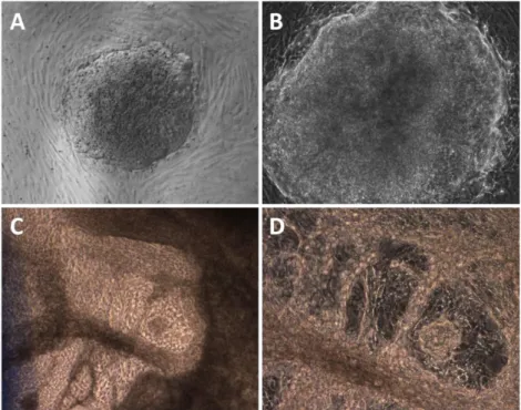
As detailed in Materials and Methods 2.7, GD iPSc colonies were separated from the underlying feeder layer without disruption and cultured in suspension for 3 days. During this time, the colonies took on a spherical morphology typical of EBs.
Figure 3.1 – Embryoid Body Differentiation Protocol. Neuronal differentiation protocol using embryoid bodies, seeding on a PA6 stromal cells layer, differentiating for 21 days under GMEM medium and treating with chaperones for 1 week.
34
When transferred to a PA6 stromal feeder layer in differentiation medium, EBs attached to the substrate as flattened discs, grew and started to differentiate (Figure 3.2). As expected, the colonies took on a heterogeneous aspect which suggested that they were composed by cells in many different stages of differentiation.
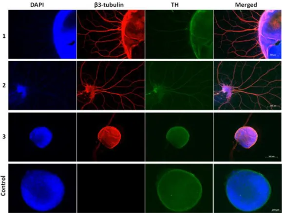
In order to characterize the differentiated cells, we analyzed the cultures for presence of neuronal markers by immunofluorescence. An anti-β3-tubulin antibody, a marker for mature/immature neurons, revealed many colonies with extended or intense signal, indicating efficient differentiation to the neuronal fate (Figure 3.3).
Figure 3.2 - Embryoid Body Differentiation Results. Embryoid bodies in the 5th day (A and B) and in the 21st day (C and D) of the neuronal differentiation. Magnifications: A,C – 50X ; B,D – 100X.
35
Although some colonies showed a positive signal for TH, the negative control (primary antibody ommitted) revealed significant unspecific binding of the secondary antibody leading us to disregard the TH immunofluorescence result. However, previous research done by our group showed that the EB formation with co-culture in PA6 stromal cells protocol yielded a high percentage of neurons with up to 35% of these being dopaminergic.
In order to manipulate β-glucocerebrosidase protein and activity levels, we chose to use chaperone compounds. In the context of GD, a chaperone is a molecule capable of interacting with β-glucocerebrosidase in a way that stabilizes its 3 dimensional structure, resulting in increased protein stability and trafficking to the lysosome. We treated the cultures with 7 different iminosugar candidate chaperones (MTD106, MG235, CVI62, MTD132, MTD131, TMB69 and TMB84) for one week, replenishing the chaperone compound every 48 hours. These pharmacological compounds were synthesized and are currently being characterized as competitive inhibitors of β-glucocerebrosidase by a collaborator group (C. Ortiz Mellet, University of Seville, Spain). Competitive inhibitors typically bind the enzyme’s active site and compete with
Figure 3.3 - Characterization of Embryoid Body Differentiation Results. Immunofluorescence against β3-tubulin (red, immature/mature neuron marker) and tyrosine hydroxylase (TH, green, dopaminergic neuron marker) and staining with DAPI (blue, nucleus marker) of GD FiPS 4F 21C embryoid bodies undergone neuronal differentiation.
36
the natural substrate with the degree of binding of each molecule determined by affinity for the binding site and concentration of the molecule. A good competitive inhibitor will have a high enough affinity to stabilize β-glucocerebrosidase in the cytoplasm, but low enough affinity to lose the competition to the natural substrate in the lysosome. Therefore, the effect of a given chaperone on protein levels and enzyme activity is not straighforward to predict. A chaperone may cause a large increase in protein stability but low increase in enzyme activity or viceversa; alternatively, protein levels and activity might be directly correlated.
In preliminar studies, the chaperones used in this work had a degree of ability to increase β-glucocerebrosidase activity in in vitro enzymatic assays, and furthermore, increase β-glucocerebrosidase protein levels and activity in fibroblasts of several GD genotypes (C. Ortiz Mellet, unpublished results). However, they have not been tested in neuronal cultures. Previous work in our laboratory had determined that a final concentration of 30 μM was the minimal amount of chaperone capable of achieving activity increases in fibroblasts. Therefore, this concentration was chosen for the experiment. A negative control (no chaperone treatment) was included (Figure 3.4).
After one week of treatment with chaperones, we harvested the differentiated cultures, extracted proteins and analyzed β-glucocerebrosidase, actin, β3-tubulin and α-synuclein protein levels through a western blot (Figures 3.5 and 3.6).
Figure 3.4 - Chaperone Treatment Protocol. Experimental scheme of GD FiPS 4F 21C differentiated cells’ treatment with chaperones.
37
We repeated the western blots and the results were variable, so we did a protein quantification using Image Lab (Bio-Rad Laboratories) of the several western blots and averaged the results. Since the protein extract comes from colonies with various cell types and actin and β-glucocerebrosidase are expressed in all types of cells, but β3-tubulin and α-synuclein are expressed only in neurons, levels of β-glucocerebrosidase
Figure 3.5 - Protein Levels of glucocerebrosidase and Actin I. Western blot analysis of β-glucocerebrosidase (GCase) and actin proteins in differentiated cells treated with chaperones. From top to bottom and from left to right: negative control (NC, no chaperone treatment) ; chaperones MTD106, MG235, CVI62 ; negative control (NC, no chaperone treatment) ; chaperones MTD132, MTD131, TMB69, TMB84.
Figure 3.6 - Protein Levels of α-synuclein and tubulin I. Western blot analysis of α-synuclein and β3-tubulin proteins in differentiated cells treated with chaperones. From top to bottom and from left to right: negative control (NC, no chaperone treatment) ; chaperones MTD106, MG235, CVI62 ; negative control (NC, no chaperone treatment) ; chaperones MTD132, MTD131, TMB69, TMB84.
38
were normalized to levels of actin but levels of α-synuclein were normalized to levels of β3-tubulin. Average results are shown in Graphs 3.1 and 3.2.
Graph 3.1 - Average GCase levels I (Fig. 3.5). Average protein levels of β-glucocerebrosidase (GCase) in differentiated cells treated with chaperones (average of 3 western blots). Normalized to actin, in relation to the negative control (no treatment). From left to right: negative control (NC, no chaperone treatment), chaperones MTD106, MG235, CVI62, MTD132, MTD131, TMB69 and TMB84. Asterisk marks statistically significant change via Student’s t-test (p < 0,05).
Graph 3.2 - Average α-synuclein levels I (Fig. 3.6). Average protein levels of α-synuclein in differentiated cells treated with chaperones (average of 5 western blots). Normalized to β3-tubulin, in relation to the negative control (no treatment). From left to right: negative control (NC, no chaperone treatment), chaperones MTD106, MG235, CVI62, MTD132, MTD131, TMB69 and TMB84. Asterisk marks statistically significant change via Student’s t-test (p < 0,05).
0 50 100 150 200 250 NC MTD106 MG235 CVI62 MTD132 MTD131 TMB69 TMB84 R atio α -sy n u cl e in /β3 -tu b u lin Samples
Average α-synuclein levels I
*
*
*
p = 0,03 p = 0,01 p = 0,01 0 100 200 300 400 500 600 NC MTD106 MG235 CVI62 MTD132 MTD131 TMB69 TMB84 R atio GCase /ac tin SamplesAverage GCase levels I
*
p = 0,00239
The large variability in β-glucocerebrosidase protein levels and lack of statistically significant differences was striking. In particular, treatment with MTD106, MG235, CVI62 resulted in unexpectedly low levels of β-glucocerebrosidase protein levels for which we have no satisfactory explanation, other than the chaperones actually destabilizing structure instead of stabilizing it in neurons, in frank contradiction with preliminar results. The high variability may be due to human error when applying the technique and/or the protein quantification by software. Even so, when analyzing the averages, it suggests that there may be a relation between β-glucocerebrosidase and synuclein, but not in the way that we hypothesized (an inverse relation), since α-synuclein levels match β-glucocerebrosidase levels. It is not possible to take any definitive conclusions regarding this result as the variability is too high. Consequence of this result, we proceeded to repeat this experiment but instead of treating with 7 chaperones, we treated the differentiated cells with only 5 chaperones (MG235, CVI62, MTD132, TMB69 and TMB84), again with a final concentration of 30 μM. After one week of treatment with chaperones we proceeded to harvest cells, extract proteins and analyze β-glucocerebrosidase, actin, β3-tubulin and α-synuclein protein levels through western blots (Figures 3.7 and 3.8).
Figure 3.7 - Protein Levels of β-glucocerebrosidase and Actin II. Western blot analysis of β-glucocerebrosidase (GCase) and actin proteins in differentiated cells treated with chaperones. From left to right: negative control (NC, no chaperone treatment) ; chaperones MG235, CVI62, MTD132, TMB69, TMB84.
Figure 3.8 - Protein Levels of α-synuclein and tubulin II. Western blot analysis of α-synuclein and β3-tubulin proteins in differentiated cells treated with chaperones. From left to right: negative control (NC, no chaperone treatment) ; chaperones MG235, CVI62, MTD132, TMB69, TMB84.