The prognostic value of the serum ferritin in a southern Brazilian cohort of
patients with Gaucher disease
Tiago Koppe
1,2; Divair Doneda
3; Marina Siebert
1,6,7; Livia Paskulin
1,2; Matheus Camargo
1; Kristiane
Michelin Tirelli
4; Filippo Vairo
4; Liane Daudt
1,5and Ida Vanessa D. Schwartz
1,2,4,61
Departamento de Genética, Universidade Federal do Rio Grande do Sul (UFRGS), Porto Alegre, RS,
Brazil.
2
Programa de Pós-Graduação em Genética e Biologia Molecular, Universidade Federal do Rio Grande do
Sul (UFRGS), Porto Alegre, RS, Brazil.
3
Laboratório de Técnica Dietética, Faculdade de Medicina, Universidade Federal do Rio Grande do Sul
(UFRGS), Porto Alegre, RS, Brazil.
4
Serviço de Genética Médica, Hospital de Clínicas de Porto Alegre (HCPA), Porto Alegre, RS, Brazil.
5Serviço de Hematologia Clínica, Hospital de Clínicas de Porto Alegre (HCPA), Porto Alegre, RS, Brazil.
6
Basic Research and Advanced Investigations in Neurosciences (BRAIN) Laboratory, Hospital de Clínicas
(HCPA), Porto Alegre, RS, Brazil.
7
Unidade de Análises Moleculares e de Proteínas (UAMP), Centro de Pesquisa Experimental, Hospital de
Clínicas de Porto Alegre (HCPA), Porto Alegre, RS, Brazil.
Abstract
The clinical utility of serum ferritin as a biomarker of disease severity and prognosis in Gaucher disease (GD) is still debated. Here, we aimed to evaluate ferritin and its relation to clinicolaboratory parameters of GD patients seen at the Reference Center for Gaucher Disease of Rio Grande do Sul, Brazil, so as to gather evidence on the utility of ferritin as a biomarker of this condition. A retrospective chart review was performed collecting pre- and post-treatment data from GD patients. Eighteen patients with ferritin levels available before and after post-treatment were in-cluded in the study. Nine of these participants were males, and seventeen had type I GD. All patients were given ei-ther enzyme replacement (n = 16) or substrate reduction ei-therapy (n = 2), and ferritin was found to decrease from 756 [318-1441] ng/mL at baseline to 521 [227-626] ng/mL (p = 0.025) after 28.8 months of treatment. Serum ferritin levels did not correlate with measures of disease severity, but showed an association with age at onset of treatment (r= 0.880;n = 18; p < 0.001). In conclusion, although serum ferritin did not correlate with disease severity, after a median 28.8 months of treatment, clinical outcomes had clearly improved, and ferritin levels had decreased.
Keywords: ferritin, biomarkers, Gaucher disease, iron metabolism.
Received: May 17, 2015; Accepted: September 28, 2015.
Introduction
Gaucher disease (GD) (OMIM #230800) is a meta-bolic disorder caused by a deficiency in lysosomal gluco-cerebrosidase activity (EC 3.2.1.45) due to mutations in the
GBA1gene (Grabowskiet al., 2014). The worldwide prev-alence of GD is about 1:40,000-200,000 (Poorthuiset al., 1999; Pintoet al., 2004), and its clinical manifestations in-clude multisystem organomegaly, pancytopenia and bone complications (Pastores and Hughes, 2000; Grabowskiet
al., 2014). Although the mechanisms by whichGBA1gene mutations influence GD phenotype remain largely un-known, the main pathogenesis of this disorder is believed to be the lysosomal accumulation of glucocerebroside in mononuclear phagocytes. This process triggers the expres-sion of cytokines and other inflammatory molecules which, in turn, engage other immune cells and ultimately lead to a systemic immune response (Baraket al., 1999; Cox, 2001; Bovenet al., 2004; Liuet al., 2012; Pandey and Grabowski, 2013; Vairoet al., 2013; Thomaset al., 2014). Several cir-culating molecules thought to be secreted by these lipid-laden macrophages have been studied as biological markers of disease burden and/or response to enzyme replacement therapy (ERT) (Aertset al., 2005; Cox, 2006). In this
re-Send correspondence to Ida V. Doederlein Schwartz. Programa de Pós-Graduação em Genética e Biologia Molecular, Universidade Federal do Rio Grande do Sul (UFRGS), Hospital de Clínicas de Porto Alegre. Rua Ramiro Barcelos 2350, Bairro Rio Branco, 90035-903 Porto Alegre, RS, E-mail: ischwartz@hcpa.ufrgs.br.
gard, although lyso-Gb1 has recently emerged as a promis-ing specific biomarker of GD (Fulleret al., 2015), the most frequent and consistently altered molecules in GD have been found to be the following: chitotriosidase (EC 3.2.1.14), angiotensin converting enzyme (EC 3.4.15.1), CCL18 (P55774), tartarate-resistant acid phosphatase (EC3.1.3.2) and serum ferritin (SF) (P02792 and P02794) (Aronson, 2005; Bootet al., 2009; Hughes and Pastores, 2013; Thomaset al., 2013, 2014). Over the past few years, several studies have described hyperferritinemia in GD pa-tients and demonstrated its reduction after the initiation of treatment. The first study to report hyperferritinemia over the course of GD was published by Morganet al.(1983), and since then these findings have been replicated in differ-ent cohorts worldwide (Pollet al., 2002; Grosboiset al., 2009; Saadiet al., 2010; Steinet al., 2010; Stirnemannet al., 2010; Stirnemannet al., 2011; Mekinianet al., 2012). The reduction in SF exhibited by patients with GD under-going ERT could be indicative of a positive clinical out-come (Stirnemann et al., 2010; Vigan et al., 2014), although no studies to date have identified a clear associa-tion between SF levels and GD severity. In Brazilian pa-tients, these findings have yet to be replicated. As such, the aim of this study was to evaluate the clinical use of SF as a biomarker of GD severity and/or response to treatment in a Brazilian cohort of GD patients.
Material and Methods
A retrospective chart review of patients with GD treated at the GD Reference Center of Rio Grande do Sul, Brazil, was performed. An extensive database of patients with GD was analyzed, and all those who had at least two measurements of SF levels - one before and the other after the onset of GD therapy (i.e., ERT or substrate reduction therapy - SRT) – were included in the study.
Ptreatment SF levels were defined as the most re-cent measurements available in the database prior to the on-set of treatment, and post-treatment SF was evaluated based on the most recent measurement available for each patient in the dataset. Hyperferritinemia was defined as SF > 322 ng/mL in males and > 291 ng/mL in females. All other vari-ables were matched to SF levels by date to determine their status as pre- or post-treatment measures. In the referral center, the diagnosis of GD is confirmed in all cases by re-duced glucocerebrosidase activity (i.e., < 10% of normal) in leukocytes. All patients received standard care, and had their clinical history, physical examinations, lab tests and medical imaging results recorded during routine medical assessments.
Asthenia, abdominal pain, bone involvement, bleed-ing, splenomegaly and hepatomegaly were evaluated based on clinical assessments or ancillary tests recommended by current guidelines. SSI (Severity Score Index) was calcu-lated on every medical visit as described in Zimranet al.
(1989). Quantitative variables were expressed as median
[25th-75th percentiles], and categorical variables were dis-played as absolute frequencies. Non-parametric tests were used to compare pre- and post-treatment variables and as-sess treatment effects. Two-tailed Wilcoxon signed-rank tests were used to analyze quantitative variables, while McNemar tests were involved in the evaluation of categori-cal data. Additionally,Spearman correlation analyses were carried out to assess the relationship between quantitative variables and SF.All tests were performed using SPSS 18 (IBM®) and statistical significance was considered whenp
< 0.05.
This study was approved by the Research Ethics Committee of the Hospital de Clínicas de Porto Alegre, Brazil.
Results
Eighteen patients with a diagnosis of GD met inclu-sion criteria for this study (male = 9, females = 9; GD type I = 17; GD type III = 1). Eleven patients had a N370S/L444P genotype, three were N370S/N370S, two were N370S/RecNciI, one was E349K/S366N and one, L444P/L444P. Median age at diagnosis and treatment onset was 39.5 years [15.7-51.2] and 41.1 years [24.6-52.9], re-spectively. Three patients had undergone splenectomy, one had cirrhosis and none had hepatocellular carcinoma. Pa-tients had no history of blood transfusions, although three female patients received a short course of iron supplementation over the follow-up and were excluded from the analysis of serum iron levels and transferrin satu-ration. At inclusion, sixteen patients were on ERT (velaglu-cerase alpha = 1; taliglu(velaglu-cerase alpha = 2; imiglu(velaglu-cerase = 13), and received a mean dosage of 21.56±12.21 UI/kg/inf every 2 weeks. Additionally, two patients were receiving SRT (eliglustat = 1; miglustat = 1). None of the 18 partici-pants had any other biochemical evidence of iron overload, such as high transferrin saturation.
18;p< 0.001) and Body Mass Index (BMI) (r= 0.713;n= 14;p< 0.005).
Discussion
This study aimed to investigate the clinical use of SF as a biomarker of disease severity and/or response to treat-ment in GD. Our results showed that hyperferritinemia is a common occurrence in GD, especially in untreated, older male patients with a higher BMI. Our results are also in agreement with findings obtained both from general and GD populations. SF levels are known to be influenced by sex and age, so that reference values differ by age and gen-der, in addition to population characteristics, such as eth-nicity and presence of other medical comorbidities (Luxton
et al., 1977; Vicenteet al., 1990; Custeret al., 1995; Koziol
et al., 2001; Pan and Jackson, 2008; Ferraroet al., 2012). Growing evidence has shown that SF is associated with BMI, obesity and insulin resistance, and nonalcoholic fatty liver disease (Guglielmiet al., 2015). In addition, several studies have found that women, regardless of age and
pres-ence of medical comorbidities, always tend to have lower SF values than men. Additionally, premenopausal women have dramatically lower SF levels than postmenopausal fe-males. The peaking of SF levels in menopause has not been associated with transferrin saturation (Koziolet al., 2001), but has been found to be related to hepcidin, the central peptide in iron metabolism regulation, whose levels are very strongly correlated with those of SF (Galeslootet al., 2011). The cutoff for hyperferritinemia varies across con-texts. In our sample, only 55% of the women were labeled as hyperferritinemic according to the cutoff points pro-vided by the laboratory where the analyses were performed. However, when the female cutoff values suggested by the American Association for the Study of Liver Diseases (15-150 ng/mL) (Baconet al., 2011) were applied to the sam-ple, the percentage of females categorized as hyperferri-tinemic was found to be higher (data not shown).
The magnitude of the increase in SF levels in patients with GD was very similar to that observed in a previous study (median 756vs.739 ng/mL, respectively) (Mekinian
et al., 2012), but lower than that reported by Stein et al.
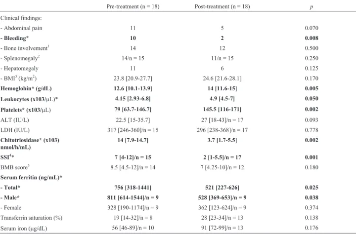
Table 1- Hyperferritinemia in patients with Gaucher disease: pre- and post-treatment data.
Pre-treatment (n = 18) Post-treatment (n = 18) p
Clinical findings:
- Abdominal pain 11 5 0.070
- Bleeding* 10 2 0.008
- Bone involvement1 14 12 0.500
- Splenomegaly2 14/n = 15 11/n = 15 0.250
- Hepatomegaly 11 6 0.125
- BMI3(kg/m2) 23.8 [20.9-27.7] 24.6 [21.6-28.1] 0.170
Hemoglobin* (g/dL) 12.6 [10.1-13.9] 14 [11.6-15] 0.005
Leukocytes (x103/mL)* 4.15 [2.93-6.8] 4.9 [4.5-7] 0.050
Platelets* (x103/mL) 79 [63.7-146.7] 145.5 [116-171] 0.002
ALT (IU/L) 22.5 [15-35.7] 27 [18-43]/n = 17 0.093
LDH (IU/L) 317 [246-360]/n = 15 296 [238-368]/n = 17 0.778
Chitotriosidase* (x103) nmol/h/mL)
14 [7.9-14.7] 3.7 [1.7-5.5] 0.002
SSI4* 7 [4-12]/n = 15 2 [1-5.5]/n = 17 0.001
BMB score5 8.5 [4.5-12]/n = 14 7 [4.25-10]/n = 12 0.180
Serum ferritin (ng/mL)*
- Total* 756 [318-1441] 521 [227-626] 0.025
- Male* 811 [614-1544]/n = 9 528 [369-653]/n = 9 0.038
- Female 328 [190-1174]/n = 9 362 [123-624]/n = 9 0.374
Transferrin saturation (%) 19 [14-32]/n = 8 28 [23-34]/n = 13 0.138
Serum iron (mg/dL) 56 [46-89]/n = 10 91 [72-99]/n = 13 0.176
Values expressed as median [25th-75th percentiles] or absolute count, unless otherwise specified;*p< 0.05 after McNemar or Wilcoxon signed-rank test; Reference values: serum ferritin (males: 22-322 ng/mL and females: 10-291 ng/mL); transferrin saturation (25-50%) and serum iron (50-170mg/dL).
1Bone involvement was considered whenever patients mentioned pain/pathological fractures without any other attributable cause, or in the presence of
imaging evidence of bone disease (e.g., osteopenia on bone densitometry).2Three patients had undergone splenectomy.3Body Mass Index (Kg/m2): < 18.5 – underweight; 18.5-24.9 – normal weight; 25-29.9 – overweight and > 30 – obesity4Clinical Severity Score Index: 0-9 – mild; 10-19 – moderate and
(2010), who found a 3.7-fold elevation above the upper limit of normality. These differences and similarities may be attributable to mean sample age, which was very similar between the present study and that performed by Mekinian
et al.(2012), but much lower in our sample than in the par-ticipants recruited by Steinet al.(2010).
Our data did not support the use of SF as a biomarker of GD severity. Previous studies have shown the same find-ings, suggesting that SF levels may not be influenced by disease severityper se, but by the number of years of dis-ease activity (i.e., older individuals). Low SF is pathogno-monic of iron deficiency and can be used to detect this condition with almost 100% specificity. However, high SF levels are not necessarily indicative of iron overload. Therefore, a transferrin saturation cutoff value > 45% has been suggested as a good predictor of iron overload (Gilles, 2013). In inflammatory conditions, such as GD, iron be-comes unavailable for erythropoiesis, despite adequate iron storage levels. This phenomenon is called functional iron deficiency and is characteristic of the anemia of chronic disorders. So, hyperferritinemia may exist in the presence or in the absence of iron overload, and recent studies have demonstrated macrophage iron accumulation in anemia of chronic disease (King and Weiss, 2014). In this regard, iron overload in inflammatory cells has also been identified in conditions such as Parkinson’s disease, multiple sclerosis and other neurodegenerative disorders using noninvasive imaging methods to quantify iron levels in different tissues (Stoll and Bendszus, 2009; Böttcheret al., 2013; Mehtaet al., 2013). The analysis of participant iron profiles showed that transferrin saturation and serum iron levels also ap-peared to increase after treatment, although this change was not statistically significant in our sample. Since iron defi-ciency and iron defidefi-ciency anemia is far more frequent in the Brazilian population than in people from developed countries and this tendency is reflected in Brazilian GD co-horts (Sobreiraet al., 2007), this lack of statistical signifi-cance could reflect a poor-iron diet. Nonetheless, taken together these findings may suggest that patients under treatment may have higher concentrations of iron available for erythropoiesis.
In conclusion, SF does not appear to be a useful biomarker of disease severity in GD, but can be used as a biomarker of diagnosis and response to treatment. Treat-ment also seemed to increase serum iron and transferrin sat-uration levels, increasing the amount of iron available for erythropoiesis. These findings need to be further evaluated in order to clarify the relation between SF and iron metabo-lism in GD patients.
Acknowledgments
Financial support for this study was provided by Con-selho Nacional de Desenvolvimento Científico e Tecnoló-gico (CNPq, Brazil), Fundo de Incentivo à Pesquisa e Eventos do Hospital de Clínicas de Porto Alegre
(FIPE/HCPA), and Fundação de Amparo a Pesquisa do Estado do Rio Grande do Sul (FAPERGS, RS, Brazil).
References
Aerts JM, Hollak CE, van Breemen M, Maas M, Groener JE and Boot RG (2005) Identification and use of biomarkers in Gaucher disease and other lysosomal storage diseases.Acta Paediatr Suppl 2005 94:43-46.
Aronson JK (2005) Biomarkers and surrogate endpoints. Br J Clin Pharmacol 59:491-494.
Bacon BR, Adams PC, Kowdlay KV, Powell LW, Tavill AS and American Association for the Study of Liver Diseases (2011) Diagnosis and management of hemochromatosis: Practice guideline from the American Association for the Study of Liver Diseases. Hepatology 54:328-343.
Barak V, Acker M, Nisman B, Kalickman I, Abrahamov A, Zimran A, Yatziv S (1999) Cytokines in Gaucher’s disease. Eur Cytokine Netw 10:205-210.
Boot RG, van Breemen MJ, Wegdam W, Sprenger RR, de Jong S, Speijer D, Hollak CE, van Dussen L, Hoefsloot HC, Smilde AK, et al. (2009) Gaucher disease: a model disorder for biomarker discovery. Expert Rev Proteomics 6:411-419. Böttcher T, Rolfs A, Meyer B, Grossmann A, Berg D, Kropp P,
Benecke R and Walter U (2013) Clinical, genetic, and brain sonographic features related to Parkinson’s disease in Gau-cher disease. J Neurol 260:2523-2531.
Boven LA, van Meurs M, Boot RG, Mehta A, Boon L, Aerts JM, Laman JD (2004) Gaucher cells demonstrate a distinct macrophage phenotype and resemble alternatively activated macrophages. Am J Clin Pathol 122:359-369.
Cox TM (2001) Gaucher disease: understanding the molecular pathogenesis of sphingolipidoses. J Inherit Metab Dis 24 (Suppl 2):106-121.
Custer EM, Finch CA, Sobel RE and Zettner A (1995) Population norms for serum ferritin. J Lab Clin Med 126:88-94. Ferraro S, Mozzi R, Panteghini M. (2012) Revaluating serum
ferritin as a marker of body iron stores in the traceability era. Clin Chem Lab Med 50:1911-1916.
Fuller M, Szer J, Stark S and Fletcher JM (2015) Rapid, sin-gle-phase extraction of glucosylsphingosine from plasma: a universal screening and monitoring tool. Clin Chim Acta. 29:6-10.
Galesloot TE, Vermeulen SH, Geurts-Moespot AJ, Klaver SM, Kroot JJ, van Tienoven D, Wetzels JF, Kiemeney LA, Sweep FC, den Heijer M and Swinkels DW (2011) Serum hepcidin: reference ranges and biochemical correlates in the general population. Blood. 117:e218-225.
Gilles A (2013) Iron’s ups and downs. Rev Med Brux 34:328-334. Grosbois B, Revest M, Decaux O (2009) Major hyperferritinemia, autoimmune thrombocytopenic purpura and lymphocytic lymphoma in Gaucher disease. Presse Med 38:2S56-2S57. Guglielmi V, D’Adamo M, Bellia A, Ciotto RT, Federici M,
Lauro D and Sbraccia P (2015) Iron status in obesity: an in-dependent association with metabolic parameters and effect of weight loss. Nutr Metab Cardiovasc Dis 25:541-547. Hughes DA and Pastores GM (2013) Haematological
manifesta-tions and complicamanifesta-tions of Gaucher disease. Curr Opin Hemato20:41-47.
Koziol JA, Ho NJ, Felitti VJ and Beutler E (2001) Reference centiles for serum ferritin and percentage of transferrin satu-ration, with application to mutations of the HFE gene. Clin Chem 47:1804-1810.
Liu J, Halene S, Yang M, Iqbal J, Yang R, Mehal WZ, Chuang WL, Jain D, Yuen T, Sun L,et al.(2012) Gaucher disease gene GBA functions in immune regulation. Proc Natl Acad Sci USA 109:10018-10023.
Luxton AW, Walker WH, Gauldie J, Ali AM and Pelletier C (1977) A radioimmunoassay for serum ferritin. Clin Chem. 23:683-689.
Mehta V, Pei W, Yang G, Li S, Swamy E, Boster A, Schmalbrock P and Pitt D (2013) Iron is a sensitive biomarker for inflam-mation in multiple sclerosis lesions. PLoS One 8:e57573. Mekinian A, Stirnemann J, Belmatoug N, Heraoui D, Fantin B,
Fain O, Charpentier A and Rose C (2012) Ferritinemia dur-ing type 1 Gaucher disease: mechanisms and progression under treatment. Blood Cells Mol Dis 49:53-57.
Morgan MA, Hoffbrand AV, Laulicht M, Luck W and Knowles S (1983) Serum ferritin concentration in Gaucher’s disease. Br Med J 286:1864.
Pan Y and Jackson RT (2008) Insights into the ethnic differences in serum ferritin between black and white US adult men. Am J Hum Biol 20:406-416
Pandey MK and Grabowski GA (2013) Immunological cells and functions in Gaucher disease. Crit Rev Oncog 18:197-220. Pinto R, Caseiro C, Lemos M, Lopes L, Fontes A, Ribeiro H,
Pinto E, Silva E, Rocha S, et al. (2004) Prevalence of lysosomal storage diseases in Portugal. Eur J Hum Genet 12:87-92.
Poll LW, Koch JA, Willers R, Aerts H, Scherer A, Häussinger D, Mödder U and vom Dahl S (2002) Correlation of bone mar-row response with hematological, biochemical, and visceral responses to enzyme replacement therapy of nonneurono-pathic (type 1) Gaucher disease in 30 adult patients. Blood Cells Mol Dis. 28:209-220.
Poorthuis BJ, Wevers RA, Kleijer WJ, Groener JE, de Jong JG, van Weely S, Niezen-Koning KE, van Diggelen OP (1999) The frequency of lysosomal storage diseases in The Nether-lands. Hum Genet 105:151-156.
Saadi T, Rosenbaum H, Veitsman E, Baruch Y (2010) Gaucher’s disease type I: a disease masked by the presence of abnormal laboratory tests common to primary liver disease. Eur J Gastroenterol Hepatol 22:1019-1021.
Sobreira E, Pires RF, Cizmarik M, Grabowski GA (2007). Pheno-typic and genoPheno-typic heterogeneity in Gaucher disease type 1: a comparison between Brazil and the rest of the world. Mol Genet Metab 90:81-86.
Stein P, Yu H, Jain D and Mistry PK (2010) Hyperferritinemia and iron overload in type 1 Gaucher disease. Am J Hematol 85:472-476.
Stirnemann J, Belmatoug N, Vincent C, Fain O, Fantin B and Mentré F (2010) Bone events and evolution of biologic
markers in Gaucher disease before and during treatment. Ar-thritis Res Ther. 12:R156.
Stirnemann J, Boutten A, Vincent C, Mekinian A, Heraoui D, Fantin B, Fain O, Mentré F, Belmatoug N (2011) Impact of imiglucerase on the serum glycosylated-ferritin level in Gaucher disease. Blood Cells Mol Dis 46:34-38.
Stoll G and Bendszus M (2009) Imaging of inflammation in the peripheral and central nervous system by magnetic reso-nance imaging. Neuroscience 158:1151-1160.
Thomas AS, Mehta AB and Hughes DA (2013) Diagnosing Gau-cher disease: an on-going need for increased awareness amongst haematologists. Blood Cells Mol Dis 50:212-217. Thomas AS, Mehta AB and Hughes DA (2014) Gaucher disease:
haematological presentations and complications. Br J Hae-matol 165:427-440.
Vairo F, Portela P, Salim PH, Jobim M, Netto C, Dorneles A, Mittlestadt S, Jobim LF and Schwartz IV (2013) KIR genes and HLA class I ligands in Gaucher disease. Gene. 516:53-57.
Vicente C, Porto G and de Sousa M (1990) Method for establish-ing serum ferritin reference values dependestablish-ing on sex and age. J Lab Clin Med 116:779-784.
Vigan M, Stirnemann J, Caillaud C, Froissart R, Boutten A, Fantin B, Belmatoug and, Mentré F (2014) Modeling chan-ges in biomarkers in Gaucher disease patients receiving en-zyme replacement therapy using a pathophysiological model. Orphanet J Rare Dis. 9:95.
Zimran A, Sorge J, Gross E, Kubitz M, West C and Beutler E (1989) Prediction of severity of Gaucher’s disease by identi-fication of mutations at DNA level. Lancet 2:349-352.
Internet Resources
Grabowski GA, Petsko GA, Kolodny EH (2014). Gaucher Dis-ease. In: Valle D, Beaudet AL, Vogelstein B, Kinzler KW, Antonarakis SE, Ballabio A, Gibson K and Mitchell G (eds), OMMBID - The Online Metabolic and Molecular Bases of Inherited Diseases, http://ommbid.mhmedical.com/con-tent.aspx?bookid = 474&Sectionid = 45374148 (accessed August 20, 2014)
Cox TM (2006) Biomarkers in lysosomal storage diseases. In: Mehta A, Beck M and Sunder-Plassmann G (eds) Fabry Dis-ease: Perspectives from 5 Years of FOS. Oxford
PharmaGenesis, Oxford,
http://www.ncbi.nlm.nih.gov/books/NBK11586/ (accessed August 20, 2014)
Pastores GM, and Hughes DA (2000) Gaucher Disease. In: Pagon RA, Adam MP, Ardinger HH,et al.(eds), GeneReviews®, http://www.ncbi.nlm.nih.gov/books/NBK1269/ (accessed August 10, 2014).,
Associate Editor: Jeremy A. Squire