INTRA OCULAR PRESSURE
IN CHRONIC USERS OF ORAL
GLUCOCORTICOIDS FOR
RHEUMATOID ARTHRITIS
Christie Graf,
Gabriele Cardoso,
Marília Barreto Silva,
A B S T R A C T
Objective: To evaluate intra ocular pressure (IOP) among rheumatoid arthritis (RA) patients who are chro-nic oral glucocorticoid users in low to moderate doses.
Material and methods: We have studied 125 subjects: 72 glucocorticoid users and 53 controls. The gluco-corticoid users were RA patients treated with 5 to 40 mg/day of prednisone or equivalent. Controls were patients with osteoarthritis or with soft tissue rheumatic syndromes who had never used glucocorticoid orally or locally. The IOP was measured three times with Perkins tonometer in both eyes and the mean va-lue was compared between groups. For statistical analysis we used the mean vava-lue between the IOP of both eyes.
Results: Among RA glucocorticoid users the mean dose was 9.7 mg of prednisone daily during a mean pe-riod of 71.1 months. The IOP of glucocorticoid users was 5.8% higher than controls. This difference did not reach statistical significance. The rise in IOP was not affected by the duration of glucocorticoid treat-ment or by the dose. No RA patient using oral glucocorticoids was found to have abnormal IOP in this study. Conclusions: Low dose glucocorticoids causes a small (5.8%), non significant increase in intraocular pres-sure.
Keywords: Glucocorticoid; Glaucoma; Eye Diseases; Rheumatoid Arthritis.
R E S U M O
Objetivo: Estudar a pressão intra ocular (PIO) em portadores de artrite reumatóide (AR) em uso de doses moderadas e baixas de glicocorticóide por via oral.
Casuística e métodos: Foram estudados 125 indivíduos: 72 em uso de glicocorticóide e 53 controles. Os usuários de glicocorticóide eram pacientes com AR tratados com doses de 5 até 40 mg/prednisona/dia ou equivalente. Os controles eram pacientes com afecções reumáticas de partes moles e portadores de os-teoartrite que nunca haviam feito uso de glicocorticóides orais ou tópicos. A PIO foi medida em ambos os olhos com tonômetro de Perkins por três vezes, sendo utilizada, para fins de estudo, uma média das três medidas. Para análise estatística considerou-se a média de PIO dos dois olhos.
Resultados: A dose média de glicocorticóide usada pelos pacientes com AR era de 9,7 mg/prednisona/ dia por um tempo médio de 71,1 meses. A PIO dos usuários de glicocorticóide era 5,8% acima daquela dos controles. A diferença não atingiu significância estatística. Nenhum dos portadores de AR em uso de gli-cocorticóide tinha PIO anormal.
Conclusões: Doses baixas de glicocorticóides, usadas oralmente, causam um aumento pequeno (5,8%) e não significante da PIO.
A R T I G O O R I G I N A L
I N T R A O C U L A R P R E S S U R E I N C H R O N I C
U S E R S O F O R A L G L U C O C O R T I C O I D S F O R
R H E U M A T O I D A R T H R I T I S
Christie Graf
*, Gabriele Cardoso
**,
Marília Barreto Silva
**,Thelma L.Skare
**Introduction
Glucocorticoids have been extensively used in rheumatology because of their immunomodula-tory and anti-inflammaimmunomodula-tory effects. Joint inflam-mation in rheumatoid arthritis (RA) patients is par-ticularly sensible to this type of drug and many RA patients use these medications in low doses for a long period of time1. Higher doses are required for
extraarticular involvement1.
Despite the positive effects of glucocorticoids in RA, patients who use them are at risk of side effects. Among such side effects is glaucoma2.
Glaucoma is a complication when patients use glucocorticoids as eye drops or sprays, but very few studies addressed this question when glucocorti-coids are used orally.
The present study examines the effects of chro-nic oral glucocorticoid use on the intra ocular pres-sure (IOP) of RA patients.
Methodology
This study was approved by the local Ethics Com-mittee and all patients gave informed consent. We studied 125 subjects: 72 RA patients on glucocorti-coids and 53 as controls. The controls were patients with osteoarthritis and soft tissue rheumatic syndromes without any glucocorticoid use (oral or local). This sample corresponds to the patients who attended the Hospital Universitário Evangélico de Curitiba during a recruitment period of 6 months and that were willing to participate in the study.
We considered chronic users patients that used oral glucocorticoids continuously for at least
6 months.
Both groups were submitted to three sequential measurements of IOP of each eye in the same day and the mean of these three measurements was the value considered for analysis. Patients were also questioned about relatives with glaucoma.
The measurement of IOP was taken by a single ophthalmologist using a Perkins tonometer. Pa-tients with inflammatory ophthalmologic disease were excluded.
The data was analyzed using the mean pressure of both eyes. As the IOP obtained values followed Gaussian distribution, we used Student’s t test to study the difference between pressure means. Pear-son’s correlation test was used for evaluation of IOP in RA glucocorticoid users according to dose and therapy duration with the help of the software Graph Pad Prism, version 4.0. The significance adopted was 5%.The sample size showed 73% po-wer to reject a type 2 error.
Results
The control group age range was 18 to 84 years with a mean of 48.9 ±16.8 years. In the glucocorticoid group patients’ age varied from 14 to 79 years with a mean of 45.8±17.02 years. One patient from the control group and two from the glucocorticoid group had first degree relatives with glaucoma. None of them had previous history of high IOP.
In the RA glucocorticoid group, the dose of this drug was between 5 and 40 mg of prednisone/day (mean of 9. 7±6.6 mg/day; median of 10 mg/day). Sixty RA patients were using up to 10mg of predni-sone/day, 9 up to 20mg and 3 more than 30mg. The
groups is present in Table II.
The mean intraocular pressure was 12.8 mm in the control group and 13.4 mm in the glucocorti-coid group corresponding this difference to a t va-lue of -1.28 (p=0.20). The difference between me-ans was of 0.58 (confident interval of 95% = -0.31 to 1.48). (Figure 1). RA glucocorticoid users showed an increase of 5.8 % in IOP (p=0.137).
No patients (RA glucocorticoid users and con-trols) had an IOP higher than 20 mm Hg, which is considered the upper limit of normal.
Analyzing the rise in intraocular pressure of RA patients and the duration of glucocorticoid use we found no correlation (r=0.027 and p= 0.824) be-tween these two variables. (Figure 2).
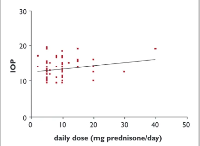
A weak relationship (r= 0.1725 and p=0.0553) was found between IOP and the daily dose of glu-cocorticoid in RA gluglu-cocorticoid treated patients. This data is depicted in Figure 3.
Discussion
The mechanisms of glucocorticoid action have been divided by Buttgereit et al in three main groups3:
1) mechanisms that causes genomic modifica-tions;
G L U C O C O R T I C O I D S A N D I N T R A O C U L A R P R E S S U R E I N R A
2) mechanisms that do not cause genomic altera-tions but are mediated by receptors;
3) physico-chemical mechanisms.
The first mechanism is the most studied one. It happens when the glucocorticoid molecule binds to intra cytoplasmatic receptors. After this the complex is transferred to the cell nucleus where it
Table I. Demographic Data in Controls and Glucocorticoid Users
Corticoid Controls Users
Number of patients 52 73
Mean age (years) 48.9±16.8 45.8±17.0
Number of patients with
relatives with glaucoma 1 2
Gender (female:male) 42:11 60:12
Table II. Distribution of Intraocular Pressure in the Studied Population
IOP 8-10mm Hg 11-12mm Hg 13-14mm Hg 15-16mm Hg 16-20mm Hg
With glucocorticoid 3 17 21 19 12
n (%) (4.17%) (23.61%) (29.17%) (26.39%) (16.67%)
Without 6 14 13 14 5
glucocorticoid n (%) (11.54%) (26.92%) (25.0%) (26.96%) (9.62%)
n = number of studied patients; IOP = intra ocular pressure; mmHg = millimeters of mercury
RA Glucocorticoid users Controls IOP 20 10 0
Figure 1. Intra ocular pressure in RA glucocorticoid
users and controls.
Months of use IOP 30 20 10 0 0 100 200 300 400
Figure 2. Intra ocular pressure in RA glucocorticoid
C H R I S T I E G R A F E C O L.
the sample size gave only a 73% chance to rejected a type 2 error.
The literature has many observations stating that locally used corticosteroid increases the intra ocular pressure with considerable morbidity7, 8, 9.
This may be explained by the fact that local use can cause higher concentrations, firing some type of mechanism other than the genomic action. As far as we know, no study has addressed the ques-tion of IOP during the oral use of this drug.
We could not find any influence of daily gluco-corticoid use in the value of IOP. However, one should note that, in this study, the median used dose was low (10mg/day of prednisone). Studies with a higher number of patients and a higher dose may be able to demonstrate this.
Another question to be taken into account is that glucocorticoid glaucoma as well as juvenile glaucoma and open angle glaucoma suffer gene-tic influence, such as allelic variations of myociclin gene (MYOC)10. In the studied population the
po-sitive history for familial glaucoma was very low, so we cannot exclude the risk of greater increase in IOP with oral corticosteroids in these families.
Conclusion
In this study all RA patients under glucocorticoid treatment had normal IOP. Low dose oral glucocor-ticoid use in RA patients causes a small, non signi-ficant increase in the IOP.
References
1. Kirwan JR. Systemic glucocorticosteroids in rheuma-tology. In Klippel JH , Dieppe PA (eds) Rheumatology, 2nd Ed , Mosby Ed , London, 1998: S3:6.1-6.
2. Lester RE, Wolwe SR, Shear NH. The risks of systemic corticosteroid use. Dermatology Clinics 1998; 16:277--88.
3. Buttgereit F, Wehling M, Burmester G-R. A new hypo-thesis of modular glucocorticoid actions. Arthritis Rheum 1998; 41:61-767.
4. Bloom J. New insights into molecular basis of gluco-corticoid action. Immunol Allergy Clin North Am 1999;19:653-670.
5. Underwood JL, Murphy CG, Chen J et al. Glucocorti-coids regulate transendotelial fluid flow resistance and formation of intercellular junctions. Am J Physiol
daily dose (mg prednisone/day)
IOP 30 20 10 0 0 10 20 30 40 50
Figure 3. Intraocular pressure in RA glucocorticoid users
according to daily use.
binds to DNA and acts by increasing or decreasing the RNA messenger transcription (RNAm). The therapeutic actions, as well as the side effects of glucocorticoids, are caused by the increased or de-creased synthesis of chemical mediators-control-led by DNA transcription into RNAm3,4.
The other mechanisms are less studied, but it is known that they require higher doses of glucocor-ticoid in order to appear. The physico-chemical mechanisms are seen only in cases of glucocorti-coid pulse therapy2. This mechanism is mediated
by changes in the passage of ions through the cyto-plasmatic membrane2.
The time required to act through each of the mechanisms follows the reverse order of the nee-ded dose2.
There are doubts on which of the mechanisms might be involved in glaucoma appearance. Ac-cording to Underwood et al glucocorticoids raise the production of ZO-1- a protein present in the in-tercellular gaps of Schlemm Canal, causing incre-ased resistance to transendotelial flow of aqueous humor5. This results from genomic action by
glu-cocorticoid which requires low doses to occur. The-re is a very inteThe-resting observation by Mc Carthy et al. referring that people with open angle glaucoma have higher levels of endogenous corticoids6.
Our findings showed that no RA patient using glucocorticoid had abnormal IOP. Corticosteroid in
acetonide injection.Am J Ophthalmol. 2004; 138:286--287.
8. Garrott HM, Walland MJ. Glaucoma from topical cor-ticosteroids to the eyelids. Clin Experiment Ophthal-mol 2004; 32: 224-226.
9. Kaushik S, Gupta V, Gupta A, Dogra MR, Singh R. In-tractable glaucoma following intravitreal triamci-nolone in central retinal vein occlusion. Am J Oph-thalmol 2004; 137: 758-760.
10. Clark AF, Steely T, Dickerson Jr JE et al. Glucocorti-coid induction of the glaucoma gene MYOC in hu-man and monkey trabecular meshwork cells and tis-sues. Invest Ophthalmol Vis Sci. 2000; 42:1769-1780.
Author for correspondence:
Thelma L. Skare
Rua João Alencar Guimarães 796 80310-420 – Curitiba - PR E-mail: tskare@onda.com.br