2 3 6 Arq Bras Oftalmol. 2015;78(4):236-40 http://dx.doi.org/10.5935/0004-2749.20150061
INTRODUCTION
Rheumatoid arthritis (RA) is a chronic, systemic, autoimmune, in -flam matory disorder that primarily affects synovial joints. Extra arti-cular manifestations are also observed, including eye involvement in RA(1). The most common manifestation of RA in the eye is dry eye disease (DED). Dry eye symptoms occur in up to 45% of RA patients, with DED being diagnosed in 2.2% to 16.3%(2). In addition to DED, cor neal involvement including stromal keratitis, sclerosing keratitis, keratolysis, marginal furrowing or guttering, and peripheral ulcerative keratitis may be observed in RA. Additionally, these corneal ulcera-tions tend to develop more commonly in the peripheral cornea(3).
The evaluation of corneal parameters is important in ophthalmic examinations. These parameters may provide valuable information ABSTRACT
Purpose: To evaluate central corneal thickness (CCT ) and peripheral corneal thick-ness (PCT ) in patients with rheumatoid arthritis (RA) and to assess the relationships among the corneal parameters, dry eye disease, and clinical variables of RA.
Methods: A total of 58 RA patients and 58 control subjects participated in this study. A detailed ophthalmological examination was performed on each subject. Dry eye evaluation was performed using Schirmer’s test, tear break-up time (TBUT ), corneal fluorescein staining, and Ocular Surface Disease Index (OSDI). Corneal thickness at the apex point, the center of the pupil, the thinnest point, and PCT (3 mm from the apex to the superior, inferior, nasal, and temporal locations) were evaluated using Scheimpflug imaging (Pentacam®). Additionally, the relative peripheral index (RPI) was calculated by dividing the PCT by the CCT. The disease severity and quality of life were evaluated with DAS28 and HAQ, respectively. The laboratory evaluation comprised ESR and CRP.
Results: The mean corneal thicknesses at the apex point, the center of the pupil, the thinnest point, and the superior, inferior, nasal, and temporal points were significantly thinner in RA patients than controls. Schirmer’s test scores and TBUT were significantly lower, and corneal staining and OSDI scores were significantly higher in RA patients. There were no significant correlations between the corneal parameters and the clinical variables of RA or dry eye tests.
Conclusion: The CCT and PCT were thinner in RA patients compared to those in control subjects. However, there were no significant correlations between the corneal parameters and the clinical variables of RA or dry eye tests.
Keywords: Rheumatoid arthritis; Corneal topography; Dry eye
RESUMO
Objetivo: Avaliar da espessura central da córnea (CCT) e espessura corneana periférica (PCT ) em pacientes com artrite reumatoide (RA). O segundo objetivo foi avaliar as relações entre os parâmetros de córnea, doença do olho seco e variáveis clínicas da RA. Método: Um total de 58 pacientes com RA e 58 indivíduos do grupo controle partici-param deste estudo. Exame oftalmológico detalhado foi realizado para cada indivíduo. Avaliação do olho seco foi realizada por meio do teste de Schirmer, tempo de ruptura do filme lacrimal (TBUT ), coloração com fluoresceína da córnea e do índice de doença da superfície ocular (OSDI). Espessura da córnea no ápice, centro da pupila e ponto mais fino, assim como PCT (três milímetros do ápice para localização superior, inferior, nasal e temporal) foram avaliadas por meio de imagens Scheimpflug (Pentacam®). Além disso, o índice periférico relativo (RPI) foi calculado dividindo-se a PCT pela CCT. A gravidade da doença e qualidade de vida foram avaliados com DAS28 e HAQ respectivamente. A avaliação laboratorial foi composta por VHS e PCR.
Resultados: As espessuras de córnea médias no ápice, centro da pupila, ponto mais fino, assim como nos pontos superior, inferior, nasal e temporal foram significa ti va-mente mais finas em pacientes com RA do que nos controles. Os resultados dos testes de Schirmer e TBUT foram significativamente menores e a coloração por fluoresceína e o OSDI foram significativamente maiores em pacientes com RA. Não houve cor-relações significativas entre os parâmetros da córnea e variáveis clínicas da RA ou testes de olho seco.
Conclusões: A espessura corneana central e periférica foram mais finas em pacientes com RA em comparação com indivíduos controle. Não houve correlações significativas entre os parâmetros da córnea e variáveis clínicas da AR ou testes de olho seco.
Descritores: Artrite reumatóide; Topografia da córnea; Olho seco
on suspected glaucoma, calculation of intraocular lens power, ke-ratoconus monitoring, and investigation of refractive disorders(4-6).
Pentacam® is a noninvasive and objective device that allows ex-tensive evaluation of the corneal structure using a three-dimensional model showing the thickness, volume, and spatial section using the Scheimpflug imaging technique(7).
In a recent study, Cingü et al.(8) investigated corneal parameters in RA patients by Pentacam; however, they did not evaluate the peri-pheral corneal thickness (PCT) nor the associations between clinical variables of RA and corneal parameters.
Therefore, the aim of this study was to evaluate the central cor-neal thickness (CCT) and PCT in patients with RA. The secondary aim was to assess the relationships among the corneal parameters, DED, and clinical variables of RA.
Evaluation of central and peripheral corneal thicknesses in patients with
rheumatoid arthritis
Avaliação das espessuras corneanas centrais e periféricas em pacientes com artrite reumatoide
Alime Gunes1, esrA erkol inAl2, levent tok1, ozlem tok1
Submitted for publication: February 13, 2015 Accepted for publication: April 15, 2015
1 Department of Ophthalmology, Faculty of Medicine, Süleyman Demirel University, Isparta, Turkey. 2 Department of Physical Medicine and Rehabilitation, Faculty of Medicine, Süleyman Demirel
University, Isparta, Turkey.
Funding: No specific financial support was available for this study.
Disclosure of potential conflicts of interest: None of the authors have any potential conflict of interest to disclose.
METHODS
The study was performed in adherence to the tenets of the De-claration of Helsinki and was approved by the local ethics committee. Written informed consent was given by all RA patients and control subjects.
This cross-sectional study was conducted between May 2014 and December 2014, and included 58 patients with RA and 58 healthy subjects. All RA patients were referred from the Physical Medicine and Rehabilitation Department of the Süleyman Demirel University for their routine eye examination. Control subjects included patients admitted to the ophthalmology outpatient clinic for routine exami-nation without any complaints.
Exclusion criteria included history of ocular trauma or surgery, presence of refractive errors more than ±4.0 diopters (D), anterior segment disease, contact lens use, active corneal infection, glauco-ma, diabetes mellitus, other chronic inflammatory diseases that may affect the eye such as ankylosing spondylitis and Behçet’s disease, and current use of topical ophthalmic medication or corneal toxic pharmaceutical drugs.
E
XAMINATIONSThe age and gender of all participants were recorded, along with the durations of disease for the patients.
The disease activity and quality of life were assessed on the same day as the ophthalmic examinations. The former was assessed using the Disease Activity Index 28 (DAS28), which was evaluated using the DAS28 online calculator of the American College of Rheumatology with respect to the number of tender and swollen joints, C-reactive protein (CRP) level, and the global health assessment of the patient using a visual analog scale(9). The quality of life was evaluated with the Health Assessment Questionnaire (HAQ)(10), which includes questions in eight categories: dressing, rising, eating, walking, hygiene, reach, grip, and usual activities. Each question is answered on a four-level scale of impairment ranging from 0 to 3: 0 = no difficulty; 1 = some difficulty; 2 = much difficulty; and 3 = inability to do so.
The laboratory evaluation tested the erythrocyte sedimentation rate (ESR) and CRP.
O
PHTHALMOLOGICEXAMINATIONSAll subjects underwent a detailed ophthalmic examination inclu-ding a medical history review, spherical equivalent (SE), best-corrected visual acuity, intraocular pressure (IOP), tear break-up time (TBUT), Schirmer’s test under topical anesthesia, corneal fluorescein staining, corneal power measurements, pachymetric measurements, and cor-neal volume measurements. The Ocular Surface Disease Index (OSDI) questionnaire was used to assess subjective discomfort related to dry eye(11).
To avoid diurnal variation in the corneal parameters, all measure-ments were performed within the same time interval (from 10 am to 2 pm), and at a temperature that ranged from 20 to 25°C and relative humidity that ranged from 35 to 45%(12).
S
CHEIMPFLUGIMAGINGCorneal parameters were performed using a high-resolution rota-ting Scheimpflug imaging system (HR Pentacam, Oculus, Germany). Both eyes of all subjects were scanned by the same ophthalmologist (AG). For the examination, the subjects sat on the chair and put their chin on the chinrest, with their forehead touching the headband. The patients were asked to look at the black spot in the middle of the blue fixation lamp. The measurement automatically started only when correct alignment and focus of the eyes were achieved. Unreasonable measurements were not evaluated, and were marked in yellow and red on the monitor. The mean of three successive measurements was used for analysis.
Cornea thickness at the apex point (regarded as the CCT), the center of the pupil, the thinnest point, and the superior, inferior,
nasal, and temporal (at 3 mm from the apex) points were evaluated automatically. Further, corneal power and corneal volume values were recorded.
In addition, the relative peripheral index (RPI) was calculated by dividing the PCT by the CCT. This index represents the rate of thicke-ning of the cornea from the center to the periphery(13).
E
VALUATIONOFDRYEYEEvaluation of DE was performed after Scheimpflug imaging to prevent inaccurate pachymetric measurements(14). The TBUT test was performed with a sterile fluorescein strip that was placed in the lower eyelid fornix. The patient was instructed to blink three times and then look straight ahead without blinking. The time interval between a complete blink and the first appearance of a dry spot in the pre-corneal tear film was measured under cobalt blue-filtered light. The mean of three consecutive TBUT test measurements were used for analysis.
Corneal fluorescein staining was evaluated under cobalt blue illumination 2.5 to 3.0 min after fluorescein instillation. The level of corneal staining was graded according to the National Eye Institute/ Industry Workshop scale(15).
Subsequently, Schirmer’s test was performed with topical anes-thesia. Three minutes after one drop of proparacaine 0.5% was instil-led, a Schirmer’s test strip was placed behind the lower lid between the temporal and middle third of the eyelid. After 5 min, the strip was removed, and the wet portion of the paper was measured in millimeters. Patients who had a Schirmer’s test score ≤5 mm/5 min were considered to have dry eye.
S
TATISTICALANALYSISSPSS statistical software, version 15.0 (SPSS, Chicago, IL, USA), was used for the statistical analyses using one randomly selected eye from each subject. The Kolmogorov–Smirnov normality test was used to determine the normal distribution of continuous variables. Parame-tric continuous variables were given as the mean ± SD. Data with non-normal distribution were presented as median and interquartile ranges (the range of values lying between the 25th and 75th percen-tiles). Categorical data were analyzed with the chi-squared test, and were presented as the number of cases and percentages. Student’s t
test was used for continuous variables with normal distribution, and the Mann-Whitney U test was used for continuous variables that were not normally distributed to determine the differences between RA patients and healthy controls. Pearson’s and Spearman’s tests were used to detect the strength of the relationships between the varia-bles. P values <0.05 were considered statistically significant.
RESULTS
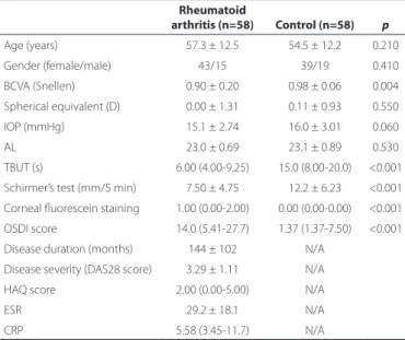
The demographic and clinical features of the study and control groups are shown in table 1. No statistically significant differences were found in terms of age, sex, visual acuity, SE, IOP, or axial length between the patient and control groups. The mean age was 57.3 ± 12.5 years for the patient group and 54.5 ± 12.2 years for the control group. The median duration of disease was 144 ± 102 months (range 6-516 months).
According to the results of Schirmer’s test, 39.3% (n=24) of the RA patients were diagnosed with DED. The results of Schirmer’s test and TBUT were significantly lower, and corneal staining and OSDI scores were significantly higher in RA patients than in controls (p<0.001, for all) (Table 1).
238 Arq Bras Oftalmol. 2015;78(4):236-40
were significantly lower in the RA group than in the control group (p<0.001, for all). The CCT was thinner than all the PCTs, followed by temporal, inferior, nasal, and superior corneal thicknesses, respecti-vely, in both groups. The mean corneal volume was also significantly lower in the RA group than in the control group (58.6 ± 3.05 mm3 versus 60.5 ± 2.85 mm3, p<0.001) (Table 2). However, no statistically significant differences in the RPI of PCTs were found between the patient and control groups (p>0.05, for all) (Table 2).
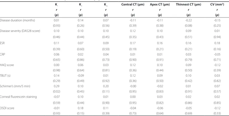
There were no significant correlations between the corneal pa-rameters and the clinical variables of RA, the dry eye tests (TBUT, Schirmer’s test, corneal fluorescein staining), and OSDI score (Table 3 and Table 4).
In addition, relationships were found between the disease duration and TBUT, and Schirmer’s test values; disease severity and OSDI score; and quality of life and Schirmer’s test, corneal fluorescein staining values, and OSDI score (Table 5).
DİSCUSSİON
To our knowledge, this is the first study to examine not only the CCT, but also the PCT, in patients with RA. In addition, dry eye tests and clinical variables of RA were also evaluated.
Ocular involvement may be quite variable in patients with RA. Dry eye and corneal involvement are common clinical ophthalmo-logic findings in patients with RA(1-3,8,16-19). The early diagnosis of DED and corneal involvement in patients with RA is crucial to provide timely management of potentially serious and vision-threatening complications.
It is well established that dry eye is frequently associated with RA, although the severity of dry eye is not associated with RA acti-vity. Therefore, it is recommended that dry eye should always be taken into consideration regardless of the RA activity(16). The results of the present study are consistent with previous reports(1,2,8,16). We found that Schirmer’s test score and TBUT were significantly lower, and corneal staining and OSDI scores were significantly higher in RA patients compared to healthy subjects. According to the results of Schirmer’s test, 39.3% of the RA patients in our study were diagnosed with dry eye. Contrary to the findings from previous studies, we found relationships between disease duration and TBUT, and Schirmer’s test values; disease severity and OSDI score; and quality of life and Schirmer’s test, corneal fluorescein staining values, and OSDI score, although these relationships need to be confirmed by larger studies. Generally, the aqueous layer of the tear film is isotonic or slightly hypotonic. There is an increase in tear osmolarity in dry eye syndrome, and a hypertonic tear film leads decreased corneal thickness(20). A chronic state of dryness and immune activation in dry eye syndrome may contribute to the measured corneal thinning(21). However, we did not find any correlation between dry eye tests and corneal parame-ters in the present study.
In addition, corneal involvement such as stromal keratitis, scle-rosing keratitis, keratolysis, marginal furrowing or guttering, and peripheral ulcerative keratitis may be observed in patients with RA(3). Ulcerative keratitis, or corneal ulceration, is a rare and late compli-cation of RA, and may cause corneal perforation after subsequent ocular surgeries(17). These corneal ulcerations tend to develop more commonly in the peripheral cornea. The peripheral cornea is well vascularized, with increased access to inflammatory cells compared to the avascular central cornea. The stem cells from abnormal B and T cell interaction and increased cytokine production may cause cor-neal ulceration in patients with RA(18). Elevated expression of these cytokines leads to an imbalance between matrix metalloproteinases (MMPs) and tissue inhibitors, especially tissue inhibitor of metallo-proteinases-1 (TIMP-1). This imbalance results in an accumulation of collagenases in the cornea, leading to destructive keratolysis(19). Smith et al.(22) reported that MMP-2, which is produced in the corneal stroma, and MMP-9, which is produced in the lacrimal glands, lead to corneal thinning, corneal ulceration, and dry eye syndrome. Addi-tionally, Prada et al.(18) reported that tumor necrosis factor alpha and interleukin-6 gene expression tend to be upregulated in keratocytes from patients with rheumatoid corneal ulcerations.
The assessment of the CCTs and PCTs is also important for analyzing the corneal biomechanical properties(23). In normal eyes, the cornea thickens from the center to the periphery due to an in crea-se in the thickness of Bowman’s layer and the stroma when reaching
Table 1. Comparison of demographic and clinical data of RA patients and controls
Rheumatoid
arthritis (n=58) Control (n=58) p
Age (years) 57.3 ± 12.5 54.5 ± 12.2 0.210
Gender (female/male) 43/15 39/19 0.410
BCVA (Snellen) 0.90 ± 0.20 0.98 ± 0.06 0.004
Spherical equivalent (D) 0.00 ± 1.31 0.11 ± 0.93 0.550
IOP (mmHg) 15.1 ± 2.74 16.0 ± 3.01 0.060
AL 23.0 ± 0.69 23.1 ± 0.89 0.530
TBUT (s) 6.00 (4.00-9.25) 15.0 (8.00-20.0) <0.001
Schirmer’s test (mm/5 min) 7.50 ± 4.75 12.2 ± 6.23 <0.001
Corneal fluorescein staining 1.00 (0.00-2.00) 0.00 (0.00-0.00) <0.001
OSDI score 14.0 (5.41-27.7) 1.37 (1.37-7.50) <0.001
Disease duration (months) 144 ± 102 N/A
Disease severity (DAS28 score) 3.29 ± 1.11 N/A
HAQ score 2.00 (0.00-5.00) N/A
ESR 29.2 ± 18.1 N/A
CRP 5.58 (3.45-11.7) N/A
N/A= not applicable; BCVA= best-corrected visual acuity; D= diopter; IOP= intraocular pressure; AL= axial length; TBUT= tear break-up time; OSDI= ocular surface disease index; DAS28= disease activity index 28; HAQ= health assessment questionnaire; ESR= erythrocyte sedimentation rate; CRP= C-reactive protein.
Table 2. Comparisons of the Pentacam indings of rheumatoid arthritis and control groups
Rheumatoid arthritis (n=61)
Control
(n=61) p
Corneal power (front)
K1 43.5 ± 1.46 43.1 ± 1.61 0.140
K2 44.5 ± 1.50 44.1 ± 1.62 0.140
Km 44.0 ± 1.42 43.6 ± 1.60 0.130
Pachymetric measurements (µm)
Central 529 ± 32.8 556 ± 27.5 <0.001
Apex 531 ± 32.8 562 ± 47.2 <0.001
Thinnest 523 ± 36.7 548 ± 28.2 <0.001
3.0 mm nasal 614 ± 38.4 640 ± 30.7 <0.001
3.0 mm temporal 592 ± 35.3 622 ± 34.6 <0.001
3.0 mm superior 634 ± 33.3 661 ± 41.2 <0.001
3.0 mm inferior 594 ± 41.2 623 ± 30.0 <0.001
Relative peripheral index
Nasal 1.15 ± 0.04 1.14 ± 0.03 0.210
Temporal 1.11 ± 0.03 1.11 ± 0.03 0.950
Superior 1.19 ± 0.05 1.18 ± 0.04 0.220
Inferior 1.11 ± 0.05 1.11 ± 0.04 0.780
Corneal volume (mm3) 58.7 ± 3.10 60.5 ± 2.95 0.001
Data are indicated as mean ± SD.
Table 3. The correlations between corneal parameters and dry eye tests and clinical variables of rheumatoid arthritis
K1 K2 Km Central CT (µm) Apex CT (µm) Thinnest CT (µm) CV (mm
3)
r r r r r r r
(p) (p) (p) (p) (p) (p) (p)
Disease duration (months) 0.01 0.14 0.07 -0.11 -0.11 -0.22 -0.15
(0.93) (0.26) (0.56) (0.39) (0.38) (0.08) (0.25)
Disease severity (DAS28 score) 0.10 0.10 0.10 0.12 0.10 0.09 0.01
(0.46) (0.44) (0.45) (0.35) (0.43) (0.51) (0.94)
ESR 0.11 0.07 0.09 0.17 0.16 0.16 0.18
(0.39) (0.60) (0.50) (0.19) (0.21) (0.21) (0.16)
CRP 0.06 0.02 0.04 0.01 0.01 0.03 -0.05
(0.65) (0.86) (0.73) (0.90) (0.91) (0.79) (0.71)
HAQ score 0.00 0.06 0.03 0.12 0.10 0.09 -0.12
(0.98) (0.64) (0.81) (0.36) (0.44) (0.50) (0.39)
TBUT (s) 0.14 -0.09 0.01 0.12 0.09 0.10 0.03
(0.29) (0.49) (0.92) (0.36) (0.50) (0.42) (0.82)
Schirmer’s (mm/5 min) 0.29 0.10 0.20 -0.00 -0.02 0.01 0.07
(0.02) (0.45) (0.11) (0.95) (0.83) (0.91) (0.57)
Corneal fluorescein staining -0.07 0.10 0.01 0.00 0.03 0.02 0.02
(0.59) (0.44) (0.90) (0.95) (0.82) (0.86) (0.85)
OSDI score -0.01 0.18 0.11 -0.04 -0.06 -0.05 -0.12
(0.93) (0.15) (0.39) (0.73) (0.64) (0.69) (0.33)
TBUT= tear break-up time; OSDI= Ocular Surface Disease Index; DAS28= Disease Activity Index 28; HAQ= Health Assessment Questionnaire; ESR= erythrocyte sedimentation rate; CRP= C-reactive protein; CT= corneal thickness; CV= corneal volume.
Table 5. The correlations between dry eye tests and clinical variables of rheumatoid arthritis
TBUT (s) Schirmer’s (mm/5 min) Corneal luorescein staining OSDI score
r r r r
(p) (p) (p) (p)
Disease duration (months) -0.29 -0.31 0.09 0.18
(0.02) (0.01) (0.46) (0.17)
Disease severity (DAS28 score) -0.12 -0.23 0.17 0.37
(0.37) (0.08) (0.21) (0.005)
ESR -0.15 -0.04 0.05 -0.01
(0.26) (0.75) (0.67) (0.93)
CRP -0.07 0.16 0.17 -0.20
(0.59) (0.23) (0.22) (0.14)
HAQ score -0.13 -0.38 0.31 0.39
(0.33) (0.005) (0.02) (0.004)
TBUT= tear break-up time; OSDI= Ocular Surface Disease Index; DAS28= Disease Activity Index 28; HAQ= Health Assessment Questionnaire; ESR= erythrocyte sedimentation rate; CRP= C-reactive protein.
Table 4. The correlations between central and peripheral corneal pachymetry values and dry eye tests
Central CT (µm) Nasal CT (µm) Temporal CT (µm) Superior CT (µm) Inferior CT (µm)
r r r r r
(p) (p) (p) (p) (p)
TBUT (s) 0.12 0.07 0.06 -0.09 0.09
(0.36) (0.59) (0.64) (0.46) (0.46)
Schirmer’s (mm/5 min) -0.00 0.06 -0.02 -0.02 0.14
(0.95) (0.61) (0.82) (0.84) (0.29)
Corneal fluorescein staining 0.00 0.00 0.07 0.12 -0.14
(0.95) (0.99) (0.56) (0.35) (0.26)
OSDI score -0.04 -0.04 -0.08 -0.03 -0.11
(0.73) (0.71) (0.51) (0.79) (0.38)
240 Arq Bras Oftalmol. 2015;78(4):236-40
the periphery of the cornea(24,25). We found that the CCT and the PCT were significantly thinner in RA patients than controls. The CCT was thinner than all the PCTs, followed by the temporal, inferior, nasal, and superior corneal thicknesses, respectively. The mean corneal volume was also significantly lower in the RA group than in the control group.
Additionally, RPI was calculated to establish how much thicker the PCT was in comparison to the CCT. Jonuscheit and Doughty des-cribed this index in 2009(13). However, we did not find any significant differences in the RPI of PCTs between patients with RA and controls.
There are several limitations to the current study. Firstly, it is a single-center study with a relatively small sample size. Secondly, the tear film thickness may affect the measured corneal thickness. Furthermore, these findings need to be confirmed in a larger patient group and compared with a similar control group in terms of dry eye test values.
In conclusion, our findings demonstrated that CCTs and PCTs were thinner in RA patients compared to control subjects. However, there were no significant correlations between the corneal parame-ters and the clinical variables of RA or dry eye tests. Evaluation of dry eye tests and corneal parameters is crucial in ophthalmologic exami-nations, and should be measured in patients with RA. The finding of peripheral corneal thinning in patients with RA could be important when assessing individuals for refractive surgery procedures or when matching donor corneas to recipients.
REFERENCES
1. Turesson C, O’Fallon WM, Crowson CS, Gabriel SE, Matteson EL. Occurrence of extra-articular disease manifestations is associated with excess mortality in a community based cohort of patients with rheumatoid arthritis. J Rheumatol. 2002;29(1):62-7. 2. Wolfe F, Michaud K. Prevalence, risk, and risk factors for oral and ocular dryness with
particular emphasis on rheumatoid arthritis. J Rheumatol. 2008;35(6):1023-30. 3. Brown SI, Grayson M. Marginal furrows. A characteristic corneal lesion of rheumatoid
arthritis. Arch Ophthalmol. 1968;79(5):563-7.
4. Shih CY, Graff Zivin JS, Trokel SL, Tsai JC. Clinical significance of central corneal thickness in the management of glaucoma. Arch Ophthalmol. 2004;122(9):1270-5.
5. Brandt JD, Beiser JA, Kass MA, Gordon MO. Central corneal thickness in the Ocular Hypertension Treatment Study (OHTS). Ophthalmology. 2001;108(10):1779-88. 6. Nemeth J, Fekete O, Pesztenlehrer N. Optical and ultrasound measurement of axial
length and anterior chamber depth for intraocular lens power calculation. J Cataract Refract Surg. 2003;29:85-8.
7. Ambrosio Jr R, Alonso RS, Luz A, Coca Velarde LG. Corneal-thickness spatial profile and
corneal-volume distribution: tomographic indices to detect keratoconus. J Cataract Refract Surg. 2006;32(11):1851-9.
8. Cingü AK, Cınar Y, Türkcü FM, Sahin M, Kaya S, Bozkurt M, Sahin A, Yüksel H, Ari S, Caça I. Evaluation of corneal parameters with scheimpflug imaging in patients with rheumatoid arthritis. Ocul Immunol Inflamm. 2013;21(5):360-5.
9. Anderson J, Caplan L, Yazdany J, Robbins ML, Neogi T, Michaud K, Saag KG, O’Dell JR, Kazi S. Rheumatoid arthritis disease activity measures: American College of Rheu-matology recommendations for use in clinical practice. Arthritis Care Res (Hoboken). 2012;64(5):640-7.
10. Küçükdeveci AA, Sahin H, Ataman S, Griffiths B, Tennant A. Issues in cross-cultural va-lidity: example from the adaptation, reliability, and validity testing of a Turkish version of the Stanford Health Assessment Questionnaire. Arthritis Rheum. 2004;51(1):14-9. 11. Schiffman RM, Christianson MD, Jacobsen G, Hirsch JD, Reis BL. Reliability and validity
of the ocular surface disease index. Arch Ophthalmol. 2000;118(5):615-21. 12. Feng Y, Varikooty J, Simpson TL. Diurnal variation of corneal and corneal epithelial
thickness measured using optical coherence tomography. Cornea. 2001;20(5):480-3. 13. Jonuscheit S, Doughty MJ. Evidence for a relative thinning of the peripheral cornea
with age in white European subjects. Invest Ophthalmol Vis Sci. 2009;50(9):4121-8. 14. Hirnschall N, Crnej A, Gangwani V, Findl O. Effect of fluorescein dye staining of the tear
film on Scheimpflug measurements of central corneal thickness. Cornea. 2012;31(1): 18-20.
15. Lemp MA. Report of the National Eye Institute/Industry workshop on Clinical Trials in Dry Eyes. CLAO J. 1995;21(4):221-32.
16. Fujita M, Igarashi T, Kurai T, Sakane M, Yoshino S, Takahashi H. Correlation between dry eye and rheumatoid arthritis activity. Am J Ophthalmol. 2005;140(5):808-13. 17. Karampatakis V, Konidaris V, Michailidou M, Gerofotis A, Daniilidis M. Peripheral
cor-neal ulceration associated with rheumatoid arthritis. Am J Case Rep. 2013;14:318-21. 18. Prada J, Noelle B, Baatz H, Hartmann C, Pleyer U. Tumour necrosis factor alpha and
in terleukin 6 gene expression in keratocytes from patients with rheumatoid corneal ulcerations. Br J Ophthalmol. 2003;87(5):548-50.
19. Goodisson LA, Bourne JT, Maharajan S. A case of bilateral peripheral ulcerative kera-titis following treatment with rituximab. Rheumatology. 2010;49(3):609-10. 20. Horwath-Winter J, Berghold A, Schmut O, Floegel I, Solhdju V, Bodner E, Schwantzer
G, Haller-Schober EM. Evaluation of the clinical course of dry eye syndrome. Arch Ophthalmol. 2003;121(10):1364-8.
21. Dayanir V, Sakarya R, Ozcura F, Kir E, Aktunç T, Ozkan BS, Okyay P. Effect of corneal drying on central corneal thickness. J Glaucoma. 2004;13(1):6-8.
22. Smith VA, Hoh HB, Easty DL. Role of ocular matrix metalloproteinases in peripheral ulcerative keratitis. Br J Ophthalmol. 1999;83(12):1376-83.
23. Jiang Z, Shen M, Mao G, Chen D, Wang J, Qu J, Lu F. Association between corneal biomechanical properties and myopia in Chinese subjects. Eye. 2011;25(8):1083-9. 24. Sanchis-Gimeno JA, Lleó-Pérez A, Alonso L, Rahhal MS, Martínez-Soriano F. Anatomic
study of the corneal thickness of young emmetropic subjects. Cornea. 2004;23(7): 669-73.