UNIVERSIDADE DA BEIRA INTERIOR
Ciências da Saúde
Estrogens and regucalcin in testicular apoptosis
and sperm function: “a matter of life and death”
Sara Carina de Lima Correia
Thesis for Doctoral Degree in
Biomedicine
(3
rdcycle of studies)
Supervisor: Sílvia Cristina da Cruz Marques Socorro, PhD
Co‐supervisor: José Eduardo Brites Cavaco, PhD
Co‐supervisor: Anna Maria Margaretha van Pelt, PhD
Ao Carlos e à minha mãe. “O caminho faz‐se caminhando” mas sempre com alguém do nosso lado.
Agradecimentos
Quero aqui expressar os meus agradecimentos a todos aqueles que contribuíram para a realização deste trabalho.
Em primeiro lugar, quero agradecer aos meus orientadores Professora Doutora Sílvia Cristina da Cruz Marques Socorro, Professor Doutor José Eduardo Brites Cavaco e Professora Doutora Ans van Pelt pela orientação científica durante a realização deste trabalho, pelo apoio, motivação e disponibilidade demonstrados. Em especial, à Professora Sílvia pelos e‐mails fora de horas, noites mal dormidas e por toda a amizade.
Aos meus colegas e amigos dos laboratórios de investigação da Faculdade de Ciências da Saúde, Cátia Vaz, Margarida Gonçalves, Luís Pedro Rato, Ricardo Marques, Henrique Cardoso, Marília Figueira, Inês Gomes e Catarina Ferreira. Com animação tudo se torna mais fácil. Em especial, à Cátia e à Margarida por toda a amizade e momentos de confidências.
A todas as pessoas do Centro de investigação em Ciências da Saúde. Não podendo deixar de referir a Sofia Duarte e a Margarida Carrilho pelo apoio e boa disposição.
Ao Carlos que viveu intensamente esta tese talvez tanto quanto eu e esteve presente em todos os momentos (mesmo quando à distância). Sem dúvida que esta tese tinha de ser dedicada a ti. Sem ti tudo não seria mais difícil, seria impossível.
Agradeço com carinho aos meus pais, Ana Maria e Joaquim Magalhães por estarem sempre presentes servindo como exemplo e apoiando.
À minha sobrinha Raquel que a dada altura do Doutoramento me disse: “Titi não te preocupes, vai tudo correr bem”. Ao meu pequenino, o meu sobrinho Santiago cujos olhinhos sorriem e fazem nascer uma vontade imensa de lutar.
À minha irmã, ao meu cunhado e aos meus avós por todo o apoio e carinho transmitido ao longo destes anos.
À Universidade da Beira Interior e a todas as pessoas que conheci que de certo modo deixaram algo.
Por último, gostaria de agradecer à FCT por financiar o meu doutoramento (SFRH/BD/60945/2009).
Publications in International Peer-Reviewed Journals
Included in the thesis:
Correia S, Cardoso HJ, Cavaco JE and Socorro S. Estrogens as apoptosis regulators in mammalian testis: angels or devils? Expert Reviews in Molecular Medicine (submitted, under revision). IF: 6.62
Correia S, Alves MR, Cavaco JE, Oliveira PF and Socorro S. Estrogenic regulation of testicular expression of SCF and c‐kit: implications in germ cell survival and male fertility. Fertility and
Sterility (2014) 102(1):299‐306. IF: 4.174
Correia S, Alves MG, Oliveira PF, Alves MR, van Pelt AMM, Cavaco JE, Socorro S. Transgenic overexpression of regucalcin leads to suppression of thapsigargin‐ and actinomycin D‐induced apoptosis in the testis by modulation of apoptotic pathways. Andrology (2014) 2(2):290‐298. IF: 3,294
Correia S, Oliveira PF, Guerreiro PM, Lopes G, Alves MG, Canário AVM, Cavaco JE, Socorro S. Sperm parameters and epididymis function in transgenic rats overexpressing the Ca2+‐binding
protein regucalcin: a hidden role for Ca2+ in sperm maturation? Molecular Human Reproduction (2013) 19(9):581‐9. IF: 4.542
Laurentino S, Correia S, Cavaco JE, Oliveira PF, Sousa M, Barros A, and Socorro S. Regucalcin, a calcium‐binding protein with a role in male reproduction? Molecular Human
Reproduction (2012) 18 (4): 161‐170. IF: 4.542
Other publications during the PhD:
Figueira MI, Cardoso HJ, Correia S, Maia CJ and Socorro S. Hormonal regulation of c‐KIT receptor and its ligand: implications for human infertility? Progress in Histochemistry and
Cytochemistry DOI: 10.1016/j.proghi.2014.09.001. IF: 5.909
Cardoso HJ, Figueira MI, Correia S, Vaz CV and Socorro S. The SCF/c‐KIT system in the male: survival strategies in fertility and cancer. Molecular Reproduction and Development DOI:10.1002/mrd.22430. IF: 2.812
Vaz CV, Maia CJ, Marques R, Gomes IM, Correia S, Alves MG, Cavaco JE, Oliveira PF, Socorro S. Regucalcin is an Androgen‐Target Gene in the Rat Prostate Modulating Cell‐Cycle and Apoptotic Pathways. The Prostate (2014) 74:1189‐1198. IF: 3.843
Oliveira PF, Alves MG, Martins AD, Correia S, Bernardino RL, Silva J, Barros A, Sousa M,Cavaco JE, Socorro S. Expression pattern of G protein‐coupled receptor 30 in human seminiferous tubular cells. General and Comparative Endocrinology (2014) 201:16‐20. IF: 2.823
Correia S and Socorro S. New Emerging Androgenic Actions in the Regulation of Sperm Production and Function. Theriogenology Insight (2014) 4(1):33‐48.
Alves MG, Martins AD, Vaz CV, Correia S, Moreira PI, Oliveira PF, Socorro S. Metformin and male reproduction: effects in Sertoli cell metabolism. British Journal of Pharmacology (2014) 171(4):1033‐42. IF: 4.99
Marques R, Maia C, Vaz C, Correia S, Socorro S. The diverse roles of calcium‐binding protein regucalcin in cell biology: from tissue expression and signalling to disease. Cellular and
Molecular Life Sciences (2014) 71(1):93‐111. IF: 5.615
Laurentino S, Pinto P, Correia S, Cavaco JE, Canário AVM, Socorro S. Structural variants of sex steroid hormone receptors in the testis: from molecular biology to physiological roles. OA
Biotechnology (2012) 1 (2):4.
Laurentino SS *, Correia S *, Cavaco JE, Oliveira PF, Rato L, Sousa M, Barros A, and Socorro S. Regucalcin is broadly expressed in male reproductive tissues and is a new androgen‐target gene in mammalian testis. Reproduction (2011) 142: 447‐456. IF: 3,451
Resumo
A espermatogénese é um processo complexo e rigorosamente coordenado, através do qual milhares de espermatozóides são produzidos diariamente nas gónadas masculinas. Para além de realizarem esta função, os testículos dos mamíferos são um órgão endócrino, e produzem as hormonas esteróides sexuais necessárias para a estimulação do processo espermatogénico e para o normal desenvolvimento do fenótipo masculino. Nos últimos anos, os estrogénios têm sido identificados como importantes reguladores da função reproductiva masculina, no entanto, a sua função na espermatogénese não tem encontrado unanimidade entre os investigadores. Alguns estudos sugerem que os estrogénios actuam como factores de sobrevivência para as células germinativas, enquanto outros, associam a acção destas hormonas à apoptose testicular e à diminuição do número de células germinativas. Para além disso, os alvos moleculares subjacentes aos efeitos dos estrogénios no testículo dos mamíferos, levando à sobrevivência ou à morte celular, permanecem por esclarecer. A regucalcina (RGN) é uma proteína de ligação ao cálcio (Ca2+) que tem um papel importante na
homeostase da concentração intracelular deste ião, e para a qual foi sugerida uma função na espermatogénese. A RGN foi identificada em diversos tecidos do tracto reproductivo masculino, sendo descrita a sua regulação pelos androgénios no testículo, bem como, a sua regulação pelos estrogénios em células de próstata de rato e humana. No entanto, são totalmente desconhecidos os efeitos dos estrogénios nos níveis de expressão da RGN no testículo. É de salientar ainda a função que tem vindo a ser atribuída à RGN ao nível do controlo da sobrevivência celular e apoptose, assim como, as suas propriedades antioxidantes. Apesar de se reconhecer que o controlo rígido da apoptose e do stress oxidativo são de importância fulcral para a função testicular, a função da RGN no testículo permanece desconhecida. É durante a passagem pelo epidídimo que os espermatozóides sofrem um processo de maturação que lhes permite adquirir motilidade progressiva e a capacidade de fertilizar o oócito. Apesar de ter sido descrita a existência de um gradiente de concentração de Ca2+ ao longo do epidídimo, os efeitos deste ião no funcionamento do
epidídimo encontram‐se insuficientemente explorados.
A presente tese tem como objectivo principal elucidar a relação existente entre os efeitos estrogénicos e a apoptose das células germinativas, incluindo igualmente os efeitos reguladores destas hormonas esteróides na expressão testicular da RGN. Em segundo lugar, será explorado o papel da RGN na regulação da apoptose e do stress oxidativo no testículo, assim como, na maturação dos espermatozóides no epidídimo. Uma dose de 100 nM de 17β‐ estradiol (E2), mimetizando as concentrações de estrogénios encontradas no testículo de
pacientes inférteis, culminou na apoptose das células germinativas e na diminuição da proliferação, o que aconteceu simultaneamente com alterações na expressão do sistema SCF/c‐kit. O tratamento com E2 foi ainda acompanhado por um aumento da expressão da
de prevenção da apoptose induzida pelo E2. Relativamente ao papel da RGN na modulação da
apoptose testicular, observou‐se que esta tem um papel importante na prevenção da morte celular induzida por estímulos nocivos. Mais ainda, verificou‐se que a sobreexpressão da RGN conduz a um aumento da protecção contra o stress oxidativo no testículo, o que confirma o papel citoprotector desta proteína. Os resultados obtidos demonstraram também a relevância da manutenção dos níveis de Ca2+ no lúmen do epidídimo e suportam o papel da RGN na
maturação dos espermatozóides.
Em suma, a presente tese demonstrou que níveis elevados de estrogénios podem destabilizar a expressão do sistema SCF/c‐kit, o que leva a uma deplecção das células germinativas devido ao aumento da apoptose e diminuição da proliferação celular. O trabalho aqui desenvolvido contribuiu para o conhecimento da base molecular de casos de infertilidade masculina associados a hiperestrogenismo. Estes resultados forneceram ainda uma base racional para explicar o sucesso do uso de inibidores da aromatase no tratamento da infertilidade masculina, sugerindo também que a manipulação dos níveis de c‐kit e da RGN poderá funcionar como um possível mecanismo de preservação da integridade das células germinativas e, consequentemente, da fertilidade. Assim sendo, a acção dos estrogénios e da RGN na fisiologia testicular e na função dos espermatozóides estabelece‐se como “uma questão de vida ou de morte”.
Palavras-chave
Testículo, Epidídimo, Espermatogénese, Espermatozóides, Estrogénios, Regucalcina, Apoptose, Stress oxidativo, Cálcio, Infertilidade masculina
Resumo Alargado
A espermatogénese é o processo complexo e rigorosamente coordenado através do qual os gametas masculinos ou espermatozóides são produzidos, e que se inicia com a diferenciação das espermatogónias estaminais. Este processo compreende três fases principais: mitose, meiose e espermiógenese. Na fase proliferativa as espermatogónias sofrem uma série de divisões mitóticas diferenciando‐se posteriormente em espermatócitos primários. Estes iniciam a divisão meiótica, para originar espermatócitos secundários, que por sua vez sofrem a segunda divisão meiótica transformando‐se em espermátides. A terceira fase, espermiogénese, envolve o rearranjo da estrutura das células e reorganização do citoplasma deferenciando as espermátides em espermatozóides. Para além de serem responsáveis pela produção dos espermatozóides, os testículos dos mamíferos são um órgão endócrino, produzindo as hormonas esteróides sexuais necessárias para a estimulação do processo espermatogénico e para o normal desenvolvimento do fenótipo masculino. As propiedades funcionais dos testículos dos mamíferos dependem de múltiplos mensageiros hormonais que agem através de vias endócrina, parácrina ou autócrina. No âmbito da regulação hormonal, destaca‐se a ação do eixo hipotálamo‐pituitária‐gónada, que uma vez activado culmina na produção de androgénios pelas células testiculares e na estimulação da espermatogénese. De facto, os androgénios são reconhecidos como os principais reguladores da função reproductiva masculina, apesar de nos últimos anos terem surgido várias evidências de que também os estrogénios têm um papel importante na regulação da espermatogénese. No entanto, este é um assunto para o qual não tem existido unanimidade entre os investigadores, havendo estudos que sugerem que os estrogénios actuam como factores de sobrevivência para as células germinativas, enquanto outros, associam a acção destas hormonas à apoptose testicular e à diminuição do número de células germinativas. Para além disso, os alvos moleculares subjacentes aos efeitos dos estrogénios no testículo dos mamíferos, levando à sobrevivência ou à morte celular, permanecem por esclarecer.
A regucalcina (RGN) é uma proteína de ligação ao cálcio (Ca2+) que tem um papel importante
na homeostase da concentração intracelular deste ião actuando através da modulação da actividade de canais e transportadores de Ca2+ na membrana celular, retículo endoplasmático
e mitocôndria. Recentemente, foi demonstrado que a RGN é expressa em diversos tecidos do tracto reprodutor masculino, e que esta apresenta um padrão de expressão alterado em casos de desordens da espermatogénese, o que sugere uma função para esta proteína no desenvolvimento das células germinativas. Para além disso, foi descrita a sua regulação pelos androgénios no testículo, bem como, a sua regulação pelos estrogénios em células de próstata de rato e humana. No entanto, são totalmente desconhecidos os efeitos dos estrogénios na regulação dos níveis de expressão da RGN no testículo. É de salientar ainda a função que tem vindo a ser atribuída à RGN ao nível do controlo da sobrevivência celular e apoptose, assim como, as suas propriedades antioxidantes reduzindo os níveis intracelulares de stress
oxidativo. Apesar de se reconhecer que o controlo rígido da apoptose e do stress oxidativo são de importância fulcral para a função testicular, a função da RGN no testículo permanece desconhecida.
O epidídimo é um órgão tubular altamente enrolado que se encontra em íntima associação com o testículo recebendo os produtos da secreção testicular, fluído e espermatozóides. É durante a passagem pelo epidídimo que os espermatozóides sofrem um processo de maturação que lhes permite adquirir motilidade progressiva e a capacidade de fertilizar o oócito. Este microambiente único do lúmen do epidídimo é gerado pela actividade específica de secreção e absorção das células epiteliais, sendo o fluído do epidídimo uma mistura complexa de iões, proteínas e outras moléculas orgânicas. No entanto, a função concreta de cada componente do fluído ainda permanece por esclarecer e apesar de ter sido descrita a existência de um gradiente de concentração de Ca2+ ao longo do epidídimo, os efeitos deste
iãono funcionamento do epidídimo encontram‐se insuficientemente explorados.
A presente tese tem como objectivo principal elucidar a relação existente entre os efeitos estrogénicos e a apoptose das células germinativas, incluindo igualmente os efeitos reguladores destas hormonas esteróides na expressão testicular da RGN. Em segundo lugar, será explorado o papel da RGN na regulação da apoptose e do stress oxidativo no testículo, assim como, na maturação dos espermatozóides no epidídimo. E, será ainda averiguada a importância da manutenção dos níveis de Ca2+ para o processo de maturação dos
espermatozóides.
Uma dose de 100 nM de 17β‐estradiol (E2), mimetizando as concentrações de estrogénios
encontradas no testículo de pacientes inférteis, culminou na apoptose das células germinativas e na diminuição da proliferação, o que aconteceu simultaneamente com alterações na expressão do sistema SCF/c‐kit. O tratamento com E2 foi ainda acompanhado
por um aumento da expressão da RGN, o que sugere que os níveis desta proteína podem estar aumentados como um mecanismo de prevenção da apoptose induzida pelo E2. Relativamente
ao papel da RGN na modulação da apoptose testicular, observou‐se que esta tem um papel importante na prevenção da morte celular induzida por estímulos nocivos. Podendo este efeito ser mediado pela prevenção do aumento do Ca2+ intracelular. Mais ainda, verificou‐se
que a sobreexpressão da RGN conduz a um aumento da protecção contra o stress oxidativo no testículo, o que confirma o papel citoprotector desta proteína. Os resultados obtidos demonstraram também a relevância da manutenção dos níveis de Ca2+ no lúmen do epidídimo
e suportam o papel da RGN na maturação dos espermatozóides.
Em suma, a presente tese demonstrou que níveis elevados de estrogénios podem destabilizar a expressão do sistema SCF/c‐kit, o que culmina na deplecção das células germinativas devido ao aumento da apoptose e diminuição da proliferação celular. O trabalho aqui desenvolvido contribuiu para o conhecimento da base molecular de casos de infertilidade masculina associados a hiperestrogenismo. Estes resultados forneceram ainda uma base racional para explicar o sucesso do uso de inibidores da aromatase no tratamento da infertilidade masculina, sugerindo também que a manipulação dos níveis de c‐kit e da RGN poderá
funcionar como um possível mecanismo de preservação da integridade das células germinativas e, consequentemente, da fertilidade. A manipulação dos níveis de expressão da RGN poderá ainda ter um impacto clínico relevante no tratamento de doenças oncológicas. Assim, a acção dos estrogénios e da RGN na fisiologia testicular e na função dos espermatozóides estabelece‐se como “uma questão de vida ou de morte” que contribuiu para aprofundar o conhecimento das bases moleculares da infertilidade masculina.
Abstract
Spermatogenesis is the intricate and coordinated process by which thousands of spermatozoa are produced daily within the male gonad. In addition, mammalian testis also serves as an endocrine organ, producing the sex steroid hormones needed for normal spermatogenesis and development of male phenotype. Over the years, estrogens have emerged as important regulators of male reproductive function, but their role in spermatogenesis has remained a matter of controversy. There are reports indicating that estrogens are survival factors for germ cells, while other strong evidences associated their actions with testicular apoptosis and diminution of germ cell number. Furthermore, the molecular targets underpinning the survival or apoptotic effects of estrogens in mammalian testis remain to be fully elucidated. Regucalcin (RGN) is a calcium (Ca2+)‐binding protein playing an important role in the
maintenance of intracellular Ca2+ homeostasis, for which a role in spermatogenesis has been
suggested. RGN was identified in male reproductive tract tissues, being described as an estrogen‐target gene in rat and human prostate cells, and as an androgen‐target gene in the testis. However, the effect of estrogens controlling the expression levels of RGN in the testis is entirely unknown. Noteworthy, it has been indicated the role of RGN in the control of cell survival and apoptosis, and its antioxidant properties also have been reported. Although a tight control of apoptosis and oxidative stress are of the paramount importance for proper testis function, the role of RGN in testicular physiology has not deserved attention yet. Sperm undergo maturation acquiring progressive motility and the capacity to fertilize oocyte only during passage through the epididymis. Although a gradient of Ca2+ along the epididymis has
been described, its effects on epididymal function remain poorly explored.
The main objective of this thesis is to disclose the relationship between estrogens and apoptosis of germ cells, including the regulatory effects of these sex steroid hormones on the testicular expression of RGN. Secondly, the role of RGN in regulating apoptosis and oxidative stress in the testis, as well as, in sperm maturation in epididymis will be explored.
A 100 nM dose of 17β‐estradiol (E2), mimicking the elevated concentrations of estrogens found
in the testis of infertile patients, induced apoptosis of germ cells and decreased cell proliferation, which was accompanied by disrupted expression of the SCF/c‐kit system. E2‐
stimulation also increased RGN expression, suggesting that the augmented expression of this protein may be a mechanism to counteract E2‐induced apoptosis. Concerning the function of
RGN in modulation of apoptosis in the testis, it was shown that RGN plays a pivotal role rescuing cells from apoptosis induced by noxious stimuli. Moreover, RGN overexpression led to increased protection against oxidative stress in the testis, which further confirmed the cytoprotective role of RGN. The results presented herein also demonstrated the importance of maintaining Ca2+ levels in the epididymal lumen and supported a role for RGN in sperm
In conclusion, the present thesis demonstrated that elevated concentrations of estrogens may unbalance the expression of SCF/c‐kit system leading to depletion of germ cells due to augmented apoptosis and decreased proliferation, which has contributed to understand the molecular basis of hyperestrogenism related male infertility. These findings also provided a rationale to explain the successful use of aromatase inhibitors to treat male infertility, and suggested that manipulation of c‐kit and RGN levels may be a possible mechanism to preserve germ cell integrity and fertility. Thus, the action of estrogens and RGN on testis physiology and sperm function has emerged as “a matter of life and death”.
Keywords
Apoptosis, Calcium, Estrogens, Epididymis, Male infertility, Oxidative stress, Sperm, Spermatogenesis, Regucalcin, Testis
Table of Contents
Chapter I ... 1
General Introduction ... 1
Brief Introduction to Mammalian Spermatogenesis ... 2
Cellular players and developmental stages ...3
Hormonal regulation...4
Non‐hormonal factors in the regulation of spermatogenesis ...5
References ...6
Estrogens as apoptosis regulators in mammalian testis: angels or devils? ... 9
Abstract ... 10
Introduction ... 10
Apoptosis in spermatogenesis ... 11
Signaling mechanisms of apoptotic cell death ... 11
Clinical findings of deregulated apoptosis in the testis of infertile men ... 13
Estrogen Receptors in Testicular Cells ... 15
The classical nuclear estrogen receptor proteins ... 15
The G protein‐coupled estrogen receptor ... 16
Overview of estrogenic actions in the male ... 17
Role of estrogens in sperm maturation and function ... 17
Experimental and clinical evidences of estrogens damaging effects for spermatogenesis ... 20
Role of estrogens controlling survival and death of testicular cells ... 21
Estrogens as survival factors for male germ cells... 21
Estrogens as apoptosis‐inducers in germ cells ... 21
Signaling pathways implicated in the estrogenic regulation of germ cell apoptosis .... 23
Conclusion ... 24
Financial support ... 25
Conflicts of interest ... 25
References ... 25
Regucalcin, a calcium-binding protein with a role in male reproduction? ... 36
Abstract ... 37
Introduction ... 37
RGN is a highly conserved X‐linked gene ... 38
RGN Expression in Male Reproductive Tract ... 40
Effects of Sex Steroids on RGN Expression ... 42
RGN Actions in Testis Physiology ... 43
Reproductive phenotype of RGN knockin and knockout models ... 45
Conclusions and Perspectives ... 46
Authors’ roles ... 46
Funding ... 47
References ... 47
Calcium homeostasis in the regulation of spermatogenesis and sperm
maturation ... 56
Abstract ... 57
Introduction ... 57
Ca
2+homeostasis in spermatogenesis ... 57
Developmental gene expression studies ... 59
Specificities of Ca2+ modulators in the testis ... 60
Perspectives from knockout animals ... 62
Clinical findings ... 63
The role of Ca
2+in sperm maturation ... 63
The calcium‐binding protein regucalcin in spermatogenesis and sperm maturation
... 66
Conclusion ... 66
References ... 67
Chapter II ... 75
Aim and outline of the thesis ... 75
Chapter III ... 78
Estrogenic regulation of testicular expression of stem cell factor, c-kit and
regucalcin: implications in germ cell survival and male fertility ... 78
Abstract ... 79
Introduction ... 79
Materials and Methods ... 80
Chemicals ... 80
Animals ... 80
Ex Vivo Culture of SeT ... 80
RNA Isolation and cDNA Synthesis ... 81
Real‐time Quantitative Polymerase Chain Reaction ... 81
Western Blot ... 82
Ki67 and c‐Kit Fluorescent Immunohistochemistry ... 82
Terminal Deoxynucleotidyl Transferase dUTP Nick‐End Labeling ... 83
Statistical Analysis ... 83
Results ... 83
E2 Treatment Decreases c‐Kit Expression while Increasing Expression of Its Ligand SCF . ... 83
Apoptosis is Favored and Proliferation Index is Decreased in Response to E2 Stimulation ... 84
Enhanced apoptosis in response to E2 is concomitant with increased expression of RGN ... 87
Discussion ... 88
Acknowledgments ... 91
References ... 91
Chapter IV ... 96
Transgenic overexpression of regucalcin leads to suppression of thapsigargin-
and actinomycin D-induced apoptosis in the testis by modulation of apoptotic
pathways ... 96
Summary ... 97
Introduction ... 97
Materials and Methods ... 98
Animals ... 98
Ex vivo culture of rat SeT ... 98
RNA isolation and cDNA synthesis ... 99
Quantitative Real‐time PCR (qPCR) ... 99
Western Blot (WB) ... 100
Caspase‐3 like colorimetric activity assay ... 101
Terminal Deoxynucleotidyl Transferase dUTP Nick‐End Labeling (TUNEL) ... 101
Statistical analysis ... 101
Results ... 101
Discussion ... 107
References ... 109
Chapter V ... 113
Role of Regucalcin modulating oxidative stress in the testis ... 113
Summary ... 114
Introduction ... 114
Materials and Methods ... 115
Animals ... 115
Total protein extraction ... 116
Glutathione S‐Transferase (GST) Assay ... 116
Superoxide dismutase (SOD) assay ... 116
Total antioxidant capacity assay ... 116
Caspase‐3‐like colorimetric activity assay ... 117
Statistical analysis ... 117
Results ... 117
GST activity ... 117
SOD activity... 118
Total antioxidant capacity ... 118
Caspase‐3 activity ... 119
Discussion ... 120
References ... 122
Chapter VI ... 125
Sperm parameters and epididymis function in transgenic rats overexpressing
the Ca
2+-binding protein regucalcin: a hidden role for Ca
2+in sperm maturation?
... 125
Abstract ... 126
Introduction ... 126
Materials and Methods ... 127
Animals and Tissue Collection ... 127
Epididymal Sperm Count and Sperm Motility ... 128
Sperm Viability and Morphology Analysis ... 128
Measurement of Epithelial Cell Height and Lumen Diameter of Epididymal Tubules .. 128
Immunohistochemistry ... 129
EF Collection ... 129
45Ca2+ influx and efflux experiments ... 129
Western Blot (WB) ... 130
Ferric Reducing Antioxidant Power (FRAP) assay ... 130
Statistical Analysis ... 131
Results ... 131
Epididymal Sperm Counts, Motility and Viability ... 131
Morphology of Epididymal Sperm ... 132
Morphology of Caput Epididymal Tubules ... 133
Ca2+ influx and efflux in the epididymis ... 134
Expression of Na+/H+exchanger (NHE3) and water channel (AQP1) ... 135
RGN expression in the distinct regions of epididymis and EF ... 136
Discussion ... 138
Acknowledgements ... 140
Author’s roles ... 140
Funding ... 141
References ... 141
Chapter VII ... 145
Summarizing Discussion and Conclusion ... 145
Summarizing Discussion ... 146
List of Figures
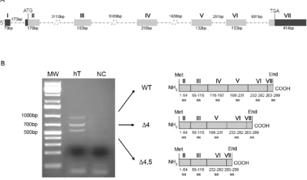
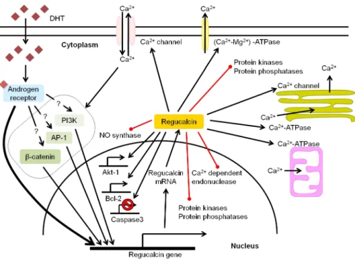
Figure I.1.1. Schematic representation of the testicular histology and mammalian spermatogenesis ... 4 Figure I.1.2. Hormonal regulation of spermatogenesis ... 5 Figure I.2.1. Extrinsic and intrinsic pathways of apoptosis ... 13 Figure I.2.2. Estrogen biosynthesis in mammalian testis ... 19 Figure I.3.1 RGN gene organization, testicular mRNA transcripts and hypothetical proteins . 39 Figure I.3.2 Multiple sequence alignment of RGN proteins and homologues in eukaryotes and prokaryotes ... 40 Figure I.3.3 Expression levels of RGN in testicular biopsies from men with obstructive azoospermia with conserved spermatogenesis, hypospermatogenesis and Sertoli cell‐only syndrome ... 42 Figure I.3.4. Schematic representation of the potential signalling pathways involved in the androgenic control of RGN expression in testis, and the possible roles of RGN protein in testicular cells ... 45 Figure I.4.1. Molecular players in intracellular Ca2+ homeostasis ... 58
Figure III.1. Effect of 100 nM E2 on mRNA and protein expression of SCF and c‐kit in rat SeT
cultured ex vivo for 24 hours ... 84 Figure III.2. Apoptosis in rat SeT cultured ex vivo for 48 hours in presence (E2) or absence
(control) of 100 nM of E2... 85
Figure III.3. Representative confocal microscopy images showing TUNEL and c‐kit positive cells in rat SeT cultured ex vivo in presence (E2) or absence (control) of 100 nM of E2 at
different experimental time‐points (0, 24, and 48 hours) ... 86 Figure III.4. Proliferation index in rat SeT cultured ex vivo for 48 hours in presence (E2) or
absence (control) of 100 nM of E2 ... 87
Figure III.5. Effect of 100 nM E2 on mRNA and protein expression of RGN in rat SeT cultured
ex vivo for 24 hours ... 88 Figure IV.1. Caspase‐3 activity and TUNEL‐positive nuclei in SeT of Tg‐RGN rats vs. Wt cultured in the presence or absence of apoptosis‐inducers Thap (10‐7 M and 10‐6 M) and Act D
Figure IV.2. Transcript levels of apoptosis‐related genes in SeT of Tg‐RGN rats vs. Wt cultured in presence (10‐6M) or absence of Thap (‐) ... 104
Figure IV.3. Protein expression of apoptosis‐related genes and Bcl‐2/Bax protein ratio in SeT of Tg‐RGN rats vs. Wt cultured in presence (10‐6M) or absence of Thap (‐) ... 105
Figure IV.4. Transcript levels of apoptosis‐related genes in SeT of Tg‐RGN rats vs. Wt cultured in presence (1 µg/ml) or absence of Act D (‐) ... 106 Figure IV.5. Protein expression of apoptosis‐related genes and Bcl‐2/Bax protein ratio in SeT of Tg‐RGN rats vs. Wt cultured in presence (1 µg/ml) or absence of Act D (‐) ... 107 Figure V.1. Glutathione‐S‐transferase (GST) activity in the SeT of Tg‐RGN rats and Wt control animals upon culture in the presence or absence of oxidant inducers TBHP (250 and 500 µM) and Cd (10 and 20 µM) ... 118 Figure V.2. Superoxide dismutase (SOD) activity in the SeT of Tg‐RGN rats and Wt control animals upon culture in the presence or absence of oxidant inducers TBHP (250 and 500 µM) and Cd (10 and 20 µM) ... 118 Figure V.3. Total antioxidant capacity (TAC) in the SeT of Tg‐RGN rats and Wt control animals upon culture in the presence or absence of oxidant inducers TBHP (250 and 500 µM) and Cd (10 and 20 µM) ... 119 Figure V.4. Caspase‐3 activity in the SeT of Tg‐RGN rats and Wt control animals upon culture in the presence or absence of oxidant inducers TBHP (250 and 500 µM) and Cd (10 and 20 µM) ... 120 Figure VI.1. Epididymal sperm counts, motility and viability in Tg‐RGN rats vs Wt ... 132 Figure VI.2. Normal and abnormal morphology of epididymal sperm in Tg‐RGN rats vs Wt .. 133 Figure VI.3. Epithelial cell height of the caput epididymis and immunolocalization of V‐ ATPase and AQP9 in Tg‐RGN rats vs Wt ... 134 Figure VI.4. Time‐course of Ca2+ influx and efflux in the epididymis of Tg‐RGN rats vs Wt .. 135
Figure VI.5. Expression of NHE3 and AQP1 in the epididymis of Tg‐RGN rats vs Wt ... 136 Figure VI.6. Expression of RGN in caput, corpus and cauda regions of rat epididymis and EF. ... 137 Figure VI.7. FRAP value (µM antioxidant potential/mg tissue) in the epididymis of Tg‐RGN rats
Figure VII.1 Integrative view of the potential actions of estrogens and regucalcin in testicular apoptosis ... 148
List of Tables
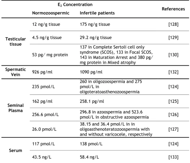
Table I.2.1. Localization of ERα, ERβ and GPER proteins in human testis ... 17 Table I.2.2. Estrogens concentrations in serum and reproductive tract of normozoospermic and infertile patients ... 20 Table I.2.3. Role of estrogens controlling apoptosis of testicular cells ... 23 Table I.3.1. Localization of RGN in male reproductive organs ... 41 Table I.3.2. Hormonal factors regulating RGN expression in reproductive and non‐ reproductive tissues ... 43 Table IV.1. qPCR primer sequences, cycling conditions, and amplicon size ... 100 Table VI.1. Epididymal caput tubule area (µm2), boundwidth, boundheight, and perimeter
List of Abbreviations
[Ca2+]
i Intracellular calcium concentration
17β-HSD 17β‐hydroxysteroid dehydrogenase Act D Actinomycin D
Apaf-1 Apoptotic protease activating factor 1 ArKO Aromatase knockout mice
BSA Bovine serum albumin c-kit Tyrosine kinase receptor Ca2+ Calcium
CaM Calmodulin
CaM-PDEs Ca2+/CaM‐dependent phosphodiesterases
CaMBP CaM‐binding protein complex
CamK4 Ca2+/Cam‐dependent protein kinase IV
cAMP Cyclic AMP
CaNBP75 Calcineurin‐binding protein CatSper Sperm‐specific Ca2+‐channel
CBPs Ca2+‐binding proteins Cd Cadmium chloride CDNB 1‐Chloro‐2,4‐dinitrobenzene cetn1 Centrin 1 DBD DNA‐binding domain DES Diethylstibestrol DHT 5α‐dihidrotestosterone DNase Deoxyribonuclease E Estrogens E2 17β‐estradiol EF Epididymal fluid ER Estrogen receptor FasL Fas ligand
FRAP Ferric Reducing Antioxidant Power FSH Follicle‐stimulating hormone GnRH Gonadotropin releasing hormone GPR30/GPER G‐protein‐coupled‐receptor 30 GST Glutathione‐S‐transferase HBSS Hank´s Buffered Salt Solution HP Hypospermatogenesis hpg Hypogonadal
IP3R Inositol 1,4,5‐trisphosphate receptors
KRb Krebs Ringer‐bicarbonate LBD Ligand binding domain LC Leydig cell
LH Luteinizing hormone MOR23 Mouse OR
NF1-A1 Nuclear factor 1‐ A1 OR Olfactory receptor
PBA PBS containing 1% (w/v) BSA PBS Phosphate‐buffered saline PCR Polymerase chain reaction PI3K Phosphatidylinositol 3‐kinase pNA p‐nitro‐aniline
qPCR Quantitative real‐time PCR Ran Ras‐related small G protein RGN Regucalcin
ROS Reactive oxygen species RyR Ryanodine receptors SC Sertoli cell
SCF Stem cell factor
SCOS Sertoli cell only syndrome SeT Seminiferous tubules
SMP30 Senescence marker protein‐30 SOD Superoxide dismutase
SSCs Spermatogonial stem cells T Testosterone
TBHP tert‐butyl hydroperoxide
Tg-RGN Transgenic rats overexpressing RGN TGF-β Transforming growth factor β Thap Thapsigargin
TNF Tumor necrosis factor
TNFR1 Tumor necrosis factor receptor 1 TRP Transient receptor potential
TUNEL Terminal deoxynucleotidyl transferase dUTP nick end labeling VDCC Voltage‐dependent Ca2+‐channels
WB Western Blot Wt Wild‐type
Chapter I
General Introduction
Brief Introduction to Mammalian Spermatogenesis
Estrogens as apoptosis regulators in mammalian testis: angels or devils?
Regucalcin, a calcium-binding protein with a role in male reproduction?
Brief Introduction to Mammalian Spermatogenesis
Cellular players and developmental stages
Mammalian spermatogenesis is a complex process involving cell division and maturation of spermatogonial stem cells (SSCs) that culminates with the production of male gametes, the spermatozoa. It is a continuous and highly regulated process that takes place in the seminiferous tubules (SeT, Figure I.1.1), the functional unit of the testis [1]. The seminiferous epithelium (Figure I.1.1) is composed by germ cells that form numerous concentric layers penetrated by a single type of somatic cell, the Sertoli cell (SC). SSCs are localized on the basal membrane of the SeTs as single cells and upon division originate daughter cells, the spermatogonia [2]. The cytoplasm of SCs extends as thin arms around all the germ cells nursing and maintaining their cellular associations throughout the several steps of spermatogenesis [3]. The presence of tight junctions between neighbouring SC forms the so‐ called blood‐testis barrier (Figure I.1.1), which divides the SeT in basal and adluminal compartments [3].
Each spermatogenic cycle in the SeT encompasses three main phases: mitosis, meiosis, and the final stage of cell differentiation, spermiogenesis [1]. Spermatogenesis (Figure I.1.1) starts with the proliferation of spermatogonia and after a species‐specific fixed number of mitotic divisions spermatogonia differentiate into primary spermatocytes [4]. These proceed to the first division of meiosis originating secondary spermatocytes, which undergo the second meiotic division and become haploid spermatids. The cellular restructure of spermiogenesis transforms round‐spermatids in elongated‐spermatids and then elongated‐spermatids into spermatozoa [1]. The output of spermatogenesis and the number of produced spermatozoa also depends of programmed cell death processes. High rates of apoptosis have been associated with the first waves of spermatogenesis [5], and in adult testis not all germ cells achieve maturity being prone to die in response to a variety of factors. It is believed that apoptosis represents a mechanism to discard excess and unfit cells maintaining the appropriate ratio of germ cells to SCs [6].
The interstitial space between SeT is mainly occupied by blood vessels and the somatic Leydig cells (LC), which are the testosterone (T) producing cells within the testis and play a crucial role in the regulation of spermatogenic process [7].
Spermatozoa leaving the testis are non‐functional gametes and it is only during passage through the long convoluted tubule of the epididymis that they acquire the ability to move progressively and the capacity to fertilize [8]. The epididymis is a highly compartmentalized organ with three distinct regions (Figure I.1.1), the caput, corpus and cauda, which sustain different functions to achieve the final goal, sperm maturation and fertilizing ability [9].
Figure I.1.1. Schematic representation of the testicular histology and mammalian spermatogenesis. Also, the anatomic relationship between testis and epididymis, as well as, the distinct functional regions of the epididymis are shown.
Hormonal regulation
Development and maintenance of successful spermatogenesis depends on assortment of a set of hormonal messengers, which exert their actions by endocrine, paracrine, juxtacrine and autocrine signaling mechanisms. The major player in the hormonal control of spermatogenesis is the hypothalamic‐pituitary‐gonadal axis [10] (Figure I.1.2). The hypothalamus releases gonadotropin releasing hormone (GnRH), which acts on the pituitary inducing the release of gonadotropins, namely, luteinizing hormone (LH) and follicle‐stimulating hormone (FSH). In the testis, LH acts on LCs stimulating the synthesis of T, while FSH acts on SCs inducing the production of several growth factors and other stimulatory factors of spermatogenesis. T diffuses into the SeT where together with FSH exerts stimulatory effects on the activity of SCs activity, which is determinant for germ cells maturation and sperm production [11, 12].
Lumen Leydig Cell Seminiferous tubule Testis Epididymis Corpus Caput Cauda Basal membrane Sertoli Cell Sertoli Cell nucleus Round spermatids Elongated spermatids Secondary spermatocytes Blood‐testis barrier Primary spermatocyte Spermatogonia Spermatozoa Basal compartment Adluminal compartment
Moreover, T regulates the spermatogenic process by a negative feedback mechanism on the hypothalamus and pituitary inhibiting, respectively, the release of GnRH and LH [10].
Other negative feedback regulatory mechanism is driven by inhibin, a member of the transforming growth factor β (TGF‐β) superfamily produced by SCs in response to FSH (Figure I.1.2). Inibin blocks the production and release of FSH by the pituitary [13, 14] controlling the output of spermatogenesis.
Although androgens and FSH are perfectly recognized as the main regulators of spermatogenesis, the last decades have witnessed the emergence of estrogens as important regulators of male reproductive function with their roles starting to be intensively discussed by the scientific community [15, 16].
Figure I.1.2. Hormonal regulation of spermatogenesis. Release of gonadotropin‐releasing hormone (GnRH) from the hypothalamus stimulates the pituitary to secrete two gonadotropins, the follicle‐ stimulating hormone (FSH) and the luteinizing hormone (LH). FSH stimulates the activity of Sertoli cells and LH acts on Leydig cells, inducing the production of testosterone (T). A negative feedback (‐) by T on the hypothalamus and pituitary regulates the levels of GnRH, LH and FSH, although its main action is to decrease secretion of LH. FSH secretion is also subject of a negative feedback (‐) by inhibin secreted by Sertoli cells.
Non-hormonal factors in the regulation of spermatogenesis
Besides sex steroids, pituitary and hypothalamic hormones, a panoply of other factors play a role in the regulation of spermatogenesis. The germ cell cycle and movement along the SeT is a process under tight control involving distinct mechanisms that include several families of kinases and phosphatases activated, for example, in response to growth factors and cytokines [17]. Many of these molecules are secreted by SCs, namely, the glial cell line‐derived neurotrophic factor, a member of the TGF‐β superfamily and the first molecule identified in the regulation of self‐renewal and differentiation of SSCs [18]. The stem cell factor (SCF) is a cytokine, also produced and secreted by SCs, which plays a crucial role controlling survival and proliferation of both SSCs and spermatogonia [19, 20]. The SCF, by interaction with its tyrosine kinase receptor the c‐KIT also seems to control the differentiation of spermatogonia
and progression into meiosis [21, 22]. Moreover, SCs produce transport or bioprotective proteins that are secreted in relative high abundance and include proteases and protease inhibitors, and metal ion transport proteins such as transferrin and ceruloplasmin [3]. Transferrin transports iron to the adluminal compartment of SeT ensuring that the developing germ cells have access to adequate and tightly regulated levels of this ion [23]. Although germ cells require considerable amounts of iron for proliferation and differentiation [23], other inorganic molecules have been implicated in spermatogenesis. This is, for example, the case of calcium (Ca2+), zinc, selenium, and copper [24].
In the context of this thesis, Ca2+ deserves particular attention and several evidences have
highlighted for its importance for spermatogenesis. A tight control of intracellular Ca2+
homeostasis has been shown to be of uttermost importance for SC function [25, 26], maintenance of SCs tight junctions and integrity of the blood‐testis barrier [27]. It also modulates the activity of enzymes that interfere in the structure of SCs [28]. A strict regulation of Ca2+ fluxes maintaining intracellular Ca2+ homeostasis also seems to be related
with the expression of the steroidogenic acute regulatory protein and LC steroidogenesis [29]. Moreover, it has been shown that administration of Ca2+ channels blockers for treatment of
hypertension causes reversible male infertility [30], which demonstrates the physiological importance of Ca2+.
References
1. Hess, R.A. and de Franca, L.R., Spermatogenesis and cycle of the seminiferous epithelium, in Molecular Mechanisms in Spermatogenesis. 2008, Springer New York. p. 1‐15.
2. de Rooij, D.G. and Mizrak, S.C. Deriving multipotent stem cells from mouse spermatogonial stem cells: a new tool for developmental and clinical research. Development, 2008. 135(13): p. 2207‐2213.
3. Griswold, M.D. The central role of Sertoli cells in spermatogenesis. Seminars in Cell and Developmental Biology 1998. 9(4): p. 411‐416.
4. Clermont, Y. Kinetics of spermatogenesis in mammals: seminiferous epithelium cycle and spermatogonial renewal. Physiological Reviews, 1972. 52(1): p. 198‐236.
5. Aitken, R.J., Findlay, J.K., Hutt, K.J., and Kerr, J.B. Apoptosis in the germ line. Reproduction, 2011. 141(2): p. 139‐150.
6. Shaha, C., Tripathi, R., and Mishra, D.P. Male germ cell apoptosis: regulation and biology. Philosophical Transactions of the Royal Society B: Biological Sciences, 2010. 365(1546): p. 1501‐1515.
7. Haider, S.G. Cell biology of Leydig cells in the testis. International Review of Cytology, 2004. 233: p. 181‐241.
8. Cornwall, G.A. New insights into epididymal biology and function. Human Reproduction Update, 2009. 15(2): p. 213‐227.
9. Robaire, B., Hinton, B.T., and Orgebin‐Crist, M.‐C., The epididymis, in Knobil and Neil´s Physiology of Reproduction. 2006, Elsevier: San Diego, CA. p. 1071‐1148.
10. Holdcraft, R.W. and Braun, R.E. Hormonal regulation of spermatogenesis. International Journal of Andrology, 2004. 27(6): p. 335‐342.
11. Walker, W.H. and Cheng, J. FSH and testosterone signaling in Sertoli cells. Reproduction, 2005. 130(1): p. 15‐ 28.
12. Walker, W.H. Molecular mechanisms of testosterone action in spermatogenesis. Steroids, 2009. 74(7): p. 602‐ 607.
13. Bilezikjian, L.M., Blount, A.L., Leal, A.M., Donaldson, C.J., Fischer, W.H., and Vale, W.W. Autocrine/paracrine regulation of pituitary function by activin, inhibin and follistatin. Molecular and Cellular Endocrinology 2004. 225(1‐2): p. 29‐36.
14. Pierik, F.H., Burdorf, A., de Jong, F.H., and Weber, R.F. Inhibin B: a novel marker of spermatogenesis. Annals of Medicine, 2003. 35(1): p. 12‐20.
15. Carreau, S. and Hess, R.A. Oestrogens and spermatogenesis. Philosophical Transactions of the Royal Society B: Biological Sciences, 2010. 365(1546): p. 1517‐1535.
16. O’Donnell, L., Robertson, K.M., Jones, M.E., and Simpson, E.R. Estrogen and Spermatogenesis Endocrine Reviews, 2001. 22(3): p. 289‐318.
17. He, Z., Kokkinaki, M., and Dym, M. Signaling molecules and pathways regulating the fate of spermatogonial stem cells. Microscopy Research and Technique, 2009. 72(8): p. 586‐595.
18. Meng, X., Lindahl, M., Hyvonen, M.E., Parvinen, M., de Rooij, D.G., Hess, M.W., Raatikainen‐Ahokas, A., Sainio, K., Rauvala, H., Lakso, M., et al. Regulation of cell fate decision of undifferentiated spermatogonia by GDNF. Science, 2000. 287(5457): p. 1489‐1493.
19. Hakovirta, H., Yan, W., Kaleva, M., Zhang, F., Vänttinen, K., Morris, P.L., Söder, M., Parvinen, M., and Toppari, J. Function of Stem Cell Factor as a Survival Factor of Spermatogonia and Localization of Messenger Ribonucleic Acid in the Rat Seminiferous Epithelium Endocrinology, 1999. 140(3): p. 1492‐1498.
20. Ohta, H., Yomogida, K., Dohmae, K., and Nishimune, Y. Regulation of proliferation and differentiation in spermatogonial stem cells: the role of c‐kit and its ligand SCF. Development, 2000. 127(10): p. 2125‐2131.
21. Guerif, F., Cadoret, V., Rahal‐Perola, V., Lansac, J., Bernex, F., Panthier, J.J., Hochereau‐de Reviers, M.T., and Royere, D. Apoptosis, onset and maintenance of spermatogenesis: evidence for the involvement of Kit in Kit‐haplodeficient mice. Biology of Reproduction, 2002. 67(1): p. 70‐79.
22. Kissel, H., Timokhina, I., Hardy, M.P., Rothschild, G., Tajima, Y., Soares, V., Angeles, M., Whitlow, S.R., Manova, K., and Besmer, P. Point mutation in kit receptor tyrosine kinase reveals essential roles for kit signaling in spermatogenesis and oogenesis without affecting other kit responses. EMBO Journal, 2000. 19(6): p. 1312‐1326.
23. Sylvester, S. and Griswold, M. The testicular iron shuttle: a “nurse” function of the Sertoli cells. Journal of Andrology, 1994. 15(5): p. 381‐385.
24. Camejo, M.I., Abdala, L., Vivas‐Acevedo, G., Lozano‐Hernández, R., Angeli‐Greaves, M., and Greaves, E.D. Selenium, copper and zinc in seminal plasma of men with varicocele, relationship with seminal parameters. Biological Trace Element Research, 2011. 143(3): p. 1247‐1254.
25. Gorczynska‐Fjalling, E. The role of calcium in signal transduction processes in Sertoli cells. Reproductive Biology, 2004. 4(3): p. 219‐241.
26. Gorczynska, E. and Handelsman, D.J. Androgens rapidly increase the cytosolic calcium concentration in Sertoli cells. Endocrinology, 1995. 136(5): p. 2052‐2059.
27. Grima, J., Wong, C.C., Zhu, L.J., Zong, S.D., and Cheng, C.Y. Testin secreted by Sertoli cells is associated with the cell surface, and its expression correlates with the disruption of Sertoli‐germ cell junctions but not the inter‐Sertoli tight junction. Journal of Biological Chemistry, 1998. 273(33): p. 21040‐21053.
28. Franchi, E. and Camatini, M. Evidence that a Ca2+ chelator and a calmodulin blocker interfere with the structure of inter‐Sertoli junctions. Tissue and Cell, 1985. 17(1): p. 13‐25.
29. Manna, P.R., Pakarinen, P., El‐Hefnawy, T., and Huhtaniemi, I.T. Functional assessment of the calcium messenger system in cultured mouse Leydig tumor cells: regulation of human chorionic gonadotropin‐induced expression of the steroidogenic acute regulatory protein. Endocrinology, 1999. 140(4): p. 1739‐1751.
30. Katsoff, D. and Check, J.H. A challenge to the concept that the use of calcium channel blockers causes reversible male infertility. Human Reproduction, 1997. 12(7): p. 1480‐1482.
Estrogens as apoptosis regulators in mammalian testis: angels or
devils?
Chapter submitted (under revision) in Correia S, Cardoso HJ, Cavaco JE and Socorro S. Estrogens as apoptosis regulators in mammalian testis: angels or devils? Expert Reviews in Molecular Medicine
Estrogens as apoptosis regulators in mammalian testis: angels or
devils?
Abstract
In the mammalian testis, spermatogenesis is the highly coordinated process of germ cell development, which ends with the release of “mature” spermatozoa. The fine regulation of spermatogenesis is strictly dependent of sex steroid hormones, which orchestrate the cellular and molecular events underlying normal development of germ cells. Sex steroids actions also rely on the control of germ cell survival, and the programmed cell death by apoptosis has been indicated as a critical process in regulating the size and quality of the germ line. Recently, estrogens have emerged as important regulators of germ cell fate. However, the beneficial or detrimental effects of estrogens in spermatogenesis are controversial, with independent reports arguing for their role as cell survival factors or as apoptosis‐inducers. The dual behavior of estrogens, shifting from “angels to devils” is supported by the clinical findings of increased estrogens levels in serum and intratesticular milieu of idiopathic infertile men. This review aims to discuss the available information concerning the role of estrogens in the control of germ cell death and summarises the signaling mechanisms driven estrogen‐induced apoptosis. The present data represent a valuable basis for clinical management of hyperestrogenism‐related infertility and provide a rationale for the use of estrogen‐target therapies in male infertility.
Keywords: 17β‐estradiol; apoptosis; aromatase; estrogens; estrogen receptor; germ cells; male infertility; sex steroids; spermatogenesis; testis
Introduction
The mammalian testis fulfills two essential functions in male reproduction: steroidogenesis and spermatogenesis. Steroidogenesis corresponds to the biosynthesis of sex steroids hormones whereas spermatogenesis is the complex cellular process that transforms spermatogonial cells into spermatozoa. Progression of spermatogenesis takes place in the seminiferous tubules, the functional units of the testis, encompassing tightly coordinated mitosis, meiosis and cell differentiation events [1]. Mitotic divisions maintain the population of spermatogonial cells and differentiation of spermatogonia originates primary spermatocytes. The first meiotic division produced secondary spermatocytes, which undergo the second meiosis originating spermatids. Spermiogenesis is the cellular restructure process that transforms round spermatids in elongated spermatids and finally in spermatozoa [2]. Nonetheless, not all germ cells achieve maturity, and the spontaneous cell death by apoptosis is a common event, particularly, in the first waves of spermatogenesis [3]. On the other hand,
deregulated apoptosis may be indicated in the etiology of male infertility, since augmented rates of apoptosis were observed in the testes of subfertile and infertile men [4‐6].
Development and maintenance of spermatogenesis depends of an assortment of hormonal messengers, which exert their actions by endocrine, paracrine, and autocrine signaling mechanisms. The sex steroids androgens are widely recognized as the main regulators of male reproductive function, and the androgenic actions are absolutely required for successful spermatogenesis [7]. Over the years, the dogma that spermatogenesis depends solely of androgens and gonadotropin actions has changed, and cumulative evidences have demonstrated the importance of estrogens on the regulation of spermatogenic process. However, discordant reports about the role of estrogens in male reproduction have been produced including those ascribing their role as survival or apoptosis‐inducer agents in male germ cells. The present review will discuss the current knowledge of estrogen‐induced apoptosis in the testis and summarize the molecular mechanisms driven apoptotic effects in male germ cells. This issue is of paramount importance for clinical management of male infertility because hiperestrogenism has been a condition associated with cases of idiopathic infertility.
Apoptosis in spermatogenesis
Signaling mechanisms of apoptotic cell death
The apoptotic cell death, characterized by several hallmarks, such as, cell shrinkage, DNA fragmentation, and externalization of phosphatidylserine at cell membrane, may be triggered by distinct pathways [8]. The receptor‐mediated (or extrinsic) and the mitochondrial (or intrinsic) are the two major pathways governing apoptosis (Figure I.2.1) [9]. In both cases, cell‐death depends of specific proteases, the so‐called caspases, which are the executioners of apoptosis. The caspases enzymes cleave serine residues and are synthesized as inactive zymogens (procaspases) becoming active in response to death stimuli [10]. Initiator caspases (caspase‐8 and ‐9) are directly activated by dimerization and, in turn, cleave the effector caspases (caspase‐3, ‐6 and ‐7) promoting their activation [10]. The extrinsic pathway (Figure I.2.1) is initiated by ligand‐binding activation of death receptors at cell membrane, namely the Fas (CD95/Apo‐1) and the tumor necrosis factor receptor 1 (TNFR1), which induce the activation of procaspase‐8 [9]. The intrinsic pathway of apoptosis (Figure I.2.1) could be activated by different stimuli, such as DNA damage, starvation, oxidative stress and autophagy. It is characterized by a decrease in the membrane potential of mitochondria and release of cytochrome c, which interact with dATP, cytosolic apoptotic protease activating factor 1 (Apaf‐1) and procaspase‐9, assembling the apoptosome complex [11]. Both extrinsic and intrinsic pathways of apoptosis converge at the activation of effector caspase‐3 (Figure I.2.1), which has been considered an end‐point of apoptotic process [8].
The apoptosis of germ cells has been shown to play an important role in controlling sperm output in many species, and massive germ cell death occurs under physiological conditions
during the earlier stages of spermatogenic process (constitutive apoptosis). The specificities of apoptosis in the first waves of spermatogenesis have been revised in recent reports [3, 12] and will not be further explored here.
In adult testis, the fine control of apoptosis is critical for maintenance of spermatogenesis and male fertility, since germ cells are very sensitive to damaging conditions, such as, heat shock, ionizing radiation, growth factor deprivation and chemotherapeutic agents. Therefore, apoptosis is a relevant mechanism for elimination of damaged germ cells avoiding passage of defects to the future generations [12].
On the other hand, male germ cells strictly depend on the physical and biochemical support of Sertoli cells (SCs), the somatic cells within the seminiferous tubules, which nourish and sustain the developing germ cells [13]. However, SCs have a limited capacity for the number of germ cells they can support [14], and it has been accepted that they play a crucial role determining germ cell fate. SCs secrete paracrine factors and establish direct cell‐to‐cell membrane contacts with adjacent germ cells, which promote germ cell survival or death maintaining the appropriate ratio of germ cell to SCs [15]. The stem cell factor (SCF), a membrane‐bound cytokine at the surface of SCs [16], and its tyrosine kinase receptor the c‐ kit, present on the surface of adjacent germ cells [17], are the main mediators of survival communication between SCs and germ cells protecting germ cells from apoptosis [18]. Perturbations in the SCF/c‐kit system by abolishment of SCF production [19], impairment of SCF binding to c‐kit [20] or disruption of c‐kit signaling mechanisms [21] have been linked to increased apoptosis and reduced proliferation of germ cells. The death communication between SCs and germ cells is established by the Fas system being accepted that SCs express the Fas ligand (FasL) while germ cells mostly express the death receptor Fas [22‐24].
Figure I.2.1. Extrinsic and intrinsic pathways of apoptosis. Extracellular ligand binding (FasL or tumor necrosis factor, TNF) to death receptors (Fas and TNF receptor, TNFR) triggers the receptor‐mediated (extrinsic) pathway resulting in the direct activation of initiator caspase‐8. The mitochondrial (intrinsic) pathway is initiated in response to apoptotic stimuli leading to the activation of proapoptotic members of the Bcl‐2 protein family, namely, Bax. The Bax protein is translocated to the mitochondria allowing the permeabilization of mitochondrial membrane with consequent release of cytochrome c, which in turn, together with apoptotic protease activating factor 1 (Apaf‐1), forms the apoptosome and activates caspase‐9. Extrinsic and intrinsic apoptosis signaling pathways converge at the activation of the executioner caspase‐3. Activation and inhibition are indicated by arrows and bar‐headed arrows, respectively. Target sites of estrogens (E) actions in the estrogen‐induced apoptosis of germ cells are highlighted by triangles.
Clinical findings of deregulated apoptosis in the testis of infertile men
Taking into account the delicate control of apoptosis in the germ line it is not surprisingly that augmented rates of apoptosis have been identified in the testes of subfertile and infertile men [4‐6, 25‐31]. Accordingly, altered expression patterns of a panoply of apoptosis‐ related genes have been described in human testes with defective spermatogenesis. The association of death receptors and the extrinsic pathway of apoptosis with male infertility has been suggested by several studies, which showed enhanced expression of FasL in cases of maturation arrest and Sertoli cell‐only syndrome, the later characterized by the absence of germ cell in the seminiferous epithelium [5, 31‐35]. Cavalcanti et al. also found an increased expression of TNF‐α family member 10 (also known as TNF‐α related apoptosis inducing
As previously stated, caspases are the key component in the apoptotic pathway and the executioner of apoptotic cell death. Increased expression of effector caspase‐6 was found in the testes of subfertile men comparatively with potentially fertile [36]. Also, the expression of effector caspase‐3 was enhanced in cases of maturation arrest and Sertoli cell‐only syndrome, the later characterized by the absence of germ cell in the seminiferous epithelium [31‐33]. In addition, increased activity of caspase‐9 and caspase‐3 was also showed in isolated spermatozoa from oligozoospermic and azoospermic patients comparatively with normozoospermic [37, 38], which is indicative of active apoptosis in spermatozoa of infertile patients. In fact, the occurrence of DNA fragmentation and the externalization of phosphatidylserine, which are late apoptotic events indicating cell commitment to apoptosis, can be detected in spermatozoa from infertile men [39, 40].
The expression of novel apoptosis regulators such as Aven, survivin and regucalcin (RGN) also was found to be altered in human testis with defective spermatogenesis [41‐43]. Aven is an apoptosis inhibitor that acts by binding both Bcl‐xL and Apaf‐1 (Figure I.2.1), enhancing the antiapoptotic function of Bcl‐xL and inhibiting the assembling of a functional apoptosome complex, thus preventing caspase activation [44]. A diminished expression of Aven was found in the testes of nonobstructive azoospermic men, which was correlated with the severity of spermatogenic defect and increased rates of apoptosis [41]. In the case of survivin and RGN, both proteins with a dual role controlling apoptosis and cell cycle progression [42, 45, 46], the balance of their expression levels should be determinant for maintenance of an appropriate germ cell number and thus, for male reproductive potential. Recently, we have found increased expression of RGN in the testis of human infertile patients with hypospermatogenesis phenotype [42]. Although the causes of idiopathic hypospermatogenesis may be diverse, accelerated apoptosis, rather than proliferative dysfunction in the mitotic phase, has been implicated as responsible for the decreased number of spermatogonia [25]. In a recent report, we demonstrated that in vivo overexpression of RGN in the testis leads to suppression of thapsigargin‐ and actinomycin D‐ induced apoptosis through modulating the expression and activity of key regulators of apoptosis [47]. This suggests that RGN may act as a protective molecule counteracting increased rates of apoptosis associated with infertility cases such as hypospermatogenesis. On the other hand, manipulation of RGN levels may represent a suitable mechanism for fertility preservation upon treatment of oncologic conditions.
Sex steroid hormones are the crucial orchestrators of the cellular and molecular events underlying normal spermatogenesis and their roles also rely on the control of germ cell survival and apoptosis. It has been shown that androgens withdrawal induces disruption of spermatogenesis with a severe reduction in the number of spermatocytes and spermatids in consequence of increased apoptosis [48, 49]. Whereas the action of androgens has been well documented and fully accepted as an essential requirement for development and survival of testicular cells [50], in the case of estrogens a dual behaviour has been assigned with independent reports describing their actions both as germ cell survival factors and as


![Figure I.4.1. Molecular players in intracellular Ca 2+ homeostasis. Resting intracellular Ca 2+ concentration ([Ca 2+ ] i ) is kept low (≈100 nM) by an orchestrated action of Ca 2+ transporters, Ca 2+ pumps, ion channels and Ca 2+ ‐binding pr](https://thumb-eu.123doks.com/thumbv2/123dok_br/18455096.897795/86.892.251.709.573.909/molecular-intracellular-homeostasis-resting-intracellular-concentration-orchestrated-transporters.webp)
