i
Dose effects on
re-irradiation of
the spinal cord
Helena Sofia Martins Alves
Master’s degree in Medical Physics
Department of Physics and AstronomyFaculty of Science, University of Porto (FCUP) [email protected]
2018
Supervisor
Isabel Bravo, PhD
Medical Physics, Radiobiology and Radiation Protection Group IPO – Porto Research Center (CI – IPOP)
Portuguese Oncology Institute of Porto (IPO – Porto) Porto, Portugal
i
Todas as correções determinadas pelo júri, e só essas, foram efetuadas. O Presidente do Júri,
iii
Declaração
Eu, Helena Sofia Martins Alves, nº 201602802, estudante do 2º ano do Mestrado em Física Médica no presente ano letivo 2017/2018, na Faculdade de Ciências da Universidade do Porto, declaro ter atuado com absoluta integridade na elaboração do texto apresentado, não apresento texto plagiado, e tomei conhecimento das consequências de uma situação de plágio.
v
Agradecimentos
Osmeus agradecimentos são para as pessoas que me acompanharam e apoiaram ao longo do meu percurso escolar.
Aos meus pais, obrigada por todos os sacrifícios que fizeram por mim e por todo o apoio em momentos mais difíceis.
À minha família, por estarmos sempre unidos em todos os momentos.
Ao Leandro pelo companheirismo e pelo apoio incondicional. Obrigada por acreditares que sou capaz e nunca me deixares desistir. Sou feliz por encontrar na mesma pessoa o meu melhor amigo e namorado.
Ao Nequinha, por sempre me fazer pensar mais longe e por todo o interesse demonstrado ao longo destes anos. O meu muito obrigada!
À minha orientadora, Dra. Isabel Bravo, por acreditar que eu seria capaz de realizar esta tese com o seu apoio. Agradeço-lhe por todo o conhecimento e interesse demonstrado ao longo destes últimos meses. Aprendi muito consigo.
A todos os meus colegas de turma, pelos momentos de diversão, trocas de ideias e por todas as sessões de estudo.
Aos meus amigos, obrigada por terem sempre uma palavra de carinho, por me motivarem e por acreditarem em mim. Estes últimos 5 anos, foram menos difíceis graças a vocês.
A todas as pessoas mencionadas acima, agradeço por terem contribuído para o meu conhecimento, enriquecimento pessoal ao longo desta jornada, que termina com a entrega deste documento.
vii Que permaneça em mim, essa gratidão por cada manhã que nasce. Essa felicidade de poder abrir os
olhos e ouvir do céu: - Hoje tudo vai dar certo. Diego Vinicius
ix
Abstract
Advances in biology and physics have allowed increased precision and accuracy in radiotherapy (RT) in order to maximize tumor damage and to minimize lesions in the dose limiting adjacent normal tissues. The spinal cord is the most critical organ at risk (OAR). Radiation myelopathy is one of the most devastating complications of clinical radiotherapy resulting in severe and irreversible morbidity. Assessment of the impact of dose and fractionation schemes on tissue tolerance has been a major area of research in radiation oncology. As a result of greater accuracy and effectiveness of cancer treatment, patient survival rates increase, and radiation oncologists are frequently faced with the problem of treatment of local recurrence or second tumors located within or close to previously treated sites. Initial dose influences different time intervals from tissue tolerance to re-irradiation as well as conditioning the recovery of radiation damage in the first treatment. It is possible to administer a higher dose in the re-irradiation if smaller doses were used at the first treatment and if the intervals between treatments were longer. Radiation myelopathy is a rare late toxicity effect in the modern era of 3-dimensional conformal conventionally fractionated RT. This devastating late effect has re-emerged as a direct result of SBRT practice, where high-dose radiation is delivered adjacent to the spinal cord to be spared. A comprehensive search was performed including relevant articles referring to “spinal cord”, “re-irradiation” and “myelopathy”. The biologically effective dose (BED) was calculated and the results are discussed considering radiobiological mechanisms.
xi
Resumo
Avanços na biologia e na física permitiram uma maior precisão e exatidão em radioterapia (RT), de modo a maximizar o dano tumoral e minimizar as lesões nos tecidos normais adjacentes, que limitam a dose. A medula espinal é denominada como o órgão mais crítico em risco (OAR – organ at risk). A mielopatia por radiação é uma das complicações mais devastadoras da radioterapia clínica, resultando em morbidade grave e irreversível. A avaliação do impacto dos esquemas de dose e fracionamento, na tolerância tecidual, tem sido uma das principais áreas de pesquisa em oncologia da radiação. Como resultado de uma maior precisão e eficácia do tratamento oncológico, as taxas de sobrevivência do paciente aumentam e os oncologistas de radiação são, frequentemente, confrontados com o problema do tratamento de recidiva local ou de segundos tumores localizados dentro ou próximos de locais previamente tratados. A dose inicial influencia diferentes intervalos de tempo, desde a tolerância do tecido à re-irradiação, bem como, condiciona a recuperação do dano por radiação no primeiro tratamento. É possível administrar uma dose maior na re-irradiação se doses menores forem usadas no primeiro tratamento e se o intervalo de tempo entre o primeiro tratamento e a re-irradiação for mais longo. A mielopatia por radiação é um raro efeito de toxicidade tardia na era moderna da RT conformacional tridimensional convencional (3D- CRT). Este devastador efeito tardio ressurgiu como um resultado direto da prática de SBRT, onde a radiação de alta dose é administrada junto à medula espinal, que terá de ser poupada. Uma pesquisa abrangente foi realizada, incluindo artigos relevantes referentes a “medula espinal”, “re-irradiação” e “mielopatia”. A dose biologicamente efetiva (BED) foi calculada e os resultados são discutidos considerando os mecanismos radiobiológicos.
xiii
Contents
Agradecimentos ... v Abstract ... ix Resumo ... xi Contents ... xiii Index of Figures ... xvIndex of Tables ... xvii
Abbreviations ... xix
Terms and Definitions ... xxi
1. Introduction ... 1
2. Theoretical concepts ... 3
2.1 The role of radiation therapy ... 3
2.2 Radiobiology: essential concepts ... 5
2.2.1 The role of radiobiology in the evolution of radiotherapy ... 5
2.2.1.1 Radiobiological mechanisms ... 5
2.2.1.2 Cell cycle control ... 8
2.2.1.3 Proliferation and differentiation ... 11
2.2.1.4 Tolerance of normal tissues to radiation ... 12
2.2.1.5 Rs of radiobiology ... 13
2.2.1.5.1 Repair of sub-lethal damage... 14
2.2.1.5.2 Redistribution of cells in the cell cycle ... 15
2.2.1.5.3 Reoxygenation ... 17
2.2.1.5.4 Repopulation ... 18
2.2.1.5.5 Radiosensitivity ... 19
2.2.2 Cell survival curves ... 19
2.2.3 Dose-response relationship ... 21
2.2.4 Biologically Effective Dose (BED) ... 23
2.2.4.1 Values of the 𝛼𝛽 ratio ... 24
2.2.4.2 Hypofractionation and hyperfractionation... 25
2.2.4.3 Dose equivalent in fractions of 2Gy (EQD2) ... 25
2.2.4.4 Incomplete repair ... 26
2.2.4.5 Time factor – repopulation ... 26
2.2.4.6 Advantages and disadvantages of BED ... 27
3. The spinal cord ... 29
xiv
3.2 Physiology ... 35
3.3 Tumors in the spinal cord ... 36
3.4 Complications after irradiation in the spinal cord ... 38
3.5 Spinal cord doses and tolerance ... 41
3.6 Radiobiology of the spinal cord ... 42
4. Re-irradiation... 45
4.1 Re-irradiation of spinal cord ... 46
4.1.1 Time interval between fractions / total treatment time ... 48
4.1.2 Fractionation ... 49
4.2 Stereotactic body radiotherapy (SBRT) ... 50
4.2.1 Late effects ... 53
4.2.2 Pathophisiology ... 59
4.2.3 Dose selection ... 61
4.2.4 Vascular damage in tumors ... 63
4.2.5 Tumor hypoxia and SBRT ... 64
4.2.6 Rs impact ... 67
4.2.7 Linear-Quadratic Model ... 69
4.2.8 Limitations and constraints of SBRT ... 70
4.2.9 Consensus guidelines ... 72
5. Conclusion and Future work ... 75
5.1 Final conclusions ... 75
5.2 Objectives achieved ... 76
5.3 Future work ... 76
Appendix A ... 77
Annex A – Framework and development of the BED formula ... 77
Appendix B ... 79
Annex B – Submission of a review article to Medical Physics AAPM ... 79
Introduction ... 80
Spinal cord doses and tolerance ... 81
Dose and fractionation ... 82
Time interval between fractions / total treatment time ... 83
Pathophisiology ... 84
Spine Stereotactic Body Radiation Therapy (SBRT) ... 86
Radiobiology: impact of repair, redistribution, repopulation, reoxygenation and radiosensitivity (5Rs) ... 88
Limitations and constraints ... 92
References ... 98
xv
Index of Figures
Figure 1 Time scale of the effects of ionizing radiation: biological changes manifest after a period of latency that can go
from minutes to weeks to years after exposure (A - early effects; B - late effects). ... 6
Figure 2 Role of cyclins and cyclin-dependent kinases (CDKs) in cell cycle regulation: The cell cycle is divided into G1, S (DNA synthesis), G2 and M (mitosis) phases. The transition between phases is controlled by cyclins and CDKs. ... 9
Figure 3 p53 Injury recognition process: under normal conditions the protein expressed by the p53 gene is responsible for the temporary stopping of the G1 cell cycle for DNA repair or, if not possible, programmed cell death (apoptosis). If the gene is mutated, the functions of p53 are not activated and the result will be uncontrolled cell proliferation. ... 10
Figure 4 Representation of the cell cycle. Adapted from [18]. ... 16
Figure 5 Cell cycle and survival curve phases. Adapted from [7]. ... 16
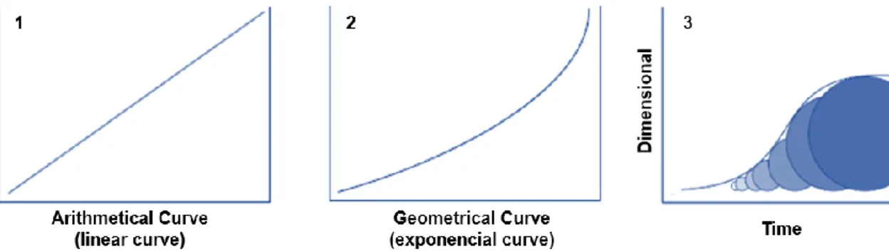
Figure 6 Curves of cellular survival: 1) arithmetic, 2) geometric and 3) Exponential increase in cell number. Adapted from [7]. ... 20
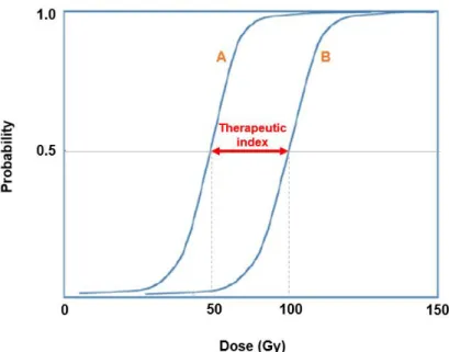
Figure 7 Principle of therapeutic index. Curve A: Probability of tumor control (TCP); Curve B: Probability of complications (NTCP). Adapted from: [21, 26]. ... 22
Figure 8 Constituents of the nervous system. ... 29
Figure 9 Constituents of the spinal cord. Adapted from [36]... 31
Figure 10 Representation of different sections of the spinal cord. ... 31
Figure 11 Spinal cord: segments and their function. Adapted from [36]. ... 32
Figure 12 Cross section of the spinal cord Adapted from [36]. ... 33
Figure 13 Diagram of transverse section of spinal cord. ... 34
Figure 14 Representative scheme of the type of tumors in the vertebral column. ... 37
Figure 15 Example of contour of the spinal cord. (Images provided by the Medical Physics Service of the IPO-Porto). 52 Figure 16 Level of lesion and extension of paralysis according to the spinal segment. ... 54
Figure 17 Hypoxic tumor. Near the blood vessel the tumor has a lot of oxygen but the greater the distance the cells to the blood vessel the lower the oxygen concentration. ... 64
Figure 18 Tumor hypoxia can occur through two different mechanisms proposed by Thomlinson and Gray and Brown. Adapted from [78]. ... 65
Figure 19 Survival curve of tumor cells as a function of dose per fraction supplied. It is assumed that daily fractionation and complete reoxygenation occurred between fractions. Adapted from [78]. ... 65
Figure 20 Iso-effect data for late response from 3 different regions, represented by □○▲, of the rat spinal cord. Where ◊ represents acute skin reactions in mice, ● for early and late murine intestinal damage. The data are plotted in a “reciprocal-dose Fe” form26 such that, if they follow an LQ relationship, the points fall on a straight line. Adapted from [33]. ... 71
xvii
Index of Tables
Table 1 Types of radiotherapy according to the type of target. Adapted from [5]. ... 3
Table 2 Description of how radiotherapy can be administered to the patient [7]. ... 4
Table 3 Phases of the cell cycle: representation and description. ... 9
Table 4 Characteristics of different types of cell death. Adapted from [9]. ... 15
Table 5 Influence of time t and T according to the "R". Adapted from: [7]. ... 19
Table 6 Types of mater. Adapted from [35]. ... 35
Table 7 Description of the functions of the different spinal nerves. Adapted from [39]. ... 36
Table 8 Description of the types of tumors that may arise in the spinal cord. Adapted from [41, 42]. ... 37
Table 9 Lesions in the spinal cord appear in different ways after irradiation. Adapted from: [46]. ... 40
Table 10 Different types of fractionation schemes and their description. Adapted from: [7]. ... 49
Table 11 Variation in BED value according to treatment schedule. ... 52
Table 12 Risk of developing myelopathy from the characteristics mentioned in the table. Adapted from [31]. ... 55
Table 13 Summary of published reports of treatments performed using re-irradiation. ... 57
Table 14 The inclusion and exclusion criteria for SBRT. Adapted from: [47, 60, 63]. ... 62
Table 15 Example, nimorazole has better results at high doses (doses being equivalent to dose used in SBRT) [78]... 66
Table 16 Impact of radiobiological mechanisms in SBRT treatment. ... 67
Table 17 Advantages and disadvantages of radiobiological mechanisms. Adapted from [81]. ... 69
Table 18 Consensus indications and contraindications. Adapted from [84]. ... 72
Table 19 Consensus and predominant practices for the delineation tumor volume for postoperative spine SBRT. Adapted from [84]. ... 73
Table 20 Clinical scenario versus reasonable dose and fractions. Adapted from [84]... 73
Table 21 Treatment planning algorithms for calculation of dose approved by RTOG. Adapted from [84]. ... 74
Table 22 Variation in BED value according to treatment schedule. ... 93
Table 23 Impact of radiobiological mechanisms in SBRT treatment. ... 94
xix
Abbreviations
3D-CRT Three-dimensional (3D) Conformal Radiation Therapy
AAPM American Association of Physicists in Medicine
ACR American College of Radiology
ALARA As Low As Reasonably Achievable
ASTRO American Society for Radiation Oncology
BED Biologically Effective Dose
BSCB Blood-Spinal Cord Barrier
Cdks Cyclin Dependent kinase
CNS Central Nervous System
CT Computed Tomography
DNA Deoxyribonucleic Acid
DSB Double – Strand Breaks
EBRT External Beam Radiation Therapy
ED50 Effect Dose 50%
ERD Extrapolated Response Dose
ETD Extrapolated Tolerance Dose
EQD2 Equivalent Dose of 2Gy
FSU Functional Subunits
Gy Gray
HR Homologous Recombination
HVL Half Value Layer
IGRT Imaged Guided Radiation Therapy
IMRT Intensity Modulated Radiation Therapy
IORT Intraoperative Radiation Therapy
xx
kVp kilo – voltage
LET Linear Energy Transfer
LD Lethal Dose
LQ Linear–Quadratic model
MRI Magnetic Resonance Imaging
NHEJ Non – Homologous End Joining NTCP Normal Tissue Control Probability
OAR Organ at Risk
OPCs Oligodendrocyte Progenitor Cells
PNS Peripheral Nervous System
RBE Relative Biological Efficacy
RM Radiation Myelopathy
RT Radiation Therapy
RTOG Radiation Therapy Oncology Group
SBRT Stereotactic Body Radiation Therapy
Sec second
SINS Spinal Instability Neoplastic Score
SRS Stereotactic Radiosurgery
SSB Such as Single Break
Sv Sievert
TCP Tumor Control Probability
VCF Vertebral Compression Fracture
xxi
Terms and Definitions
The glossary of terms and definitions has been adapted from: the book "Radiobiology for the Radiologist", the Council Directive 2013/59/EURATOM of 5 December 2013, the International Atomic Energy Agency (IAEA) TRS-398 and the online dictionary “The Free Dictionary” [1, 2, 3, 4]. This glossary has specific terms used in radiotherapy, radiobiology, radiological quantities and some clinical terms.
Absorbed dose – Measure of the energy imparted per
unit mass by ionizing radiation to matter at a specific point. The SI unit of absorbed dose is joule per kilogram (J/kg). The denomination of this unit is gray (Gy). The previously used unit of absorbed dose, the rad, was defined to be an energy absorption of 100 erg/g. Thus, 1 Gy = 100 rad.
Absorption – Way in which the energy of a photon is
taken up by matter, typically the electrons of an atom. Removal of x-rays from a beam.
Accelerated fractionation – The treatment schedule,
in this case, exceeds the equivalent of 10Gy per week, in fractions of 2 Gy.
Acute hypoxia – Tumor region characterized by low
oxygen concentration associated with changes in blood flow through the blood vessels may also be called perfusion limited hypoxia.
Adjuvant therapy – Type of treatment to combat
cancer, used in addition to primary therapy. Usually, radiotherapy is used as an adjuvant for surgery or chemotherapy.
ALARA (as low as reasonably achievable) – Principle
adopted to limit the dose of radiation to patients exposed to levels as low as reasonably possible, considering economic and social factors.
Angiogenesis – Denomination for the process of
formation of new blood vessels.
Anterior – The ventral portion of a structure.
Apoptosis – A mode of rapid cell death after irradiation
in which the cell nucleus displays characteristic densely staining globules and at least some of the DNA is subsequently broken down into internucleosomal units.
Bone marrow – The soft, organic, sponge like material
in the cavities of bones; called also medulla ossium.
Cancer – Characterized by uncontrolled growth of the
cells in the human body and the ability of these cells to migrate from the original site and spread to distant sites. If the spread is not controlled, cancer can result in death.
Cdks (cyclin-dependent kinases) – Proteins that
complex with their cyclin regulatory subunits to phosphorylate proteins necessary for progression through the cell cycle.
Cell cycle checkpoint – Mechanism of control that acts
to verify if each phase of the cell cycle was completed correctly before progression to the next phase.
Cells – Cells are the structural and functional units of
living organisms. Each cell plays a specialized role in the body. Groups of cells are arranged together to form tissues. Tissues are organized to form organs in the body.
Central nervous system (CNS) – The portion of the
nervous system consisting of the brain and spinal cord.
xxii
Dose limit – Limit on dose that is applied for exposure
of individuals to prevent the occurrence of deterministic effects and to limit the probability of stochastic effects.
Dose rate – Radiation dose delivered per unit time and
measured, for example, in grays per hour.
ED50 (effect dose 50%) – Dose that produces the
desired effect in 50 per cent of a population.
Effective dose – (E) is the sum of the weighted
equivalent doses in all the tissues and organs of the body from internal and external exposure. The unit for equivalent dose is the sievert (Sv).
Equivalent dose – (HT) is the absorbed dose, in tissue
or organ (T) weighted for the type and quality of radiation (R). The unit for equivalent dose is the sievert (Sv).
Fractionation – The daily dose of radiation based on
the total dose divided into several daily treatments.
Free radical – A fragment of an atom or molecule that
contains an unpaired electron, which, therefore, make it very reactive.
Function subunits (FSU’s) – Many tissues can be
thought of as consisting of discrete FSUs. These may be arranged in series as in the spinal cord, or in parallel as in the kidney.
Gray (Gy) – The special name for the SI unit of
absorbed dose, kerma, and specifi c energy imparted equal to 1 J/kg. The previous unit of absorbed dose, rad, has been replaced by the gray. One gray equals 100 rad.
Grey matter – Part of the central nervous system
consisting mainly of nerve cell bodies. The grey matter of the brain includes the outer layer (the cortex) and several centrally placed masses called nuclei. In the spinal cord, the grey matter occupies the central axis.
Homeostasis – The state of equilibrium, balance
between opposing pressures, in the body with respect to various functions and to the chemical compositions of the fluids and tissues.
Hyperfractionation – The dose per fraction is less than
2Gy.
Hypofractionation – The dose per fraction is greater
than 2Gy.
IMRT (Intensity Modulated Radiation Therapy) –
Type of radiation treatment characterized with highly conformal dose distribution around the target using non-uniform beam intensities, which is possible using static or dynamic segments.
In vivo – Occurring in an artificial environment.
In vitro – Occurring within the living body of an
organism.
Irradiation – Exposure to radiation, as in a nuclear
reactor.
Late responses – Radiation-induced normal tissue
damage that in humans is expressed months to years after exposure. The α/β ratio tends to be small, normally, > 5Gy.
Lethal dose (LD) – Dose of ionizing radiation enough
to cause death. LD50 or MLD is the median lethal dose,
what is the dose required to kill, within a specified period, half the individuals in a large group of organisms similarly exposed. For humans, LD50/60 is about 4Gy. Linear energy transfer (LET) – LET of charged
particles in a medium is the quotient De/dl, where dE is the average energy locally imparted to the medium by a charged particle of specified energy in traversing a distance of dl [keV/µm].
Linear-quadratic model (LQ) – Used to describe the
cell survival curve.
Metastasis – Occurs when cancerous cells invade
surrounding tissues, enter the circulatory system and establish new malignancies in body tissues distant from the site of the original tumor.
Metastatic cancer – The stage of cancer is advanced
in which cells from the primary site have spread, i.e., metastasized.
Misrepair (error prone repair) – Reconstitution with a
loss of information (e.g., deletion caused by the loss of a fragment of the molecule or mutation or translocation).
Mitosis – Replication of a cell to form progeny cells with
identical number (sets) of chromosomes.
Mitotic death – Cell death related with a post-irradiation
mitosis.
Mitotic delay – As a result of treatment, delayed input
into mitosis may occur due to accumulation of cells in the G2 phase.
xxiii
Myelopathy – Any neurologic deficit related to the
spinal cord. If its due to trauma, it is known as spinal cord injury. If it is inflammatory, it is known as myelitis. Disease that is vascular in nature is known as vascular myelopathy.
Oligonucleotide – DNA polymer composed of only a
few nucleotides.
Oncologist – A physician who specializes in the study
and treatment of neoplastic diseases, particularly in the treatment of cancer.
Oncology – Science dealing with the physical,
chemical, and biological features of neoplasms, including causation, pathogenesis, and treatment.
Pathology – The branch of medicine treating of the
essential nature of disease, especially of the changes in body tissues and organs that cause or are caused by disease. The study of diseased is realised both by gross and by microscopic examination of tissues removed during surgery and post-mortem.
Peripheral nervous system (PNS) –
Part of the vertebrate nervous system constituting the nerves outside the central nervous system and includin g thecranial nerves, spinal nerves, and sympathetic an d parasympathetic nervous systems.
Posterior – Also called dorsal. Situated in the back.
Opposite of the previous denomination.
Probability – A mathematical ratio of the number of
times something will occur to the total number of possible occurrences.
Protocol – A detailed written set of instructions to guide
the care of a patient or to assist the practitioner in the performance of a procedure developed specifically for tumors.
Radiation – Electromagnetic radiation consists of wave
motion of electric and magnetic fields. The photons have neither mass nor charge and have an energy inversely proportional to the wavelength of the wave. The electromagnetic spectrum is divided into radio waves, infrared light, visible light, ultraviolet light, and x-rays, according to increasing photon energy and decreasing wavelength.
Radiation (ionizing) – Energy transferred in the form
of particles or electromagnetic waves of a wavelength of 100 nanometres or less (a frequency of 3 x 10¹⁵ hertz or more) capable of producing ions directly or indirectly.
Radiation dose – The quantity of radiation what is
absorbed for an irradiated object. It is expressed in gray (Gy), defined to be 1 J/kg.
Radiation quality – For high-energy photons produced
by clinical accelerators the beam quality (Q) is specified by the tissue–phantom ratio, TPR20,10. This is the ratio of the absorbed doses at depths of 20cm and 10cm in a water phantom, measured with a constant source– chamber distance of 100cm and a field size of 10cm x 10cm at the plane of the chamber.
Radiation therapy – Use of ionizing radiation or any
other type of radiation for the treatment of diseases. It is also called radiotherapy or actinotherapy.
Radiobiology – Study of the scientific principles,
mechanisms, and effects of the interaction of ionizing radiation on living matter. Also called as radiation biology.
Radio-oncologist – Specialist physician with training in
the use of radiotherapy, in order to reduce or cure patients with neoplasia.
Radiosensitivity – Susceptibility of cells, tissues,
organs or organisms to the effects of radiation, such as x-ray or other radiation. Result of radiation effect.
Radiosensitizer – A chemical used to increase the
radiosensitivity of cells to radiation. This substance mimics oxygen in fixing free radical damage.
Radiotherapy – Type of treatment to combat a
neoplasia using ionizing radiation. This type of treatment has as main objective to give an optimal dose of radiation in the place of interest, to cause the smaller possible damages to the normal tissues. Also called radiation therapy.
Redistribution – Cells may exhibit different sensitivity
depending on the phase of the cell cycle they are in. At the mitosis phase cells are more sensitive to DNA damage and late S phase are more resistant. With several dose fractions, there is progress in the cells through a new phase of the cell cycle.
Reoxygenation – Occurs only in tumor cells.
Phenomenon where hypoxic cells become oxygenated after a dose of radiation.
Repair – Refers to the repair of the sublethal lesion. It
occurs more efficiently in normal tissues, since tumor cells usually have more mitoses than the normal cells that generated them, uncontrolled cell cycle and
xxiv activation of the checkpoints for repair. Thus, by
fractionating the treatment of the patient there is the possibility of repairing the normal tissues.
Repopulation – Growth capacity of tumor cells that escaped radio-induced death.
Save dose – is the maximum dose related to the body
mass of a pharmacological agent that can be administered within 24 hours.
SBRT (Stereotactic Body Radiation Therapy) –
Involves stereotactic localization techniques combined with delivery of multiple small photon fields in a few high–dose fractions, for extracranial treatments, leading to a highly conformal dose delivery with steep dose gradients. Stereotactic localization techniques may include the use of relocatable rigid frames, image– guidance techniques, and other positioning tools.
Spinal cord – Part of the central nervous system, along with the brain. It is characterized as being a thick cord of nervous tissue within the spinal canal. In humans it gives rise to 31 pairs of spinal nerves.
Stem cells – Non-specialized human cells that can
produce all types of specialized cells in a lineage.
Syndrome – Combination of characteristics and
symptoms that are indicative of a disease or disorder.
TCD50 – Radiation dose indicating that there is a 50%
probability of tumor control.
Therapeutic index (therapeutic ratio) – Tumoral
response for a permanent level of normal-tissue damage.
Tolerance – Maximum radiation dose prescribed by the
therapist which is indicated as acceptable. It depends on several factors such as time between fractions, fractions indicated, field sizes and treatments previously performed.
Tumor – Growth of cells abnormally. Tumors can be
benign or malignant (cancerous).
White matter – Constituted by myelinated nerve fibers.
1
Chapter 1
1. Introduction
Radiotherapy (RT) is a cancer treatment based on the use of ionizing radiation, where a major evolution occurred over the years. Advances in medical imaging, dose planning and treatment delivery allowed to maximize tumor damage and to minimize the damage in the adjacent normal tissues. Radiobiology has achieved a prominent role over the years, which has contributed to evolution of radiotherapy.
With improved delivery of radiotherapy treatments, survival rates have been increasing in many patients. This increase allowed the development of new tumors and local or regional recurrences, often within or near the previously irradiated site. When these situations occur, re-irradiation is a possibility that presents new challenges to radiation oncologists [5, 6].
One of the most important challenges posed by irradiation is the tolerance of organs at risk (OARs). Given that radiation has previously occurred, there are several factors to be considered so that complications in normal tissues do not overlap with the benefit that a new irradiation brings to the tumor. The spinal cord is considered an OAR because it is characterized by late complications that come from its irradiation, as is the case of radiogenic myelopathy. Since the spinal cord is the most dose-limiting organ in radiotherapy, it is important to understand what factors should be considered with re-irradiation. The tolerance of spinal cord irradiation depends on the irradiated volume, total dose, dose per fraction, time between treatments and the spinal cord region involved. In this way it is expected to control the occurrence of late complications associated to this organ that can give rise to devastating functional deficits [5, 6].
In this work, we first present a review of the actual knowledge about the spinal cord, such as its anatomy and physiology. A special attention is given to radiobiology and its role in a course of radiotherapy. With the advancement of medical technology and with a higher life expectancy of patients, local and regional recurrence often appears,
2 and it is necessary to prescribe re-treatments. Thus, this work intends to answer several questions, such as: what maximum dose can be given in the first treatment so that a re-treatment can be performed, what maximum dose can be prescribed in a re-treatment without causing severe late effects, what is the time interval between treatments, and which type of RT treatment best applies to the patient's condition. Stereotactic body radiotherapy (SBRT) is an innovative RT treatment that aims to deliver with maximum precision the maximum possible radiation dose, in a small number of fractions (1 to 5 fractions) compared to conventional RT.
3
Chapter 2
2. Theoretical concepts
2.1 The role of radiation therapy
Radiation therapy (RT) is a common treatment for various cancers and uses ionizing radiation to damage the deoxyribonucleic acid (DNA) of malignant cells, which usually replicate at a faster rate than normal cells in the body. Ionizing radiation deposits energy in the material along its path, being absorbed by the same material through various interactions. When radiation interactions occur, it breaks the molecular bonds of the DNA of the cells thus altering its structure. Through this mechanism, radiotherapy can prevent the replication of abnormal cells causing cellular dead [7, 8]. The different types of radiotherapy are shown in (Table 1).
Table 1 Types of radiotherapy according to the type of target. Adapted from [5].
Radiotherapy may be used as a single or neoadjuvant treatment (treatment given prior to any other treatment) or adjuvant (treatment given after any other type of treatment such as surgery and chemotherapy) [7].
RT can be administered in the following ways (Table 2): internal radiotherapy (brachytherapy, intraoperative radiotherapy (IORT)) and external radiotherapy (stereotactic body radiotherapy (SBRT), three-dimensional conformal radiotherapy (3D-CRT), radiotherapy modulated by intensity (IMRT), image-guided radiotherapy (IGRT) and radiosurgery).
Types of radiotherapy
Preventive radiotherapy Curative radiotherapy Palliative radiotherapy Prevention of possible metastases or
recurrences through the application of radiotherapy.
Tissue–tumor ratio is such that curative doses of radiation can be used without unduly harming normal tissue.
Radiation therapy can be administered to relieve pain in cancer patients.
4
Table 2 Description of how radiotherapy can be administered to the patient [7].
The main objective of radiotherapy is to administer the prescription dose in the target volume, saving as much adjacent normal tissues as possible [7].
Type of treatment Description
Brachytherapy
Uses radioactive sources (temporary or permanent) placed near the target volume. These sources can be
inserted directly into the tumor or placed through applicators previously inserted into a body cavity.
Intraoperative radiotherapy (IORT)
Administered under intraoperative conditions, usually by electron beams or low energy x-rays. It is used after resection of the primary tumor and external radiation
therapy is usually necessary. External radiotherapy
It involves the use of a linear accelerator to administer, outside the patient's body, radiation beams focused on
the target volume to be treated.
Stereotactic body radiotherapy (SBRT)
Administered through multiple beams that are focused on a three-dimensional target. For tumors of the central
nervous system (CNS) a thermoplastic mask is used, whereas for extracranial sites a body frame may or may
not be used.
Three-dimensional conformal radiotherapy (3D-CRT)
Technique used where dose volume is made to fit the target using 3D anatomical data acquired from computed
tomography (CT) or magnetic resonance imaging (MRI) imaging modalities. The goal is to apply the maximum dose to the target and save neighbouring structures as
much as possible with the help of advanced computer hardware and software.
Radiotherapy modulated by intensity (IMRT)
It provides a highly conformal dose distribution around the target using non-uniform beam intensities, which is possible using static or dynamic segments. The isodose
distribution can then be closely monitored by the target, modulating the intensity.
Image-guided radiotherapy (IGRT)
It uses various radiological and functional imaging techniques to perform high-precision radiotherapy. The main goals are to reduce the configuration and internal margins, and account for changes in target volume during radiation therapy, such as decreased tumor volume or weight loss (adaptive radiotherapy).
Radiosurgery
Can be performed by all kinds of advanced radiotherapy techniques, including IMRT, IGRT, synchronized respiratory radiotherapy, tumor-tracking radiotherapy
5
2.2 Radiobiology: essential concepts
In radiation oncology, radiobiology is defined as the science that investigates the interactions between ionizing radiation and living systems, as well as the consequences of these interactions [7].
2.2.1 The role of radiobiology in the evolution of radiotherapy
Radiobiology has allowed creation of new ideas and identification of potentially exploitable mechanisms in radiotherapy. According to experimental and theoretical studies in radiobiology, it was possible to verify that this area contributed through three different levels for the development of radiotherapy [9]:
▪ Conceptual basis for radiotherapy: identifying the mechanisms and processes underlying the response of tumors and normal tissues to irradiation, which lead to the explanation of observed phenomena. The knowledge about the 5 Rs of radiotherapy is an example of the knowledge acquired through the conceptual basis;
▪ Treatment strategies, allowing the development of new approaches in radiotherapy. Examples are the discovery of hypoxic cell sensitizers, high linear energy transfer (LET) radiotherapy and hyperfractionation;
▪ Protocols, providing a more diverse range of treatment schemes in clinical radiotherapy.
These three levels used by radiobiology are a starting point that provide insight into new options. Many treatment strategies produced through radiological investigation do not produce demonstrable clinical benefits. Creating protocols through experiments is a slow process. The ability of laboratory science to guide a radiation oncologist in the choice of specific protocols is limited by the inadequacy of theoretical and experimental models to clinical practice [9].
2.2.1.1 Radiobiological mechanisms
In the exposure of living tissues to ionizing radiation absorption of photon energy by cells occurs. The radiation through the biological material will trigger a series of events, interact with the atoms and molecules of the medium, with the consequent transfer of energy.
There is strong evidence that DNA is the target of the biological effects of ionizing radiation, namely cell death, loss of clonogenic capacity, genetic mutations and
6 chromosomal aberrations, with consequent somatic, hereditary, teratogenic and carcinogenic effects.
Ionizing radiation causes damage to cells or tissues by depositing energy in a sequence of events. Different types of radiation have distinct abilities to cause biological damage. All these lesions are caused at the cellular or molecular level. The biological effects subsequently manifested are due to the impact of these lesions on millions of cells in an organ or tissue. Considering that the nature of the damage is molecular or cellular, it is therefore essential to understand the mechanisms involved.
Ionizing radiation, used in radiotherapy, causes a cascade of events, which begins immediately after its emission. The initial ionization (physical phase) is followed by immediate damage of vital macromolecules at the cellular level – direct effect, or indirectly through interaction with water molecules, resulting in free radicals of oxygen, highly reactive at the molecular level (physical–chemical phase) – indirect effect.
The energy deposited initially by ionizing radiation is used in the formation of free radicals. Since the cells are mainly composed of water, most of the lesions caused by x-rays to biological molecules - about 2/3, are mediated through free radicals, namely hydroxyl radicals. The free radicals resulting from these reactions initiate complex
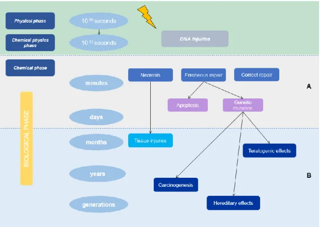
Figure 1 Time scale of the effects of ionizing radiation: biological changes manifest after a period of latency that can go from minutes to weeks to years after exposure (A - early effects; B - late effects).
7 chemical reactions, which can lead to the destruction or inactivation of vital molecules in the cell. Chemical phase follows in which sub-lethal damage repair can occur. Then, during the biological phase, the manifestation of unrepaired lesions in the DNA occurs. These 3 different phases described previously differ in time scale (Figure 1) [9]:
1) Physical phase (about 10-18sec) – phase where photon energy deposition occurs
to the orbital electrons, exciting or ionizing them;
2) Physical–chemical phase (about 10-13sec) – the breakdown of chemical bonds
caused by excited or ionized atoms occurs. These atoms can cause damage to important molecules, such as DNA, in a direct way or through the formation of free radicals;
3) Chemical phase (seconds to years) – constituted by the reaction of the cells to the damages caused by the radiation. Damage to DNA can be repaired by specific enzymes, but some are irreparable leading to cell death. Cell death is not immediate occurring in most cell division after irradiation (and may occur up to 5 or 6 cycles after).
The main function of radiobiology is to observe the phenomena that occur with irradiation of the tumor and normal tissues, suggesting improvements to the existing therapeutic options. The tumor response to irradiation is called regression and may be followed by recurrence. If tumor recurrence does not occur during the patient's life, local tumor control may be considered to exist [9].
Thus, when a cell absorbs radiation 4 possibilities can occur:
▪ Absorption of the radiation may have no adverse effects, or if injury occurs, it may be repaired without any trace of exposure occurring; if not repaired, the apoptosis pathway may be activated;
▪ Cell can suffer lethal, irreparable and irreversible damage, leading to cell death;
▪ Cell may lose its clonogenic capacity;
▪ Gene mutations may occur, with distinct consequences depending on the type of cell where they occur.
The most frequent cause of radiation-induced cell death is the inability to correct double lesions in the DNA strand and manifests itself when the cell attempts subsequent cell division. In this way, more proliferative cells manifest damage or die much earlier than cells with longer proliferation times. Thus, biological changes in cells and tissues due to ionizing radiation occur only after a latency period, which may range from minutes to
8 weeks or even years (as a function of dose, dose rate, cell kinetics, control of cycle regulating genes cell phone, etc.).
Genetic and biochemical constitution is a determining factor in the molecular response to radiation, and several molecules are already identified through which cells detect radio-induced lesions.
Recognition of these lesions activates signal transduction pathways suitable for cellular response to injury. This process is influenced by internal cell signalling processes as well as external factors such as hypoxia, cytokines, intercellular contact and extracellular matrix. The result of these interactions may promote cell survival or death, cell cycle arrest or blockage, and DNA repair or genetic instability, depending on how the cells respond to radio-induced lesions.
Thus, while initial energy deposition and subsequent events occur in 10-18–10-13 sec, the
chain of biological effects that begins by inducing programmed cell death or repairing sublethal and potentially lethal damage, leading to tissue repair and remodelling can take minutes, hours, days, months or years to express these effects.
There are also several factors that affect the cell response to radiation: physical factors (dose, dose rate, fractionation, LET - linear energy transfer - and RBE - relative biological efficacy); chemical factors (radiosensitizers, radioprotectants, O2 tension) and biological
factors (proliferative state, cell cycle phase, physiological or metabolic state, genetic constitution of the cell) - reviewed in [10].
2.2.1.2 Cell cycle control
The concept of cell cycle is essential for the knowledge of all cellular radiobiology processes. It is a necessary process that involves great fidelity and of extreme importance for the propagation of organisms (Figure 2).
The cell cycle consists of a succession of events that lead to duplication of genetic material and other cellular components and eventually cell division (Table 3).
9
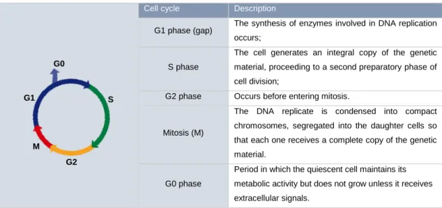
Table 3 Phases of the cell cycle: representation and description.
Cell cycle Description
G1 phase (gap) The synthesis of enzymes involved in DNA replication occurs;
S phase
The cell generates an integral copy of the genetic material, proceeding to a second preparatory phase of cell division;
G2 phase Occurs before entering mitosis.
Mitosis (M)
The DNA replicate is condensed into compact chromosomes, segregated into the daughter cells so that each one receives a complete copy of the genetic material.
G0 phase
Period in which the quiescent cell maintains its metabolic activity but does not grow unless it receives extracellular signals.
For cell cycle progression to occur, a DNA check system is required to ensure complete duplication of the genome in an orderly and highly faithful manner. Changes in normal cell cycle control lead to genetic instability, a major factor in carcinogenesis. This is why the existence of checkpoints for the integrity and state of the replication of the genetic material that allows the stop of the cell cycle progression in case of damage or mutation of the DNA, so that it is evaluated and repaired, ensures the complexity and irreversibility of the cell cycle, as well as the processes of activation, expression and degradation of the proteins involved. On the other hand, physiological cell death in self-regulating tissues, such as the skin, intestine and bone marrow, is necessary to give rise to the cells that are constantly formed: it is programmed cell death also called apoptosis. In this
Figure 2 Role of cyclins and cyclin-dependent kinases (CDKs) in cell cycle regulation: The cell cycle is divided into G1, S (DNA synthesis), G2 and M (mitosis) phases. The transition between phases is controlled by cyclins and CDKs. G1 G0 G2 S M
10 process, where several regulatory genes and a family of proteases, the caspases, are involved, the cells are fragmented into membrane-bound corpuscles, with consequent phagocytosis by neighbouring cells, with no inflammatory response.
Failure to induce apoptosis contributes to carcinogenesis by allowing the occurrence of genetic instability and the deregulation of the activity of the genes involved in cell cycle control and their checkpoints. During the regulation processes, the cell activates and inactivates the proteins by addition by the kinases or removal by the phosphatases of the phosphate groups, respectively. For faster and more effective kinases and phosphatases can physically bind to the protein they modify, with formation of multiprotein complexes [11].
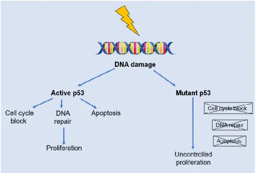
The p53 gene, known as the "guardian of the genome", is a tumor suppressor gene that encodes a nuclear phosphoprotein that, because of its physiological functions, ensures cellular genetic integrity: cell cycle regulation, apoptosis control and DNA repair (Figure 3).
When DNA damage occurs, the p53 protein is responsible for the temporary stopping of the cell cycle in G1 for repair or, when this is not possible, the process of cell apoptosis begins. Thus, if p53 mutation occurs, the functions of the protein may not be activated, and the consequence will be uncontrolled cell proliferation. Mutations of this gene are described in more than 50% of human tumors, conferring a proliferative advantage on
Figure 3 p53 Injury recognition process: under normal conditions the protein expressed by the p53 gene is responsible for the temporary stopping of the G1 cell cycle for DNA repair or, if not possible, programmed cell death (apoptosis). If the gene is mutated, the functions of p53 are not activated and the result will be uncontrolled cell proliferation.
11 cells carrying the mutation. Apoptosis is one of the best studied processes in cell death by ionizing radiation and depends on p53. Cells with mutations in this gene do not undergo apoptosis after treatment with ionizing radiation, unlike in cells where the p53 gene is active [12].
2.2.1.3 Proliferation and differentiation
The concept of cell proliferation and its influence on response to treatment is crucial in RT. Cell proliferation in normal tissues, unlike in tumors, is very well organized through homeostatic control: there is a balance between cell production and the loss of more differentiated cells. Radiation is a unique cytotoxic agent because lesions caused to DNA can remain inactive for hours, days, weeks or months. This occurs because in the clear majority of normal and tumor tissues, cell death only occurs when the cell attempts the next division, which depends on the proliferative characteristics of the tissue. In cells of the intestinal mucosa, in which cell cycle time (Tc) is short (12-24h), cell death occurs after a few hours: in the skin (Tc = 4 days), may take a week; in the kidney (Tc indeterminate) can take months [13].
As at the cellular level there are preferential targets for the action of radiation, also at the tissue level there are cells or groups of cells more sensitive or whose death depends on the changes observed in the body after irradiation. The different organs or tissues of the living organism are composed of several cell types and the response of an organ or tissue to the radiation depends on the intrinsic sensitivity of the different cellular populations in that organ or tissue and the proliferative characteristics of each population. The Law of Rubin and Casarett differentiates tissue sensitivity – being that this is essentially a function of the type of cell that constitutes the tissue – in five distinct groups. In the first group are the most sensitive cells, as they are those that are more metabolically active, divide more quickly and more undifferentiated. In the second group are the differentiated, intermittent cells, the result of the cell division of the cells of the previous group, which are still very active and relatively undifferentiated mitotically. In the intermediate group, which includes connective tissue cells, we find vascular endothelial cells and fibroblasts. In the following groups cell differentiation increases and proliferation decreases until the last classification, which includes fixed postmitotic cells, where we find examples of the most resistant cells [14].
12
2.2.1.4 Tolerance of normal tissues to radiation
Cellular organization in proliferative and functional compartments has important consequences for tissue response to radiation. In this way, the tissues can be divided into 2 categories.
In the first category - hierarchical cell organization - tissues that have a clear separation between the proliferative compartment (including the population of stem cells, capable of unlimited self-renewal; the amplification compartment - cells that proliferate rapidly but only over a limited number of cell divisions), and the differentiated cell compartment, responsible for organ / tissue functions. As examples we have the most proliferative tissues such as the hematopoietic system, the cells of the basal layer of the epidermis and the lining of the gastro-intestinal system and the spinal cord.
The begin of the acute reactions of these tissues to the radiation is correlated with the life span of the differentiated functional cells, and the intensity of these reactions reflects the ratio between the rate of destruction of the stem cells and the rate of regeneration of the surviving clonogenic cells.
In the second category - flexible cell organization - tissues are included in which there is no clear separation between the two compartments but in which some differentiated cells also exhibit self-renewal capacity. In this type of organ/ tissue with a low proliferation rate (kidney, lung), the relationship between cell death and tissue response is less evident because organ damage can occur due to changes in vascular, connective or parenchymal tissue.
Late effects are not only restricted to these slow cell renovation tissues. For example, in epithelial tissue, late lesions - fibrosis, atrophy and telangiectasia - may occur in addition to early reactions. Thus, different types of lesions may occur sequentially in an organ or tissue, resulting from distinct mechanisms and cellular interactions.
The difference between acute and late effects can be explained by their progression: while the acute effects are quickly repaired by the high proliferation of stem cells and can be completely reversible, the late effects can be attenuated but never completely repaired as they result from the association of vascular lesions with loss of parenchymal cells.
This distinction has relevant biological consequences: as acute reactions occur during conventional RT treatment, it is possible to make the necessary changes to allow the survival of stem cells that will repopulate and ensure cell proliferation. Late effects, which
13 occur months or years after RT, are much more sensitive to changes in fractionation than early effects.
Tolerance of normal tissues may also be influenced by treatment-related variables (total dose and dose per fraction, dose rate, total time of treatment, energy and volume irradiated, use of concomitant chemotherapy), patient (age, comorbidity associated with diabetes, vascular disease), or with the organ in question (development of radiation toxicity, variation in intrinsic radiosensitivity of the organ) - reviewed in [15].
2.2.1.5 Rs of radiobiology
The complexity of the response to RT increases with the characteristics of the surrounding normal tissues: while cells and tissues may respond differently to the same dose of radiation, the response of a tissue is strongly determined by the rate of cell proliferation and tissue repopulation capacity in addition to the molecular and cellular factors that determine intrinsic cellular radiosensitivity.
Tumor responses show greater variability than normal tissues and these biological differences are explored in dose fractionation in radiotherapy, where the protocols are derived empirically but exploit differences in biological response between normal and tumor tissues at the same dose of radiation. The biological factors that influence the response of normal and tumor triglycerides to RT were summarized by Withers (1975) as the radiotherapy Rs [16].
Radiotherapy given in a single, high dose fraction is ineffective for tumor control and has serious side effects. To reduce these effects, radiotherapy was given in small fractions daily and at low doses and this type of treatment was referred to as fractional radiotherapy [7].
Fractional radiotherapy is based on five main features known as the "Rs of radiobiology". These are described then in order of occurrence, i.e., the first biological mechanism observed is the repair of sub-lethal damage, followed by redistribution of cells in the cell cycle, reoxygenation, and finally repopulation [7].
The classical fractionation principles, i.e., the Rs of radiobiology, explain the effects of high doses of ionizing radiation on tumors and adjacent normal tissues. The outcome of standard clinical radiation treatment is determined by the Rs of radiobiology.
14
2.2.1.5.1 Repair of sub-lethal damage
In the literature, this mechanism can be called in several ways: "repair", "repair of DNA damage" or "repair of sub-lethal damage".
Repair of sublethal DNA damage: normal cell are more effective than tumor cells in this
process as observed from cell recovery in the 2 hour period after exposure to ionizing radiation. Radiation randomly interacts with molecules in the cell, but DNA is the main target molecule for the biological effects of radiation, including cell killing, carcinogenesis and mutation. In radiotherapy radiation damage is primarily manifested by the loss of reproductive capacity. Radiation causes a wide range of lesions in DNA such as single (SSB) and double-strand breaks (DSB) in the sugar-phosphate backbone of the DNA molecule. SSB can be readily repaired using the undamaged chromatid as a template. The most deleterious lesion induced by ionizing radiation is DSB, a break in both strands of 10 base pairs or less. There are several mechanisms to repair DSBs, which indicate the importance and difficulty of repairing this type of DNA injury. The most important are non-homologous end joining (NHEJ) and homologous recombination (HR). Homologous recombination provides greater repair accuracy than NHEJ, the major pathway to repair DSB throughout all the phases of the cell cycle [17].
Radiotherapy cause lethal damage to tumor cells and sublethal damage to normal tissue cells. Sub-lethal damage can be repaired if enough time is given between exposures to radiation. If the cell is exposed to radiation before the repair occurs, the damage can become lethal, i.e., the sum of the damages caused leads to the repair not being viable and cell death occurs (Table 4) [7].
Normal tissue cells that have a late response to radiation can repair faster than tumor cells, if there is an ideal range between fractions of 6-12h [7].
Different types of cells have distinct abilities to correct radiation-induced damage, and some cells having been verified to be faster compared to others to repair sub-lethal damage [9].
15
Table 4 Characteristics of different types of cell death. Adapted from [9].
Morphological changes
Types of cell death Nucleus Cell membrane Cytoplasm Biochemical features
Apoptosis
Condensation of chromatin, nuclear fragmentation; DNA
laddering
Blebbing Fragmentation Caspase-dependent
Autophagy Partial chromatin
condensation; Blebbing Autophagic vesicles
Caspase-independent
Necrosis Degradation of
nuclear DNA Swelling and rupture
Organelle degeneration and mitochondrial dilatation - Senescence Distinct heterochromatic structure - increased granularity
and flattening SA-β-gal activity
Mitotic catastrophe nuclear fragmentation and dicentric chromosomes. - - Caspase-independent (at early stage)
It is relevant to distinguish two processes that are commonly accepted as the same: repair and recovery. While the first refers to the method in which the cell corrects a radiation-induced error, recovery is understood as the ability of a tissue, not a cell, to increase its cellular survival or decrease the damage caused if it has sufficient time for this process to occur, such as the recovery of an erythema.
Repair is due to the correction of sub-lethal damage and can be measured, for example by a sequence of irradiances separated by variable time interval. On the other hand, recovery may involve the recruitment of cells that are in a non-division phase, G0, to enter the cell cycle and thus compensate for cell death [9].
2.2.1.5.2 Redistribution of cells in the cell cycle
Cell cycle consists of four distinct and consecutive phases (Figure 4) [9]:
▪ Phase G1 and G2, which are periods of apparent inactivity (gap), where the G1 phase occurs before S phase and the G2 phase between synthesis and mitosis; ▪ Phase M corresponds to the phase of mitosis;
▪ Phase S, which is the period where the synthesis of genetic material occurs to proceed to the division.
16
Redistribution: when radiotherapy is given to a heterogenous cell population, cells may
be in different phases of the cell cycle. Cells in S phase are more radioresistant and cells in late G2 and M phases are more sensitive. A small dose of radiation will destroy the more sensitive cells, and a resistant cell population that is now synchronized survives. As fractionated radiotherapy treatment continues, the resistant surviving cells will continue throughout the cell cycle and when a new dose is delivered some of these cells have moved from a resistant to a more sensitive stage and will then be killed more easily (Figure 5) [19].
Figure 4 Representation of the cell cycle. Adapted from [18].
17
2.2.1.5.3 Reoxygenation
As tumor volume increases through tumor cell proliferation, vascularization does not accompany this tumor growth, becoming insufficient to meet its requirements and hypoxic regions begin to appear in tumor tissue. Hypoxic cells are 2-3 times more resistant to radiation, and well-oxygenated cells are eliminated during treatment of fractionated radiotherapy. Since oxygen supply is constant, hypoxic cells gradually gain vascularity and oxygenation, and the output radiosensitivity increases [7].
Reoxigenation: The level of oxygenation in a tumor is a major determinant of the
effectiveness of radiotherapy. Tumor cell microenvironment presents areas with decreased pH, lack of nutrients and hypoxia. Oxygen concentration (pO2) varies between 10- and 80-mm Hg in normal tissues but in tumors these values can be lower than 5 mm Hg in some areas. This anomaly is due to the development of abnormal vasculature during tumor angiogenesis. A significant proportion of tumor cells is hypoxic, showing great heterogeneity that is not correlated with standard prognostic factors such as size, stage and histological type [20].
Hypoxia may have a crucial role in treatment outcome and may also influence metastatic capacity of tumor cells resulting from genetic changes such as those involving blood oxygen transport or inducing vascularization. As a result of prolonged exposure to hypoxia, cells can acquire genetic resistance to apoptosis suggesting that hypoxia can favour tumor progression through clonal selection of cells with more aggressive phenotypes [21].
After an initial dose of radiotherapy, the more sensitive oxygenated cells are killed; during reoxygenation, surviving tumor hypoxic cells can increase their oxygen supply thus increasing their sensitivity to radiation. Biological efficacy of ionizing radiation relies on oxygen interacting with cells and making DNA lesions permanent [22].
Prolonged exposure to hypoxia can induce tumor death by apoptosis, as cells with mutations in the p53 gene acquire genetic resistance to hypoxia-mediated apoptosis, suggesting that hypoxia may favour tumor progression by selecting cells with mutations in p53 [19]. Other studies suggest that cells in hypoxia may develop genomic instability or that these cells may reduce the functionality of proteins involved in DNA repair [23]. After a first dose in the treatment of RT, the more sensitive oxygenated cells are eliminated; the surviving tumor cells are in hypoxia but later, during treatment, their O2
18 ionizing radiation depends on the presence of oxygen, which reacts with the cells making the DNA lesions permanent [24].
The division of the dose into several fractions spares the normal tissue due to the occurrence of repair of the sublethal damages and the cellular repopulation that occurs between the fractions. Simultaneously, fractionation allows for greater damage to the tumor caused by reoxygenation of hypoxic cells and the redistribution of cells to more radiosensitive phases of the cell cycle.
To oxygenate a tumor in the hypoxic state [7]:
▪ If the haemoglobin value is low, a blood transfusion can be given to the patient; ▪ High pressure oxygen may be applied during radiotherapy;
▪ The patient may be prevented from using hypoxic materials, such as cigarettes, during radiation therapy;
▪ Hypoxic radiosensitizers may be used, such as metronidazole.
2.2.1.5.4 Repopulation
Repopulation: Some time after irradiation an increase in cell division is seen in normal
and malignant cells. Repopulation occurs at different rates depending on the tissues and represents cell proliferation that aims at compensating the cell population that was killed. This homeostatic response to cell loss occurs in situations other than irradiation and is related to specific cell-cycle time: as a result of radiotherapy cell death occurs after irradiated cells attempt mitosis and thus highly proliferative tissues (and tumors) show damage much faster than slowly proliferative tissues.
Normal and tumor cells continue to proliferate when exposed to radiation. This proliferation is a physiological response of tissues to a decrease in cell numbers. The proliferation of the cells leads to two main consequences, such as the increase in the number of tumor cells to be destroyed, which is against the stipulated treatment, and the increase in the number of normal cells after irradiation, which is in favour of the treatment [7].
![Figure 4 Representation of the cell cycle. Adapted from [18].](https://thumb-eu.123doks.com/thumbv2/123dok_br/15835992.1083696/46.892.253.634.121.419/figure-representation-cell-cycle-adapted.webp)

![Figure 9 Constituents of the spinal cord. Adapted from [36].](https://thumb-eu.123doks.com/thumbv2/123dok_br/15835992.1083696/61.892.187.697.146.534/figure-constituents-spinal-cord-adapted.webp)
![Figure 11 Spinal cord: segments and their function. Adapted from [36].](https://thumb-eu.123doks.com/thumbv2/123dok_br/15835992.1083696/62.892.153.734.550.995/figure-spinal-cord-segments-function-adapted.webp)
![Figure 12 Cross section of the spinal cord Adapted from [36].](https://thumb-eu.123doks.com/thumbv2/123dok_br/15835992.1083696/63.892.160.727.669.999/figure-cross-section-spinal-cord-adapted.webp)
