A
rt
ic
le
J. Braz. Chem. Soc., Vol. 20, No. 6, 1095-1102, 2009. Printed in Brazil - ©2009 Sociedade Brasileira de Química 0103 - 5053 $6.00+0.00
*e-mail: [email protected]
Acanthoic Acid and other Constituents from the Stem of
Annona amazonica
(Annonaceae)
Maria Lúcia B. Pinheiro,*,a Clahildek M. Xavier,a Afonso D. L. de Souza,a Diego de Moura Rabelo,a
Cristiane L. Batista,b Regiane L. Batista,b Emmanoel V. Costa,b Francinete R. Campos,b
Andersson Barison,b Rodrigo H. Valdez,c Tânia Ueda-Nakamurac and Celso V. Nakamurac
aDepartamento de Química, Universidade Federal do Amazonas, 69077-000 Amazonas-AM, Brazil
bDepartamento de Química, Centro Politécnico, Universidade Federal do Paraná, 81530-900 Curitiba-PR, Brazil
cDepartamento de Análises Clinicas, Universidade Estadual de Maringá, 87020-900 Maringá-PR, Brazil
O presente trabalho descreve o isolamento, a partir do caule de Annona amazonica, do ácido
acantóico, um diterpeno do tipo pimaradieno que possui várias e importantes atividades biológicas descritas na literatura. Neste estudo foi verificado que este composto apresenta significante atividade
tripanocida contra as formas epimastigotas de Trypanosoma cruzi. Também foi constatado que este
diterpeno é o constituinte majoritário da planta, encontrado em cerca de 65% do extrato hexânico,
demonstrando que A. amazonica é uma nova fonte natural renovável desta substância. Além
do ácido acantóico, a investigação química resultou no isolamento dos alcalóides liriodenina e cassiticina, entre outros compostos, tais como terpenos, esteróides e ácidos graxos. Adicionalmente,
é descrita a completa e inequívoca atribuição dos deslocamentos químicos de RMN de 1H e 13C
da cassiticina.
The present work reports the isolation of acanthoic acid, a promising pimaradiene-type diterpene with several important biological activities described in the literature, from the stems of Annona amazonica. We found that acanthoic acid has significant trypanocidal activity against
the epimastigote forms of Trypanosoma cruzi. This diterpene is the major constituent of the plant,
comprising at least 65% of the hexane extract, demonstrating that A. amazonica is a new renewable
natural source for this compound. The chemical investigation also resulted in the isolation of the alkaloids liriodenine and cassythicine, and other compounds including terpenes, sterols, and fatty
acids. Additionally, the complete and unequivocal 1H and 13C NMR chemical shift assignments
for cassythicine are provided.
Keywords: Annona amazonica, Annonaceae, acanthoic acid, cassythicine, trypanocidal activity
Introduction
The family Annonaceae, comprised of tropical and subtropical species with about 135 genera and more than 2500 species and widely distributed in South and Central America, Africa, Asia, and Australia,1 is known for its edible fruits and
the medicinal properties of several species.2 In Brazil, there
are 26 genera with about 260 species, including the genus
Annona, which contains approximately 120 species.3
Chemical investigations of species of Annonaceae have revealed their high chemical diversity in terms of secondary metabolites, such as alkaloids, acetogenins,
terpenoids and lactones. These compounds have shown important biological activities, including antiparasitic, in particular against Leishmania sp., Plasmodium falciparum, and Trypanosoma cruzi.4-12
Annona amazonica R.E. Fries is a tropical tree that grows up to 20-25 m tall and 35-60 cm in diameter, and is found from Panama to South America. In Brazil this species commonly occurs in the Amazon, mainly in the states of Amazonas and Pará.13 To the best of our knowledge, only one
previous phytochemical study has described the isolation and identification of cyanogenic constituents from this species.14
Acanthoic Acid and other Constituents from the Stem of Annona amazonica (Annonaceae) J. Braz. Chem. Soc. 1096
promising compound with several important biological activities described in the literature. This compound was found to be the principal component of A. amazonica, and showed trypanocidal activity against epimastigote forms of Trypanosoma cruzi, the causative agent of Chagas’ disease. In addition, the full NMR analyses of the alkaloid cassythicine (3) are included.
Experimental
General procedures
1D and 2D NMR experiments were recorded on a Bruker AVANCE 400 spectrometer operating at 9.4 T, equipped with a 5 mm multinuclear direct detection probe with z-gradient, observing 1H and 13C at 400.13
and 100.61 MHz, respectively. 1H-13C correlation
(HSQC and HMBC) experiments were performed with the average coupling constant 1J(C,H) and LRJ(C,H)
optimized for 140 and 8 Hz, respectively. The 1D nOe experiments were obtained by selective excitation of each 1H NMR frequency and gradient selection using the
double-pulsed field gradient spin-echo (DPFGSE)-nOe experiment with constant mixing time of 500 ms. IR spectra were recorded in KBr pellets on a Perkin-Elmer Spectrum 2000 spectrometer. Low resolution ESI-MS and ESI-ESI-MS/ESI-MS data were taken in positive ion mode, on a Micromass Quatro LC mass spectrometer, equipped with an ESI/APCI “Z-spray” ion source. GC-MS analyses were performed on an Agilent Technologies 6896 N instrument equipped with a HP-5MS (29.6 m × 0.25 mm; 0.25 µm film thickness) fused-silica capillary-column. The following conditions were used: temperature program: 70 oC for 5 min; 5 oC min-1
until 300 oC; 300 oC for 20 min; injector temperature:
250 oC; carrier gas: helium, adjusted to a linear rate of
32 cm s-1 (measured at 100 oC); injection type: split flow,
adjusted to give a 1:20 ratio; septum sweep constant at 1.0 mL min-1; EI-MS: electron energy at 70 eV. Melting
points were obtained on a Quimis Q-340S23 micromelting apparatus. Optical rotations were measured on a Rudolph Research Autopol III polarimeter. Gravity-column chromatography (CC) was carried out on silica gel 60 (70-230 mesh, Merck), while analytical and preparative thin-layer chromatography (TLC) was run on 0.25 mm thick aluminum-backed silica-gel 60 plates type F/UV254 and 366 (Merck) and 2 mm thick glass-backed silica gel 60 plates type P/UV254 (Macherey-Nagel), respectively. The spots were detected by exposure to UV light at 254 or 366 nm, as well as by spraying with Dragendorff’s reagent or 5% H2SO4 in EtOH and then heating on a hot plate.
Botanical material
The stems of A. amazonica weres collected in May 2003 in the Adolpho Ducke Forest Reserve (coordinates: 02º54’26’’ to 03º00’22”S; 59º52’40” to 59º58’40”W), situated 26 km northeast of the city of Manaus in the state of Amazonas, and identified by a specialist in Annonaceae, Prof. Dr. A.C. Webber. A voucher specimen (# 1796) was deposited in the Herbarium of the Instituto Nacional de Pesquisas da Amazônia (INPA).
Extraction and isolation
The powdered air-dried stems of A. amazonica (1.7 kg) were extracted successively with n-hexane (4 × 2 L), CH2Cl2 (4 × 2 L), and MeOH (4 × 2 L), at room temperature. Removal of the solvents under reduced pressure gave hexane (10.5 g), CH2Cl2 (6.0 g), and MeOH (30.0 g) extracts.
Hexane extract: isolation of 1 and GC-MS analysis
The hexane extract yielded large crystals in very high quantity. These were manually collected, washed in hexane, and submitted directly to NMR and other spectroscopic investigations. They were identified as a pimaradiene-type diterpene, (-)-ent-pimara-9(11),15-dien-19-oic acid or acanthoic acid (1, 6.9 g). After the acanthoic acid crystals were removed, 10 mg of the remaining hexane extract was methylated with diazomethane and submitted to GC-MS analyses according to the conditions described in the general procedures, above. The chemical constituents were identified on the basis of their GC retention indices with reference to a homologous series of C8-C26n-alkanes, as well as by matching their mass spectra with those from the NIST 98 MS and Wiley 7n MS libraries, and by comparing the fragmentation patterns of the mass spectra with those reported in the literature.15
Dichloromethane extract: isolation of 1-10
Pinheiro et al. 1097 Vol. 20, No. 6, 2009
The alkaloid fraction (0.7 g) was initially subjected to silica-gel column chromatography (CC), previously treated with a 10% NaHCO3 solution9 and eluted with increasing
concentrations of CH2Cl2 in hexane, followed by EtOAc in CH2Cl2, and MeOH in EtOAc. The eluted fractions (30 mL) were evaluated and pooled by TLC analysis, affording 10 subfractions. Subfraction 5 (200 mg) was further purified by silica gel CC using the same methodology as above, and subsequently on preparative TLC eluted with CH2Cl2-MeOH (95:05, v/v), resulting in the oxoaporphine alkaloid liriodenine (2, 4 mg), and in the aporphine alkaloid cassythicine (3, 6 mg).
The neutral fraction (4.5 g) was initially subjected to silica-gel CC, and eluted with increasing concentrations of CH2Cl2 in hexane, followed by EtOAc in CH2Cl2 and MeOH in EtOAc. The eluted fractions (10 mL) were evaluated and pooled by TLC analysis, giving 18 groups (A-M). Group F (700 mg) was purified by silica-gel CC eluted with increasing concentrations of CH2Cl2 in hexane, to give five subfractions (If-Vf). Subfraction IIf yielded a mixture of three methyl esters of the fatty acids, oleic, linoleic, and linolenic (4, 5
and 6, 44 mg). Subfractions IIIf-Vf afforded acanthoic acid (1, 385 mg), and the sesquiterpene, caryophyllene oxide (7, 11 mg) after being purified by preparative TLC eluted with hexane-EtOAc (90:10, v/v). Group K (300 mg) was further purified by silica-gel CC eluted with the same gradient system described previously, to give seven subfractions
(Ik-VIIk). Subfraction IIIk was purified by preparative TLC eluted with hexane-EtOAc (80:20, v/v) to give a mixture of two steroids, β-sitosterol and stigmasterol (8 and 9, 66 mg). Subfraction IVk yielded a mixture containing acanthoic acid (1, 22 mg). Group N (558 mg) was further purified by using silica-gel CC eluted with the same gradient system described previously, to give eight subfractions. Subfractions Vn and VIIIn yielded one monounsaturated fatty acid, oleic acid (10, 36 mg), and in a mixture of three methyl esters of the fatty acids, oleic, linoleic, and linolenic (4, 5, and 6, 36 mg), respectively. Additionally, all fractions that showed the presence of high amounts of acanthoic acid as evidenced by 1H NMR analysis were pooled and purified
according to the conditions described above, to give 1.8 g of this compound.
In vitro trypanocidal activity assay
Parasite
The epimastigote form of Trypanosoma cruzi strain Y was grown in liver infusion tryptose (LIT) supplemented with 10% fetal-calf serum (FCS, Gibco) at 28 ºC for 96 h.
Cell culture
Acanthoic Acid and other Constituents from the Stem of Annona amazonica (Annonaceae) J. Braz. Chem. Soc. 1098
FCS, and 50 mg L-1 gentamycin, and buffered with sodium
bicarbonate.
Antiproliferative activity on epimastigote form
The epimastigote form of T. cruzi in the logarithmic phase was used for this assay. Acanthoic acid was redissolved in DMSO and LIT medium to obtain concentrations of 3, 16, 33, 165, and 331 µmol L-1 at the well. The final concentration
of DMSO did not exceed 1%. For each experiment, there was a growth control with and without DMSO.
Cells at a density of 1×10⁶ epimastigotes mL-1 were
cultured in a 24-well microplate to obtain a final volume of 1 mL. The cells were incubated at 28 ºC and their growth was determined by counting the parasites with a hemocytometer chamber after 96 h. The IC50 value (50% inhibition concentration) was determined using linear regression analysis from the inhibition percentage. These tests were performed in triplicate on separate occasions.16
Cytotoxicity assay
The cellular toxicity was evaluated against the LLCMK2 cell line in 96-well plates. The cells were seeded onto microplates at a concentration of 2.5 cells × 105 cells mL-1,
and incubated in DMEM supplemented with 10% FCS. The monolayer obtained was treated with different concentrations of acanthoic acid (20; 41; 82; 165; 331; 1655; 3311 µmol L-1). DMSO was used as the negative
control. After incubation at 37 °C with 5% CO2 for 96 h, cell viability was evaluated by the sulforhodamine B technique.17 The absorbance was read at 530 nm in a
microplate spectrophotometer (Biotek-Power wave XS). The CC50 of the substance (the concentration that lysed 50% of the cells) was calculated. These tests were performed in duplicate on separate occasions.
Results and Discussion
Chemical studies
Members of the genus Annona have a good ability to produce alkaloids, a class of compounds typically found in species of the family Annonaceae. However, A. amazonica produces as its major secondary metabolite, the pimaradiene-type diterpene acanthoic acid (1), as evidenced by the surprising and extraordinary appearance of large glassy prism crystals in very high quantities, practically 65% of the hexane extract, which allowed their direct collection and investigation by spectrometric methods, mainly by NMR analysis, without the need of any additional purification procedure. Compound 1 showed an
uncorrected melting point at 134-135 oC, [α]25
D of −50.4 o
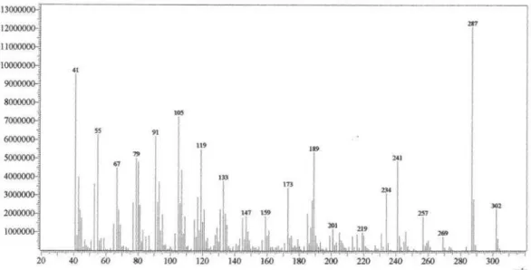
(c = 1.25, CHCl3), IR bands (KBr) at 3200-3081, 1692, 1636, and 908 cm-1 and EI-MS ion peaks at m/z (rel.
int. %) 302 (18) [M]+., 287 (100) [M-CH 3]
+., 241 (41);
and 234 (26). The 1H NMR spectra showed four olefinic
signals whose coupling constants allowed the assignment of a monosubstituted terminal double bond, related to the signals at d 4.93 (dd, J 17.5, 1.4 Hz), 4.86 (dd, J 10.7, 1.4 Hz), and 5.81 (dd, J 17.5, 10.7 Hz), and a trisubstituted double bond (signal at d 5.40, dd, J 5.4, 4.8 Hz) that is characteristic for the ring C position ∆9(11) of
pimaradiene-type diterpenes.18 These findings, confirmed by 13C NMR
data as well as by one-bond and long-range heteronuclear
1H-13C correlations from HSQC and HMBC experiments,
along with the MS fragmentation pattern, mainly by the ion at m/z 234, attributed to an elimination of 2-methyl-butadiene by a retro-Diels-Alder process, allowed us to establish the structure of 1 as acanthoic acid.18 Additionally,
all relative configurations in 1 were established by 1D nOe experiments, as follows. Selective irradiation of the resonance frequency of H-20 caused a nOe enhancement in the signal of H-8, and selective irradiation of the resonance frequency of H-18 showed a nOe intensification in the signal of H-5, but not in the signal of H-17. On the other hand, selective irradiation of the resonance frequency of H-17 did not cause any nOe enhancement in the signals of H-5, H-8, and H-18, indicating that H-5 and H-8 are located on the same side of the molecule as methyl groups C-18 and C-20. These results are in full accordance with those previously described for acanthoic acid.18 Moreover,
almost all of the investigated fractions of the CH2Cl2 extract contained high amounts of 1, as evidenced by 1H
NMR analysis. Acanthoic acid (1) was first isolated from
Acanthopanax koreanum (Araliaceae)18 and has also been
reported from Mikania sp. (Asteraceae),19Rollinia pittieri
and R. exsucca (Annonaceae).20 Therefore, this is the fifth
description of 1 in the literature, the first in the genus
Annona, and the third in the family Annonaceae, which is important from a chemotaxonomic point of view.
Several important in vitro and in vivo biological activities against a wide range of diseases and medical conditions, mainly antitumor and anti-inflammatory, have been described in the literature for 1.21-33 These findings clearly
show the medicinal potential of 1. However, almost all of the biological activities described in the literature for 1 have been shown by the compound extracted from Acanthopanax
koreanum, which resulted in the protection of the process for its extraction from this plant by an international patent,21
and has attracted interest in its synthetic preparation and modification.26,29,34,35 Therefore, according to the results
Pinheiro et al. 1099 Vol. 20, No. 6, 2009
acanthoic acid (1) from Annona amazonica has been demonstrated, which can advance research for new drugs based on this compound.
After the acanthoic acid (1) crystals were removed from the hexane extract, GC-MS analyses also allowed us to identify several other constituents that were present in lower concentrations: sesquiterpenes, β-cubebene (0.73%), α-cubebene (0.90%), α-humulene (1.24%),
α-amorphene (1.54%), α-copaene (2.08%), caryophyllene oxide (3.12%), α-muurolene (4.56%), trans-caryophyllene (4.70%), d-cadinene (7.22%), γ-cadinene (11.11%), and germacrene D (12.43%);15 fatty acids: hexadecanoic acid,
methyl ester, or methyl palmitate (1.09%), (Z,Z)-9,12-octadecadienoic acid, methyl ester, or methyl linoleate (0.12%), and (Z)-9-octadecenoic acid, methyl ester, or methyl oleate (0.28%).36
The chemical investigation of the alkaloid fraction of the CH2Cl2 extract resulted in the isolation and identification of the oxoaporphine alkaloid liriodenine (2) as yellow needles (mp 280-281 oC) and the aporphine alkaloid cassythicine
or N-methylactinodaphnine (3) as white needles (mp 204-205 oC). Liriodenine (2) is frequently found in species of
Annonaceae4,9,37-42 and therefore can be considered as a
chemotaxonomic marker of this family, since it can be found in most of the genera. On the other hand, cassythicine (3) is more commonly found in species of Lauraceae, a family phylogenetically close to the Annonaceae, particularly in the genera Cassytha, Laurus, Litsea, Stephania, and
Neolitsea; however, it is also found in some species of the genus Annona, such as A. glabra.37-40,43 Additionally, the
neutral fraction of the CH2Cl2 extract yielded acanthoic acid (1),17 three methyl esters of fatty acids 4, 5, and 6,4,36
one sesquiterpene 7,44 a mixture of the steroids 8 and 9,45
and one monounsaturated fatty acid 10,36 described for
the first time in this species. They were characterized on the basis of chromatographic and spectroscopic methods, such as GC-MS, IR, ESI-MS, and NMR (1D and 2D), and comparison with data from the literature.
Because cassythicine (3) was isolated a long time ago, only partial 1H NMR data are described in the
literature.46 Therefore, this study reports the complete and
unequivocal 1H and 13C NMR chemical shift assignments
for cassythicine 3, as well as their heteronuclear 1H-13C
correlations, nOe observations, and accurate 1H-1H scalar
coupling constants (Table 1). The NMR data for 3 were initially collected in CDCl3 as the solvent. However, in this solvent it was too difficult to observe both 1H and 13C signals of the aliphatic system, probably due to a
dynamic process, mainly for the aliphatic moiety, and consequently it was too difficult to identify all the signals, measure the 1H-1H coupling constants, and recognize
the heteronuclear 1H-13C correlations in the HSQC and
HMBC experiments. For this reason, cassythicine (3) was redissolved in methanol-d4 and resubmitted to NMR analysis. In contrast to CDCl3, the signals were much more evident in both 1H and 13C{1H} NMR spectra
obtained in methanol-d4 and it was possible to recognize all signals in the spectra as well as to perform all NMR chemical shift assignments and accurately measure the
1H-1H coupling constants. Moreover, by taking the NMR
measurements of 3 in methanol-d4 it was possible to observe the nuclear Overhauser effect, which was essential for the relative 1H NMR chemical shift assignments of the
aliphatic hydrogens. Selective irradiation of the resonance frequency of H-8 caused a nOe enhancement only in the signal of H-7 at 3.38 ppm. Selective irradiation of the resonance frequency of H-7 at 3.38 ppm showed a nOe enhancement in the signals of H-8, H-6a, and CH3-N, whereas selective irradiation of the resonance frequency of H-7 at 2.82 showed no nOe enhancements in the signal of H-6a, nor in the signal of CH3-N. These findings are supported by the selective irradiation of the resonance frequency of CH3-N, which showed nOe intensification in the signals of H-7 at 3.38 ppm, H-6a, and both H-5s (Table 1). As for H-8, selective irradiation of the resonance frequency of H-3 also showed nOe intensification only in the signal of H-4 at 2.99 ppm, and irradiation of H-4 at 2.99 ppm caused a nOe enhancement in the signals of H-3 and both H-5. Selective irradiation of the resonance frequency of H-5 at 3.49 ppm showed nOe enhancement in the signals of H-4 at 2.99, CH3-N, and H-6a (Table 1), indicating that H-7 at 3.38, H-6a, CH3-N, H-5 at 3.49 and H-4 at 2.99 are located on the same side of the molecule. This conclusion was supported by exploring the coupling constants (Table 1). In contrast, in the 1H NMR spectrum
obtained in methanol-d4 the HO-9 signal was not evident as in CDCl3. Here, we report all NMR data obtained in both CDCl3 and methanol-d4 for cassythicine (3), which can aid in further elucidation of the chemical structure (Table 1).
Biological studies
Acanthoic acid (1) showed anti-proliferative activity against epimastigote forms of Trypanosoma cruzi. After 96 h this compound considerably reduced the number of parasites, by causing progressive injury compared with untreated cells. A dose-dependent effect was observed, and at this time the IC50 value was 59 µmol L-1 (Figure 1), whereas the standard
drug benznidazole showed an IC50 of 7 µmol L-1.47 Other
Acanthoic Acid and other Constituents from the Stem of Annona amazonica (Annonaceae) J. Braz. Chem. Soc. 1100
Table 1.13C {1H}, 1H, 1H-13C HMBC, and nOe data for cassythicine (3)a
Position 1H d mult. Jb 1H d mult. Jc 13C{1H} db 13C{1H} dc 1H-13C HMBCb nOed
1 150.1 147.6
1a 118.1 116.9
2 144.2 142.3
3 6.65 s 6.52 s 107.4 106.7 1, 1a, 2, 3a, 3b, 4 and 6a Ha-4
3a 125.2 124.2
3b 121.1 122.8
4 a 2.99 ddd 12.8;4.6;1.1 b 3.27 ddd 12.9;12.8;6.0
a 2.73 brs b 3.38 brs
27.2 27.1 3, 3a, 3b and 5
3a and 5
H-3, Hb-4, Ha-5 and Hb-5 Ha-4 and Hb-5
5 a 3.49 ddd 12.9;12.5;4.6 b 3.78 ddd 12.5;6.0;1.1
a 2.92 brs b 3.35 brs
54.1 52.8 4 and 6a
3a, 4, 6a and CH3-N
Ha-4, Hb-5, H-6a and CH3-N Ha-4, Hb-4, Ha-5 and CH3-N 6a 4.30 dd 14.2;4.5 NO 64.0 61.9 3a, 3b, 7, 7a and CH3-N Ha-5, Hb-7 and CH3-N 7 a 2.82 dd 14.2;13.8
b 3.38 dd 13.8;4.5
a 2.96 brs b 3.11 brs
31.9 32.3 3b, 6a, 7a, 8 and 11a 3b, 6a, 7a, 8 and 11a
Hb-7
H-6a, Ha-7, H-8 and CH3-N
7a 126.4 127.2
8 6.82 s 6.80 s 116.4 114.5 1a, 7, 9, 10, 11 and 11a Hb-7
9-OH NO 8.54 brs 148.2 145.6 8*, 9* and 10*
10 148.6 145.7
11 7.68 s 7.60 s 112.0 110.0 1a, 7, 7a, 8, 9, 10 and 11a
11a 122.8 122.5
CH3-N 3.17 s 2.76 s 42.0 42.1 5 and 6a Ha-5, Hb-5, H-6a and Hb-7
CH3-O 3.87 s 3.90 s 56.6 56.2 10 H-11
O-CH2-O a 6.00 d 1.1 b 6.15 d 1.1
a 5.94 d 1.4 b 6.10 d 1.4
102.9 101.0 1 and 2
1 and 2
aThe experiments were performed at 295 K at 400 and 100 MHz for 1H and 13C, and all NMR chemical shifts are given in ppm related to TMS signal at 0.00 ppm as internal reference and coupling constants (J) in Hz. bData obtained in CD
3OD.
cData obtained in CDCl 3.
d1D nOe obtained by selective excitation of each hydrogen in the second table column. NO = Not Observed. *Correlations observed in the experiment obtained in CDCl3.
Figure 1. Effect of acanthoic acid (1) on the proliferation of epimastigote forms of Trypanosoma cruzi. The parasite was cultured in LIT medium at 28 °C for 96 h. Values are the mean of three independent experiments.
Figure 2. Toxic effect of acanthoic acid (1) on the LLCMK2 cell line after 96 h of treatment. The cells were cultured in DMEM medium. Values are the mean of two independent experiments.
(Asteraceae), that showed IC90 between 10-205 µmol L-1
against different strains of T. cruzi.48
The potential toxic effect of 1 on the LLCMK2 cell line was also evaluated. After 96 h of treatment, the 50% cytotoxic concentration was 347 µmol L-1 (Figure 2). Therefore, by
the selectivity index (SI) ratio (CC50 for LLCMK2/IC50 for
parasite), acanthoic acid (1) was more selective against the parasite than against mammal cells, with a SI of 5.9.
Pinheiro et al. 1101 Vol. 20, No. 6, 2009
therapeutic potential of compounds derived from natural sources.16,48-49 Several synthetic compounds have also
shown trypanocidal activity.50,51
Acanthoic acid (1) at 3 µmol L-1 (the lowest concentration
tested) displayed a growth inhibition of parasites of 13.6%, while at the highest concentration (331 µmol L-1), over 90%
of the cells were affected. Therefore, the 50% inhibition concentration was determined at 59 µmol L-1. In contrast,
the toxic effect on mammalian cells occurs only in high concentrations, with a CC50 of347 µmol L-1, showing that
1 is 5.9 times more toxic to the epimastigote forms of
T. cruzi than the LLCMK2 cell line.
Conclusions
Our results demonstrate that Annona amazonica, in addition to containing alkaloids, a class of compounds typically found in species of the family Annonaceae, produces as its major secondary metabolite, the pimaradiene-type diterpene, acanthoic acid (1). Therefore, this plant is a new renewable natural source of this promising compound for drug development. Knowledge of the medicinal potential of 1 was augmented by demonstrating its trypanocidal activity, together with its low toxicity. In addition, the complete and unequivocal 1H and 13C NMR chemical shift
assignments for cassythicine are now available.
Acknowledgments
The authors are grateful to Prof. Dr. A.C. Webber from the Universidade Federal do Amazonas (UFAM), Brazil, for botanical identification; as well as to CAPES, CNPq, FAPESP, FINEP, and the RENOR Project for financial support and fellowships.
Supplementary Information
Supplementary information containing 1D and 2D NMR and MS data is available free of charge at http://jbcs.sbq.org.br as a PDF file.
References
1. Chatrou, L. W.; Rainer, H.; Maas, P. J. M. Annonaceae (Soursop Family). In: Smith, N.; Mori, S. A.; Henderson, A.; Stevenson, D. W.; Heald, S. V. (eds.). Flowering Plants of the Neotropics. The New York Botanical Garden, New York, pp. 18-20, 2004. 2. Corrêa, M. P.; Dicionário das Plantas Úteis do Brasil e das
Exóticas Cultivadas, IBDF: Rio de Janeiro, Brasil, 1984. 3. Maas, P. J. M.; Kamer, H. M.; Junikka, L.; Mello-Silva, R.;
Rainer, H.; Rodriguésia2001, 52, 61.
4. Leboeuf, M.; Cavé, A.; Bhaumik, P. K.; Mukherjee, B.; Mukherjee, R.; Phytochemistry 1982, 21, 2783.
5. Rupprecht, J. K.; Hui, Y. -H.; McLaughlin, J. L.; J. Nat. Prod. 1990, 53, 237.
6. Likhitwitayawuid, K.; Angerhofer, C. K.; Chai, H.; Pezzuto, J. M.; Cordell, G. A.; J. Nat. Prod. 1993, 56, 1468.
7. Alali, F. Q.; Liu, X. -X.; McLaughlin, J. L.; J. Nat. Prod. 1999, 62, 504.
8. Campos, F. R.; Batista, R. L.; Batista, C. L.; Costa, E. V.; Barison, A.; Santos, A. G.; Pinheiro, M. L. B.; Biochem. Syst. Ecol. 2008, 36, 804.
9. Costa, E. V.; Pinheiro, M. L. B.; Xavier, C. M.; Silva, J. R. A.; Amaral, A. C. F.; Souza, A. D. L.; Barison, A.; Campos, F. R.; Ferreira, A. G.; Machado, G. M. C.; Leon, L. L. P.; J. Nat. Prod. 2006, 69, 292.
10. Costa, E. V.; Teixeira, S. D.; Marques, F. A.; Duarte, M. C. T.; Delarmelina, C.; Pinheiro, M. L. B.; Trigo, J. R.; Maia, B. H. L. N. S.; Phytochemistry2008, 69, 1895.
11. Boyom, F. F.; Ngouana, V.; Zollo, P. H. A.; Menut, C.; Bessiere, J. M.; Gut, J.; Rosenthal, P. J.; Phytochemistry 2003, 64, 1269.
12. Queiroz, E. F.; Roblot, F.; Cavé, A.; Paulo, M. Q.; Fournet, A.; J.Nat. Prod. 1996, 59, 438.
13. Maas, P. J. M.; Maas, H.; Miralha, J. M. S.; Junikka, L.; Rodriguésia2007, 58, 617.
14. Thomsen, K.; Brimer, L.; Bot. J. Linnaeus Soc. 1997, 124,273. 15. Adams, R. P.; Identification of Essential Oil Components by
Gas Chromatography/Quadrupole Mass Spectroscopy, Allured Publ. Corp.: Carol Stream, IL, USA, 2001.
16. Luize, P. S.; Nakamura-Ueda, T.; Dias-Filho, B. P.; Cortez, D. A. G.; Nakamura, C. V.; Biol. Pharm. Bull. 2006, 10, 2126. 17. Skehan, P.; Storeng, R.; Scudiero, D.; Monks, A.; McMahon,
J.; Vistica, D.; Warren, J. T.; Bokesh, H.; Boyd, M. R.; J. Natl. Cancer Inst. 1990, 82, 1107.
18. Kim, Y. H.; Chung, B. S.; J. Nat. Prod. 1988, 51, 1080. 19. Nunez, C. V.; Amêndola, M. C.; Lago, J. H. G.; Roque, N. F.;
Biochem. Syst. Ecol. 2004, 32, 233.
20. Jayasuriya, H.; Herath, K. B.; Ondeyka, J. G.; Guan, Z.; Borris, R. P.; Tiwari, S.; Jong, W.; Chavez, F.; Moss, J.; Stevenson, D. W.; Beck, H. T.; Slattery, M.; Zamora, N.; Schulman, M.; Ali, A.; Sharma, N.; MacNaul, K.; Hayes, N.; Menke, J. G.; Singh, S. B.; J. Nat. Prod. 2005, 68, 1247.
21. Pyun, K. H.; Choi, I.; Kang, H. S.; Lee, J. J.; Kim, Y. H.; PCT Int. Appl. WO 1995034300, 1995.
22. Kang, H. S.; Kim, Y. -H.; Lee, C. S.; Lee, J. J.; Choi, I. Pyun, K. H.; Cell. Immunol.1996, 170, 212.
23. Camussi, G.; Lupin, E.; Drugs1998, 55, 613.
24. Cai, X. F.; Sehn, G.; Dat, N. T.; Kang, O. H.; Kim, J. A.; Lee, Y. M.; Lee, J. J.; Kim, Y. H.; Chem. Pharm. Bull.2003, 51, 605. 25. Szekanecz, Z.; Koch, A. E.; Kunkel, S. L.; Strieter, R. M.; Drugs
Acanthoic Acid and other Constituents from the Stem of Annona amazonica (Annonaceae) J. Braz. Chem. Soc. 1102
26. Ling, T.; Chowdhury, C.; Kramer, B. A.; Vong, B. G.; Palladino, M. A.; Theodorakis, E. A.; J. Org. Chem.2001, 66, 8843. 27. Park, E. -J.; Zhao, Y. -Z.; Kim, Y. H.; Lee, J. J.; Sohn, D. H.
Planta Med.2004, 70, 321.
28. Kim, J. -A.; Kim, D. -K.; Tae, J.; Kang, O. -H.; Choi, Y. -A.; Choi, S. -C.; Kim, T. -H.; Nah, Y. -H.; Choi, S. -J.; Kim, Y. -H.; Bae, K. -H.; Lee, Y. -M.; Clin. Chim. Acta2004, 342, 193. 29. Suh, Y. -G.; Lee, K. -O.; Moon, S. -H.; Seo, S. -Y.; Lee, Y. -S.;
Kim, S. -H.; Paek, S. -M.; Kim, Y. -H.; Lee, Y. -S.; Jeong, J. M.; Lee, S. J.; Kim, S. G.; Bioorg. Med. Chem. Lett.2004, 14, 3487.
30. Kang, O. -H.; Choi, Y. -A.; Park, H. -J.; Kang, C. -S.; Song, B. -S.; Choi, S. -C.; Nah, Y. -H.; Yun, K. -J.; Cai, X. -F.; Kim, Y. -H.; Bae, K. -H.; Lee, Y. -M.; J. Ethnopharmacol.2006, 105, 326.
31. Na, M.; Oh, W. K.; Kim, Y. H.; Cai, X. F. Kim, S.; Kim, B. Y.; Ahn, J. S.; Bioorg. Med. Chem. Lett.2006, 16, 3061. 32. Jung, H. J.; Shim, J. S.; Suh, Y. –G.; Kim, Y. –M.; Onu, M.;
Kwon, H. J.; Cancer Sci.2007, 98, 1943.
33. Traves, P. G.; Hortelano, S.; Zeini, M.; Chao, T. -H.; Lam, T.; Neuteboom, S. T.; Teodorakis, E. A.; Palladino, M. A.; Castrillo, A.; Bosca, L.; Mol. Pharmacol. 2007, 71, 1545.
34. Lam, T.; Ling, T.; Chowdhury, C.; Chao, T.–H.; Bahjat, F. R.; Lloyd, G. K.; Moldawer, L. L.; Palladino, M. A.; Theodorakis, E. A.; Bioorg. Med. Chem. Lett. 2003, 13, 3217.
35. Palladino, M. A.; Teodorakis, E. A.; Macherla, V. R. R.; Chao, T. -H.; Suh, Y. G.; PCT Int. Appl. WO 2007015757, 2007. 36. Wele, A.; Ndoye, I.; Badiane, M.; Nig. J. Nat. Prod. Med. 2004,
8, 62.
37. Guinaudeau, H.; Leboeuf, M.; Cavé, A.; Lloydia1975, 38, 275.
38. Guinaudeau, H.; Leboeuf, M.; Cavé, A.; J. Nat. Prod. 1979, 42, 325.
39. Guinaudeau, H.; Leboeuf, M.; Cavé, A.; J. Nat. Prod. 1983, 46, 761.
40. Guinaudeau, H.; Leboeuf, M.; Cavé, A.; J. Nat. Prod. 1994, 57, 1033.
41. Harbone, J. B.; Baxter, H.; Moss, G. P.; Phytochemical Dictionary: A Handbook of Bioactive Compounds from Plants, 2nd ed., CRC Press: London, 1999.
42. Jumana, S.; Hassan, C. M.; Rashid, M. A.; Biochem. Syst. Ecol. 2000, 28, 483.
43. Guinaudeau, H.; Leboeuf, M.; Cavé, A.; J. Nat. Prod. 1988, 51, 389.
44. Moreira, I. C.; Roque, N. F.; Contini, K.; Lago, J. H. G.; Rev. Bras. Farmacogn. 2007, 17, 55.
45. Chang, Y. -C.; Chang, F. -R.; Wu, Y. -C.; J. Chin. Chem. Soc. 2000, 47, 373.
46. Tewari, S.; Bhakuni, D. S.; Dhar, M. M.; Phytochemistry1972, 11, 1149.
47. Izumi, E.; Morello, L. G.; Ueda-Nakamura, T.; Yamada-Ogatta, S. F.; Dias-Filho, B. P.; Cortez, D. A. G.; Ferreira, I. C.; Morgado-Díaz, J. A.; Nakamura, C. V.; Exp. Parasitol.2008, 118, 324.
48. Fournet, A.; Muñoz, V.; Roblot, F.; Hocquemiller, R.; Cavé, A.; Phytother. Res. 1993, 7, 111.
49. Dantas, A. P.; Salomão, K.; Barbosa, H. S.; Castro, S. L.; Mem. Inst. Oswaldo Cruz2006, 101, 207.
50. Bisaggio, D. F. R.; Adade, C. M.; Souto-Padrón, T.; Int. J. Antimicrob. Agents2008, 31, 282.
51. Valdez, R. H.; Tonin, L. T. D.; Ueda-Nakamura, T.; Filho, B. P. D.; Morgado-Diaz, J. A.; Sarragiotto, M. H.; Nakamura, C. V.; Acta Trop.2009, 110, 7.
S
u
p
p
le
m
e
n
ta
ry
In
fo
rm
a
ti
o
n
J. Braz. Chem. Soc., Vol. 20, No. 6, S1-S15, 2009. Printed in Brazil - ©2009 Sociedade Brasileira de Química 0103 - 5053 $6.00+0.00a
*e-mail: [email protected]
Acanthoic Acid and other Constituents from the Stem of
Annona amazonica
(Annonaceae)
Maria Lúcia B. Pinheiro,*,a Clahildek M. Xavier,a Afonso D. L. de Souza,a Diego de Moura Rabelo,a
Cristiane L. Batista,b Regiane L. Batista,b Emmanoel V. Costa,b Francinete R. Campos,b
Andersson Barison,b Rodrigo H. Valdez,c Tânia Ueda-Nakamurac and Celso V. Nakamurac
aDepartamento de Química, Universidade Federal do Amazonas, 69077-000 Amazonas-AM, Brazil
bDepartamento de Química, Centro Politécnico, Universidade Federal do Paraná, 81530-900 Curitiba-PR, Brazil
cDepartamento de Análises Clinicas, Universidade Estadual de Maringá, 87020-900 Maringá-PR, Brazil
Figure S1. 1H NMR spectrum of acanthoic acid in CDCl
Acanthoic Acid and other Constituents from the Stem of Annona amazonica (Annonaceae) J. Braz. Chem. Soc. S2
Figure S2.13C{1H} and DEPT 135 NMR spectra of acanthoic acid in CDCl
3 at 100 MHz.
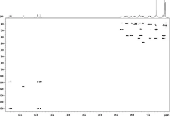
Figure S3.1H-1H correlation map from the COSY NMR experiment on acanthoic acid in CDCl
Pinheiro et al. S3 Vol. 20, No. 6, 2009
Figure S5.1H-13C long-range correlation map from the HMBC NMR experiment on acanthoic acid in CDCl
3 at 400 and 100 MHz.
Figure S4.1H-13C one-bond correlation map from the HSQC NMR experiment on acanthoic acid in CDCl
Acanthoic Acid and other Constituents from the Stem of Annona amazonica (Annonaceae) J. Braz. Chem. Soc. S4
Figure S6. EI-MS of acanthoic acid.
Figure S7.1H NMR spectrum of liriodenine in MeOD-d
Pinheiro et al. S5 Vol. 20, No. 6, 2009
Figure S9.1H-13C long-range correlation map from the HMBC NMR experiment on liriodenine in MeOD-d
4 at 400 and 100 MHz.
Figure S8.1H-13C one-bond correlation map from the HSQC NMR experiment on liriodenine in MeOD-d
Acanthoic Acid and other Constituents from the Stem of Annona amazonica (Annonaceae) J. Braz. Chem. Soc. S6
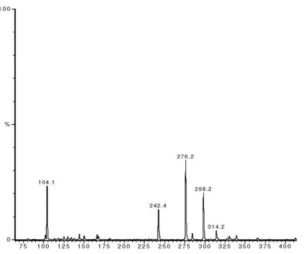
Figure S10. ESI-MS of liriodenine in MeOH, positive mode.
Pinheiro et al. S7 Vol. 20, No. 6, 2009
Figure S13.13C{1H} NMR spectrum of cassythicine in MeOD-d
4 at 100 MHz.
Figure S12.1H NMR spectrum of cassythicine in MeOD-d
Acanthoic Acid and other Constituents from the Stem of Annona amazonica (Annonaceae) J. Braz. Chem. Soc. S8
Figure S14.1H-13C one-bond correlation map from the HSQC NMR experiment on cassythicine in MeOD-d
4 at 400 and 100 MHz.
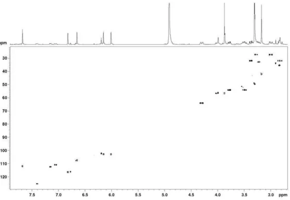
Figure S15.1H-13C long-range correlation map from the HMBC NMR experiment on cassythicine in MeOD-d
Pinheiro et al. S9 Vol. 20, No. 6, 2009
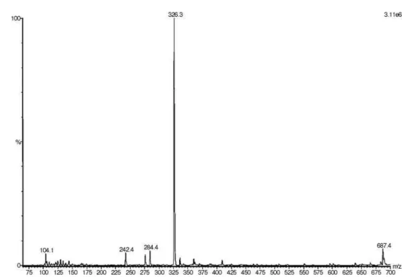
Figure S16. ESI-MS of cassythicine in MeOH, positive mode.
Acanthoic Acid and other Constituents from the Stem of Annona amazonica (Annonaceae) J. Braz. Chem. Soc. S10
Figure S18.1H NMR spectrum of the mixture of three methyl esters of the fatty acids, oleic, linoleic, and linolenic in CDCl
3 at 400 MHz.
Figure S19.13C{1H} spectrum of the mixture of three methyl esters of the fatty acids, oleic, linoleic, and linolenic in CDCl
Pinheiro et al. S11 Vol. 20, No. 6, 2009
Figure S20.1H NMR spectrum of caryophyllene oxide in CDCl
3 at 200 MHz.
Figure S21.13C{1H} and DEPT 135 NMR spectra of caryophyllene oxide in CDCl
Acanthoic Acid and other Constituents from the Stem of Annona amazonica (Annonaceae) J. Braz. Chem. Soc. S12
Figure S22.1H-1H correlation map from COSY NMR experiment of caryophyllene oxide in CDCl
3 at 400 MHz.
Figure S23.1H-13C one-bond correlation map from the HSQC NMR experiment on caryophyllene oxide in CDCl
Pinheiro et al. S13 Vol. 20, No. 6, 2009
Figure S24.1H-13C long-range correlation map from the HMBC NMR experiment on caryophyllene oxide in CDCl
3 at 400 and 100 MHz.
Figure S25.1H NMR spectrum of β-sitosterol and stigmasterol mixture in CDCl
Acanthoic Acid and other Constituents from the Stem of Annona amazonica (Annonaceae) J. Braz. Chem. Soc. S14
Figure S26.13C{1H} NMR spectrum of the β-sitosterol and stigmasterol mixture in CDCl
3 at 100 MHz.
Figure S27.1H NMR spectrum of oleic acid in CDCl
Pinheiro et al. S15 Vol. 20, No. 6, 2009
Figure S28.13C{1H} and DEPT 135 NMR spectra of oleic acid in CDCl