A
r
t
ic
le
0103 - 5053 $6.00+0.00
*e-mail: [email protected]
Chemical and Biological Evaluation of Essential Oils with
Economic Value from Lauraceae Species
Jefferson R. de A. Silva,*,a Dominique F. M. do Carmo,a Érika M. Reis,a Gérzia M. C. Machado,b
Leonor L. Leon,b Bianca O. da Silva,c José Luiz P. Ferreirac,d and Ana Claudia F. Amaralc
aDepartamento de Química, Universidade Federal do Amazonas, 69077-000 Manaus- AM, Brazil
bDepartamento de Imunologia, Fundação Oswaldo Cruz, 21040-360 Rio de Janeiro-RJ, Brazil
cLaboratório de Plantas Medicinais e Derivados, Fundação Oswaldo Cruz, 21041-250 Rio de Janeiro-RJ, Brazil
dFaculdade de Farmácia, Universidade Federal Fluminense, 2421-000 Niterói-RJ, Brazil
Este trabalho compara pela primeira vez a composição química do óleo essencial das folhas de Licaria canella coletadas em duas estações climáticas. Os resultados foram comparados com os obtidos para o óleo essencial das folhas de outra espécie da família Lauraceae, Aniba canelilla, coletada no mesmo período. Ambos os óleos essenciais foram analisados por CG-DIC e CG-EM, e os resultados indicaram uma grande quantidade de benzenóides, sendo o principal constituinte em L. canella o benzoato de benzila e para A. canelilla, o 1-nitro-2-feniletano. A comparação das atividades biológicas mostrou que o óleo de L. canella (IC50 19 µg mL
-1) foi mais ativo contra as
cepas de Leishmania amazonensis e menos citotóxico em cultura de macrófagos do que o de A. canelilla (IC50 40 µg mL-1). Por outro lado, o óleo de L. canella exibiu uma maior citotoxicidade
contra Artemia salina com uma concentração letal (CL50) igual a 5,25 µg mL-1.
This work compares the chemical composition of the essential oils from the leaves of Licaria canella collected in two different seasons. The results of this investigation were compared with the leaf essential oils of other species of the Lauraceae family, Aniba canelilla, collected at the same time. Both essential oils were analyzed by GC-FID and GC-MS. The results demonstrated a larger predominance of benzenoids, being the main constituent benzyl benzoate for L. canella and 1-nitro-2-phenylethane for A. canelilla. The comparison of the biological activities showed that L. canella (IC50 19 µg mL-1) was more active against Leishmania amazonensis strains and
less cytotoxic in macrophage cultures than A. canelilla (IC50 40 µg mL-1). On the other hand, the
L. canella oil displayed a higher cytotoxicity against Artemia salina with a lethal concentration (LC50), equal to 5.25 µg mL
-1.
Keywords: Licaria canella, Aniba canelilla, essential oil, Leishmania amazonensis, cytotoxicity
Introduction
The Lauraceae family contains about 50 genera and approximately 2500-3500 species distributed in tropical to subtropical areas, with a few occurrences in temperate regions and is among those with the greatest number of specimens in different regions of the Amazon.1
Twenty-two genera are found in Brazil, distributed in rain forests as well as in restingas and cerrados.2 The floristic
mapping held in the Adolpho Ducke Reserve (Amazonas
State, Brazil) cataloged so far 13 genera and 99 species.3
only rubber and chestnut, that rank first and second, respectively.4Licaria canella (Meissner) Kostermans is
a botanical species popularly known as “louro-pirarucu”. Within the ethnic group Tacana of the Amazonian region, this species has the same name and use as Anibacanelilla (H.B.K.) Mez, probably due to their aromatic barks.5
The barks of both species have ethnopharmacological use to alleviate abdominal pain, intestinal cramps or discomfort, without diarrhea.6 Phytochemical
studies of the trunk wood of L. canella described the presence of dillapiol, elemicin and the neolignans canellins A-C.7 The ethanol extract of the bark of this
species showed activity in vitro against chloroquine sensitive Plasmodiumfalciparum (IC50 = 3.8 µg mL-1)
and also resistant strains (IC50 = 3.2 µg mL-1).6 The
extract of the stem demonstrated low activity against RPMI 8226 cancer cells.8
Aniba canelilla, known as “casca-preciosa” (precious bark), was the target of an ancestral confusion in the search for the “country of cinnamon”.9 The cinnamon flavor
was responsible for the confusion between A. canelilla and the Cinnamomum species. In the case of the bark of
C. zeylanicum Blume, the major principle of cinnamon
odor is cinnamic aldehyde,10 while in A. canelilla, it is the
1-nitro-2-phenylethane.11 Previous works on A. canelilla
described the isolation of benzyltetrahydroisoquinoline alkaloids from the stem bark.12 The chemical compositions
of the essential oilsand hexane extract from the leaves and stem bark were identified by spectroscopic methods.13,14
Crude extracts of the stem bark from A. canelilla were active at 100 µg mL-1 invitro against Leishmania spp.
and Trypanosoma cruzi.15 The bark oil of this species
showed cardiovascular effects in normotensive rats.16 A
higher antioxidant activity using DPPH radical scavenging (EC50 = 4.41 µg mL-1) was reported for methanol extracts
of the wood. The brine shrimp bioassay performed with the trunk oils of A. canelilla (LC50 = 21.61 µg mL-1)
showed cytotoxicity higher than the wood extracts (LC50 = 91.38 µg mL-1).17 1-Nitro-2-phenylethane
produced antinociceptive effects but its mechanism of action was not elucidated.18
This report presents the chemical composition of the essential oils of L. canella and relates it to the oil of A. canelilla, collected in two different seasons. The emergence of parasite resistance to current therapies highlights the importance of essential oils as novel antiparasitic agents. In this context, the oils collected in October 2007 were tested in vitro on promastigote forms of Leishmania amazonensis. Additionally, toxicity evaluation of these oils with non-parasite macrophages and brine shrimp (Artemia salina) were performed.
Experimental
Plant Material
The leaves of L. canella and A. canelilla were collected from four individuals at the Adolpho Ducke Reserve, near the city of Manaus, Amazonas state, Brazil. Voucher specimens were deposited in the Herbarium of INPA (Amazonas state) under numbers 226360 and 220094, respectively. These species were collected in the dry (October 2007) and rainy seasons (February 2008). Flowers were observed in February, without fruits in either month. Data of pluviometric precipitation were obtained at the site of the National Institute of Meteorology and these maps showed no atypical differences in these seasons.19
Extraction of essential oil
The leaves collected from L. canella and A. canelilla (600 g) were dried at room temperature for 3 days, minced and submitted to hydrodistillation for 4 h in a Clevenger-type apparatus.
Essential oil analysis
The identification of compounds was performed by comparison of their retention indices and mass spectra with those reported in the literature or stored in the Wiley data system library.20,21 The retention indices were calculated
for all volatile constituents using n-alkane homologous series. GC analyses were performed using a HP 5890 gas chromatograph equipped with a DB-5 capillary column (30 m × 0.25 mm, film thickness 0.25 µm) and a FID detector. The oven temperature was programmed from 60 °C to 290 °C at a rate of 3 °C min-1, then isothermal at
290 °C for 10 min, using H2 as the carrier gas (1.0 mL min-1).
Injector and detector temperatures were 230°C and 280 °C, respectively.
GC-MS analyses were performed using a HP 6890 gas chromatograph interfaced with a HP 5873 Mass Selective Detector (ionization voltage 70 eV), equipped with a DB-5MS capillary column (30 m × 0.25 mm, film thickness 0.25 µm), using He as the carrier gas (1.0 mL min-1). Oven
and injector temperatures were as described before.
In vitro antileishmanial assay
with 10% fetal calf serum (FCS), pH 7.2. Parasites were harvested from the medium on day 4, when a high percentage of infective forms (metacyclic promatigotes) were found. After being harvested, the parasites were counted in a Neubauer’s chamber and adjusted to a concentration of 4 × 10⁶ promastigotes mL-1 using the
supernatant of each culture as diluent. The samples were dissolved in DMSO (the highest concentration was 1.4%, which was not hazardous to the parasites) and added to parasite suspensions in final concentrations between 0.156 to 320 µg mL-1. After 24 h of incubation, the parasites
were counted and compared to the controls, containing only DMSO and parasites. Pentamidine isethionate was used as the reference drug. The sample concentration corresponding to 50% of parasite growth inhibition was expressed as the IC50.
Cytotoxicity assay
In order to evaluate the toxicity of the sample for the host cell, mice peritoneal macrophages were isolated in RPMI 1640 medium (Sigma Cell Culture, St. Louis, MO, USA), containing 200 UI mL-1 penicillin, 200 µg mL-1 of
streptomycin, 1 mmol L-1 sodium piruvate, 1 mmol L-1
of L-glutamine and 1 mol L-1 HEPES buffer (Sigma Cell
Culture, St. Louis, MO, USA). Cells were counted in a Neubauer’s chamber using Erythrosine B as vital dye (Sigma Cell Culture, St. Louis, MO, U.S.A.) and adjusted to a concentration of 4 × 10⁶ mL-1. After that, the cells
were cultured in a 96 well culture plate (Falcon, New Jersey, U.S.A.), at 37 °C and in an atmosphere of 5% CO2. The sample was added to the medium in a concentration equivalent to IC50 and 2 × IC50 of the in vitro activity assay from L. amazonensis species. The sample and pentamidine isethionate (reference drug) were added to the cultures and after 24 hours the viability of treated cells were compared to the control without drugs, through the MTT methodology.22,23
Toxicity testing against brine shrimp
The brine shrimp (Artemia salina) lethality test was performed by the method of Meyer et al.24 with
some modifications. The samples were dissolved in dimethylsulfoxide (DMSO) and diluted serially in seawater. In each case three replicates of each concentration were assayed. Survivors were counted after 24 h and the LC50 values in µg mL-1 were determined by Probit analysis.25
Saline solution with DMSO was used as negative control (LC50 > 1000 µg mL-1), while lapachol was used as a positive
control (LC50 = 23.0 µg mL-1).
Results and Discussion
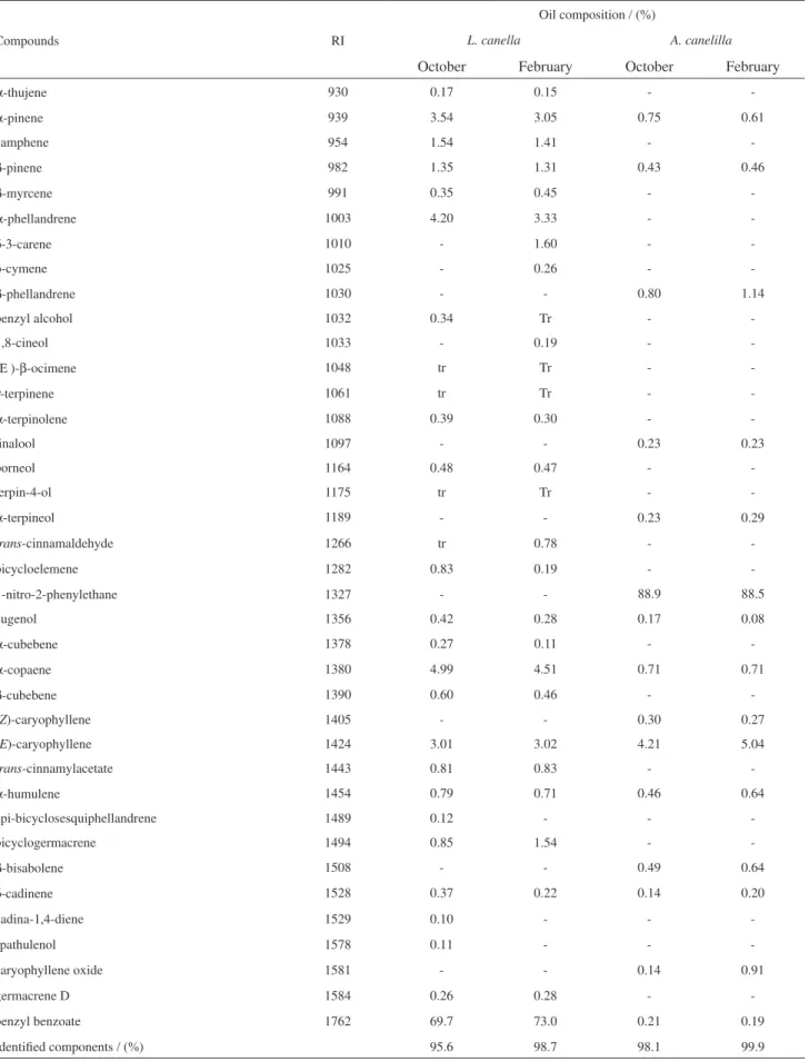
The yield of the oils was 1.2% (October, dry season) and 1.3% (February, rainy season) for L. canella leaves. The extraction resulting from A. canelilla leaves was 0.8% (October) and 0.9% (February). As shown in Table 1, compounds of the essential oils of L. canella and A. canelilla were identified by GC-FID and GC-MS analyses.
The results showed that the essential oils of these species were rich in benzenoid compounds, with minor constituents represented by mono and sesquiterpenoids (Table 2).
The major constituent of the leaf essential oils from L. canella (October 2007, 69.7% and February 2008, 73.0%) was benzyl benzoate. For A. canelilla it was 1-nitro-2-phenylethane (October 2007, 88.9% and February 2008, 88.5%). The percent content of this last compound, which was recently isolated and identified by NMR spectral data,17 is in agreement with previous studies published on
this species collected in the rainy season.26On the other
hand, in contrast with the results described for the species collected in the State of Pará,26 the collection related to
the dry season (October 2007) did not show a significant difference in its content of 1-nitro-2-phenylethane when compared to the rainy season. Additionally, the presence of methyleugenol, a component considered as a marker of the oils from Pará State,26 was not identified in the analyses
of the essential oils of A. canelilla found in Amazon State. The qualitative comparison and the percent content of the oils from the two collections showed similarity, (E )-caryophyllene being the second most abundant constituent in both collections. With respect to the essential oils from the leaves of L. canella, there is no report on the description of its chemical composition, which presented almost the same profile in both extractions, with benzyl benzoate being the main constituent. The other more abundant constituents were: α-pinene, α-phellandrene, α-copaene and (E )-caryophyllene. p-cymene, δ-3-carene and 1,8-cineol were detected only in the essential oils of the rainy season.The percent differences are listed in Table 1.
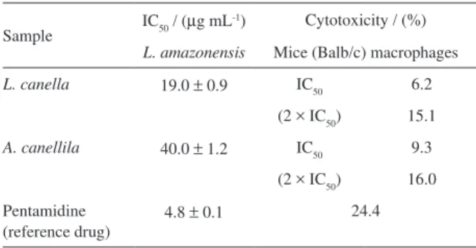
The evaluation of the antileishmanial activity of the essential oils (October 2007) of L. canella (IC50 = 19 µg mL-1), and A. canelilla (IC
50 = 40 µg mL -1)
Table 1. Chemical composition of the leaf essential oils of L. canella and A. canelilla
Compounds RI
Oil composition / (%)
L. canella A. canelilla
October February October February
α-thujene 930 0.17 0.15 -
-α-pinene 939 3.54 3.05 0.75 0.61
camphene 954 1.54 1.41 -
-β-pinene 982 1.35 1.31 0.43 0.46
β-myrcene 991 0.35 0.45 -
-α-phellandrene 1003 4.20 3.33 -
-δ-3-carene 1010 - 1.60 -
-p-cymene 1025 - 0.26 -
-β-phellandrene 1030 - - 0.80 1.14
benzyl alcohol 1032 0.34 Tr -
-1,8-cineol 1033 - 0.19 -
-(Ε )-β-ocimene 1048 tr Tr -
-γ-terpinene 1061 tr Tr -
-α-terpinolene 1088 0.39 0.30 -
-linalool 1097 - - 0.23 0.23
borneol 1164 0.48 0.47 -
-terpin-4-ol 1175 tr Tr -
-α-terpineol 1189 - - 0.23 0.29
trans-cinnamaldehyde 1266 tr 0.78 -
-bicycloelemene 1282 0.83 0.19 -
-1-nitro-2-phenylethane 1327 - - 88.9 88.5
eugenol 1356 0.42 0.28 0.17 0.08
α-cubebene 1378 0.27 0.11 -
-α-copaene 1380 4.99 4.51 0.71 0.71
β-cubebene 1390 0.60 0.46 -
-(Z)-caryophyllene 1405 - - 0.30 0.27
(E)-caryophyllene 1424 3.01 3.02 4.21 5.04
trans-cinnamylacetate 1443 0.81 0.83 -
-α-humulene 1454 0.79 0.71 0.46 0.64
epi-bicyclosesquiphellandrene 1489 0.12 - -
-bicyclogermacrene 1494 0.85 1.54 -
-β-bisabolene 1508 - - 0.49 0.64
δ-cadinene 1528 0.37 0.22 0.14 0.20
cadina-1,4-diene 1529 0.10 - -
-spathulenol 1578 0.11 - -
-caryophyllene oxide 1581 - - 0.14 0.91
germacrene D 1584 0.26 0.28 -
-benzyl benzoate 1762 69.7 73.0 0.21 0.19
Identified components / (%) 95.6 98.7 98.1 99.9
In this context, the results obtained in the evaluation with Artemia salina indicated high cytotoxicity for L. canella, with a lethal concentration (LC50) equal to 5.25 µg mL-1,
while in the case of A. canelilla, the LC50 observed was about 13 times greater (68.37 µg mL-1), being thus in
agreement with the greater activity against L. amazonensis strains found for the oils of L. canella.
Conclusions
The main constituent of the essential oils of L. canella is commercially used as a topical medication against several parasitoses, which suggests a potential use of this oil for this purpose. The leaves of L. canella and A. canelilla may be qualified as two natural, abundant and ecologically renewable sources of benzyl benzoate and 1-nitro-2-phenylethane, respectively, with commercial value for to medicinal purposes in the case of L. canella28,29
and as an aroma for the cosmetic and food industries regarding A. canelilla.
Acknowledgments
The authors are grateful for the financial support provided by CNPq and by the FAPEAM / PIPT Program and thank Ms. June Justa for revising the manuscript.
References
1. Eklund, H.; Bot. J. Linn. Soc. 2000, 132, 397. 2. Quinet, A.; Acta Bot. Bras. 2005,19, 563.
3. Ribeiro, J. E. L. S.; Hopkins, M. J. G.; Vicentini, A.; Sothers, C. A.; Costa, M. A. S.; Brito, J. M.; Souza, M. A. D.; Martins, L. H. P.; Lohmann, L. G.; Assunção, P. A. C. L.; Pereira, E. C.; Silva, C. F.; Mesquita, M. R.; Procópio, L. C.; Flora da Reserva Ducke. Guia de Identificação das Plantas Vasculares de uma Floresta de Terra-Firme na Amazônia Central, INPA, DFID: Manaus, AM, Brasil, 1999.
4. Marques, C. A.;Floresta e Ambiente2001, 8, 195.
5. Hocking, G. M.; A Dictionary of Natural Products. Terms in the Field of Pharmacognosy Relating to Natural Medicinal and Pharmaceutical Materials and the Plants, Animals, and Minerals from which they are Derived, 2nd ed., Plexus
Publishing: Medforf, 1997, p. 992.
6. Deharo, E.; Bourdy, G.; Quenevo, C.; Muñoz, V.; Ruiz, G.; Sauvain, M.; J. Ethnopharmacol. 2001, 77, 91.
7. Giesbrecht, A. M.; Franca, N. C.; Gottlieb, O. R.; Rocha, A. I.;
Phytochemistry1974, 13, 2285.
8. Suffredini, I. B.; Paciencia, M. L. B.; Varella, A. D.; Younes, R. N.; Fitoterapia2007, 78, 223.
9. Pinto, A. C.; Quim. Nova1995, 18, 608.
10. Senanayake, U. M.; Lee, T. H.; Wills, R. B. H.; J. Agric. Food Chem. 1978, 26, 822.
11. Gottlieb, O. R.; Magalhães, M. T.; J. Org. Chem.1959, 24, 2070.
12. Oger, J. M.; Fardeau, A.; Richomme, P.; Fournet, A.; Guinaudeau, H.; Can. J. Chem. 1993, 71, 1128.
13. Lima, M. P.; Silva, T. M. D.; Silva, J. D.; Zoghbi, M. G. B.; Andrade, E. H. A.; Acta Amaz.2004, 34, 329.
14. Oger, J. M.; Richomme, P.; Guinaudeau, H.; Bouchara, J. P.; Fournet, A. ; J. Essent. Oil Res. 1994, 6, 493.
15. Fournet, A.; Barrios, A. A.; Muñoz, V.; J. Ethnopharmacol. 1994, 41, 19.
16. Lahlou, S.; Magalhães, P. J. C.; Siqueira, R. J. B.; Figueiredo, A. F.; Interaminense, L. F. L.; Maia, J. G. S.; Sousa, P. J. C.;
J. Cardiovasc. Pharmacol. 2005, 46, 412.
17. Silva, J. K. R.; Sousa, P. J. C.; Andrade, E. H. A.; Maia, J. G. S.; J. Agric. Food Chem.2007, 55, 9422.
18. Lima, A. B.; Santana, M. B.; Cardoso, A. S.; Silva, J. K. R.; Maia, J. G. S.; Carvalho, J. C. T.; Sousa, P. J. C.; Phytomedicine 2009, 16, 555.
19. http://reia.inmet.gov.br/html/clima.php?lnk=/conab/climatologia/ dimap/selecao_spi_X.php, accessed in January 2009.
20. Adams R. P.; Identification of Essential oil Components by Gas Chromatography/Quadrupole Mass Spectroscopy, Allured Publishing Corp.: Carol Stream, IL, 2001.
21. Lin, L.-Y; Peng, C.-C; Liang, Y.-J; Yeh, W.-T; Wang, H.-E; Yu, T.-H.; J. Agric. Food Chem. 2008, 56, 4435.
Table 2. The chemical class distribution of the essential oils of L. canella
and A. canelilla
Compound class
L. canella / (%) A. canelilla / (%)
October February October February
Monoterpenoids 12.0 12.5 2.4 2.7
Sesquiterpenoids 12.3 11.2 6.4 8.4
Benzenoids 71.3 74.9 89.3 88.8
Table 3. In vitro activity of essential oils of L. canella and A. canelilla
against L. amazonensis and macrophages
Sample IC50 / (µg mL
-1) Cytotoxicity / (%)
L. amazonensis Mice (Balb/c) macrophages
L. canella 19.0 ± 0.9 IC50 6.2
(2 × IC50) 15.1
A. canellila 40.0 ± 1.2 IC50 9.3
(2 × IC50) 16.0
Pentamidine (reference drug)
22. Mosmann, T.; J. Immunol. Methods1983, 65, 55.
23. Ferrari, M.; Fornasiero, M. C.; Isetta, A. M.; J. Immunol. Methods1990, 131, 165.
24. Meyer, B. N.; Ferrigni, N. R.; Putnam, J. E.; Jacobsen, L. B.; Nichols, D. E.; McLaughlin, J. L.; Planta Med. 1982,45, 31. 25. Finney, D. J.; Probit Analysis, 3rd ed.,Cambridge University
Press: Cambridge, 1971.
26. Taveira, F. S. N.; de Lima, W. N.; Andrade, E. H. A.; Maia, J. G. S.; Biochem. Syst. Ecol. 2003, 31, 69.
27. Amato Neto, V.; Tuon, F. F.; Bacha, H. A.; Neto, V. A.; Nicodemo, A. C.; Acta Tropica2008, 105, 1.
28. Oladimeji, F. A.; Orafidiya, O. O.; Ogunniyi, T. A. B.; Adewunmi, T. A.; Onayemi, O.; Int. J. Aromather.2005, 15, 87.
29. Oladimeji, F. A.; Orafidiya, L. O.; Ogunniyi, T. A. B.; Adewunmi, T. A.; J. Ethnopharmacol.2000, 72, 305.