Angiogenic Properties of Natural Rubber Latex Biomembranes and The Serum Fraction of
Hevea
brasiliensis
Mariselma Ferreira∗
Departamento de F´ısica e Matem´atica FFCLRP-USP and Centro de Ciˆencias Naturais e Humanas CCNH-UFABC
Ricardo Jos´e Mendonc¸a and Joaquim Coutinho-Netto
Departamento de Bioqu´ımica FMRP-USP
Marcelo Mulato
Departamento de F´ısica e Matem´atica FFCLRP-USP
(Received on 5 May, 2009)
The angiogenic properties of natural rubber were evaluated in this work. We have used the chick chorioal-lantoic membrane assay (CAM) as a model to investigate the influence of the heating on biological activity in rubber membranes and in non rubber fraction as well. Results showed that natural rubber membranes can induce vascularization. It was observed that angiogenesis activity was maximum when membranes were heated in tem-peratures between 65oC and 85oC, considering a range from 55oC to 105oC. The same behavior was observed for non rubber fraction and it indicates that this serum fraction may be responsible for angiogenesis. When infrared spectroscopy was performed in the cast films of non rubber fraction samples, as a function of heating, no structural changes was observed. The results obtained shown that natural rubber latex films produced by casting induce the vessel growth in the CAM and it can be considered as a potential biomaterial.
Keywords: Biomatherials; Hevea brasiliensis; Natural Latex; Rubber; Angiogenesis
Introduction
Angiogenesis is fundamental for living organisms in pro-cesses such as organ growth, reproduction and wound re-pair. It is characterized by in situformation of blood ves-sels, thus permitting proliferation, migration, regulation and differentiation of vascular cells. When well regulated, an-giogenesis occurs for a given period of time until the phys-iological aim is achieved. On the other hand, angiogenesis is also involved in the development of diseases, including tumors and arthritis. The mechanism of angiogenesis regula-tion is still a matter of debate. Some angiogenic molecules, referred to as Growth Factors, are known to trigger prolif-eration of endothelial cells [1,2], while the so-called anti-angiogenic factors inhibit cell propagation [3]. Though some of these molecules have been identified and investigated for more than two decades [1], many questions remain as how they activate and regulate the normal and abnormal angio-genesis [4]. Folkman and collaborators [1] classified 8 an-giogenic polypeptides according to their biological activity, which included the polypeptides FGF (fibroblast growth fac-tor) and VEGF (vascular endothelial growth facfac-tor). The first occurs in two forms, the acid and basic fibroblast growth fac-tor (aFGF and bFGF respectively), being mitogenic for vari-ous cell types. VEGF is specific for vascular endothelial cells and increases the vascular permeability [5,6].
Angiogenesis has also been an important topic in can-cer research, wound healing and tissue engineering [7-13]. Materials have been developed in tissue engineering, which incorporate angiogenic molecules in the matrix (therapeutic angiogenesis). These bioactive materials are generally
devel-∗Electronic address:[email protected]
oped as coatings to enhance cell proliferation or as curatives to promote wound healing [10, 11], by inducing localized growth of blood vessels in the tissue. Biopolymeric delivery matrices for angiogenic growth factors may be obtained from natural and synthetic polymers and macromolecules such as polysaccharides (chitosan, hyaluronan, agarose) [7]. The matrices are required to be immunogenic and must have no inflammation reactions with the body environment. In addi-tion, the immobilized growth factors must remain functional during material fabrication and after its implantation [7]. In their review paper, Zisch and coworkers emphasized as a key feature for the matrices the controlled release of the growth factor, because unregulated diffusion may induce formation of dense, unstable vessels [7]. A methodology to improve cell seeding and growth was based on the immobilization of the polysaccharide heparin and bFGF in a collagen ma-trix [12], with heparin binding to FGFs and improving their immobilization and prolonged release. In fact, heparin-like molecules help maintaining the biological activity of growth factors [14]. Heparin was also used with aFGF immobilized in layer-by-layer (LbL) films, which included cell culture tests to verify bioactivity of the films [15].
The studies mentioned above all shared the goal of chang-ing an inert surface (made of synthetic or natural polymer) into a bioactive material to promotein situangiogenesis and enhance cell proliferation.
rubber [17,18]. More recently, natural rubber has been re-ported as potential biomaterial [19-22], and a material based on natural latex showed induction of wound healing in the esophageal wall of dogs, attributed to a marked increase in vascularization (angiogenesis) [20]. A similar material was used to treat leg ulcers with clear signs of granulation accom-panied by reduction in the patient’s pain [19]. The latter work was the first to demonstrate the potential of natural rubber la-tex for tissue regeneration, and has generated a commercial curative [23] already approved by ANVISA, the Brazilian government authority responsible for regulating health de-vices. As it has been suggested [19] that NR latex may con-tain vascular growth factors that act on human tissue, it can enhance local vascularization and induce angiogenesis.
Natural rubber (Hevea brasiliensis)
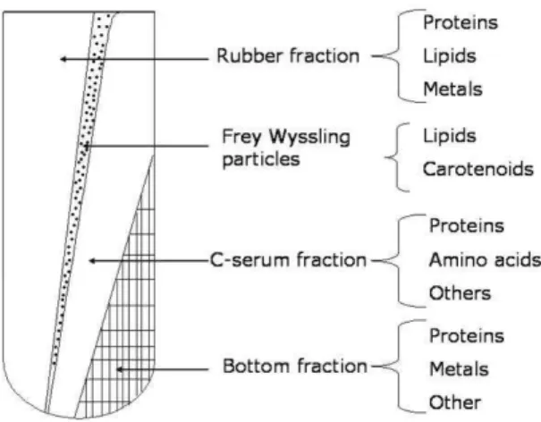
Natural rubber (NR) latex extracted from the Hevea brasiliensis is a polydisperse system containing 30-45% weight of rubber molecules (cis-polyisoprene), 4-5% weight of non-rubber constituents such as protein, lipids, carbohy-drates and sugar and 50% of water [24, 25]], Figure 1. The latex composition depends on the season of the year for the extraction and age of the tree [26].
FIG. 1: Fractions of natural rubber latex after centrifugation [19]
Natural rubber is a naturally occurring form ofcisisoprene, Figure 2, and exhibits elastomeric behavior upon intermolec-ular crosslinking (i.e. vulcanization). The rubber particles are surrounded by protein anions and are effectively nega-tively charged. When exposed to air, latex coagulation oc-curs as proteins are decomposed rapidly by bacteria and en-zymes, while crosslinking within the rubber particles leads to degradation of the rubber chains. Natural rubber is an im-portant raw material for a large range of industrial applica-tions such as tires, automobile, shoes and aircrafts. As the living cytoplasm of laticeferous cells, Hevea brasiliensis la-tex is a rich blend of organic substances that include many different proteins comprising about 1% to 1.5% of this la-tex system. The bulk of these proteins is removed when the latex is processed into its products, remaining only a small fraction in the product denominated residual extractable pro-teins (EPs). Yipet al. showed that high EP levels are associ-ated with positive skin prick test responses [27,28]. EP level
lower than 400µg/gof glove tested in individuals with latex allergy showed that 60% did not have a positive response, and in levels of about 100 g/g and less, the percentage of non responders reached about 100%. A few of these pro-teins are recognized allergens by the International Union of Immunological Societies (IUIS). Allergic reactions to rub-ber products, made from both natural and a wide variety of synthetic rubbers, have been known for many years, but the vast majority of these reactions (usually known as Type IV allergies) can be traced to the residues from accelerators and other compounding ingredients. Natural rubber latex prod-ucts had been marketed on a relatively large scale for the last fifty years without any serious suspicion of health risk to the users. Examples of products include baby bottle teats, elas-tic thread, gloves, condoms, foam rubber mattresses, pillows and adhesives.
A systematic study of the angiogenic NR is lacking, how-ever, and this is the topic addressed in the present paper. We use the chick chorioallantoic membrane assay as a model and investigate the influence from membrane fabrication param-eters (heating) on the biological activity. Furthermore, we separated the serum from the rubber fraction and studied the effects from heating in the angiogenic activity of nonrubber components as well.
C
C
CH
2H
H
2C
CH
3n
FIG. 2: Cispolyisoprene monomer
Materials and Methods
Sample Preparation
assay during 15 months, totalizing 15 experiments. For each experiment we investigated 4 samples in six distinct tempera-tures (45oC, 55oC, 65oC, 75oC, 85oC, 95oC). Considering this range of data, we used the Excel software for determine the Average and Standard Deviation of the data, which were showed in the figures 4 and 6.
We also investigated the angiogenic response of the non-rubber constituents of natural non-rubber, ie. the serum fraction in the absence of rubber particles. The latex stabilized with ammonium was submitted to a coagulation process by addi-tion of acetic acid (2.0% v/v). The coagulaaddi-tion takes place when pH falls below 5.0, and the latex is separated into two parts: the rubber fraction and the serum fraction (nonrubber constituents). The serum fraction was purified in a column of DEAE-cellulose (5×40 cm) eluted with stepwise gradi-ent ofNH4HCO3/NaCl. From this purification, three
frac-tions were obtained by exclusion based on the charge affin-ity with the DEAE column. Each fraction was dialyzed and lyophilized.
The first fraction (FrHB1) eluted from the separation col-umn had the most evident angiogenic effects in the CAM assays [29]. Therefore, we submitted this most active frac-tion (FrHB1) to the thermal treatment and evaluated the an-giogenic response. The lyophilized (FrHB1) fraction was weighted and dissolved in distilled water (2.5mg/mL). Then, a 3µLdroplet of this solution was pipetted on the filter disc papers (0.5 cm diameter). These discs were left to dry at room temperature and after been dried the testing samples were heated in an oven in temperatures ranging from 45-105oC to simulate the thermal treatment performed with the NR membranes. After the heating process, the sample con-taining serum fraction was placed on chorioallantoic mem-brane for evaluation of the angiogenic activity.
Chorioalantoic membrane assay
The chick embryo choriolantoic membrane (CAM) was first described by embryologists, being the most widely used system for studying angiogenesis [30]. Fertilized eggs were purchased from Pena Branca Company (S˜ao Carlos, SP) and placed in an incubator at 38oC with 65-70% relative humid-ity. The incubator (Brasmatic Ind. Com. LTDA, So Paulo, Brazil) was equipped with a temperature controlling system. On the fifth day of incubation the eggs were cleansed with 70% ethanol and opened using a circular drill (Dremel Multi-pro, Breda, USA). The cap was removed and the shell mem-branes were moisturized with a sterilized saline solution to help the complete detachment of the shell from the CAM. Eggs were then incubated again and at the thirteenth day of incubation the test samples were applied on the CAM. At day 17 (of incubation), the embryos were sacrificed by ad-dition of formaldehyde 10%. Digital images of the CAM were captured using an optical microscope with a CCD video camera (Sony Model DXC-107A). Image data were captured and analyzed with commercial software (PixelViewStation v5.19TV). The amount of vessels was quantified using other commercial software (ImageJ1, 32J, NH, USA) commonly used for quantifying pixels in digitalized images. The re-sults presented in Figures 4 and 6 (circles) show an
aver-age of amount of vessels obtained for data acquired in fifteen months. All the experimental manipulations were done with sterile instruments (glassware and tweezers), but we did not manipulate the opened eggs in a tissue-culture hood.
FTIR
The FTIR measurements were carried out with a Nico-let equipment in the cast films fabricated by dropping 20 L of a 2.5mg/mL serum fraction solution onto gold substrates. The substrates were heated to temperatures between 45oand 105oC. The solution was prepared using Milli-Q water.
Results and Discussion
The effects from thermal treatment on the angiogenic ac-tivity of natural rubber are illustrated in Figures 3.a)-f), which show the membranes placed in the CAM for heating temperatures ranging from 55-105◦C. A qualitative exami-nation of the CAM images shows no change in the vessels density for temperatures between 55oand 65oC. In contrast, for temperatures between 75◦and 85oC, a red region marked by an arrow appears in Figures 3 e)-g), which is indicative of rubber angiogenesis. The difference is clear upon com-paring Figures 3d and 3e with the control membranes. For membranes heated from 95o-105oC no changes in the vessel density could be observed in the CAM companying with the control membranes.
The angiogenic activity may be evaluated quantitatively by counting the number of vessels in the graph of Figure 4. The amount of vessels presented shows an average of the fif-teen experiments performed for each temperature. The error bars on Figure 4 show the standard deviation (SD) of data. In spite of the dispersion in the data points (to be discussed be-low), the activity for the thermally-treated NR membranes in Figure 3 is always higher than the control sample made with synthetic material (black dashed line). An optimized temper-ature for treating the membranes is between 65oC and 85oC, where angiogenesis activity was maximum.
The large dispersion in the data (i.e. large error bars) may have several sources. First, NR does possess different prop-erties depending on the period of collection (seasonal vari-ation) and storage time [24,26,31]. Indeed, an inspection of our results points to differences in the activity for membranes stored for different periods of time. This may be due to the poor stability of non-rubber constituents of NR, especially in the presence of ammonium [31]. Most affected are the lipids, which are major components surrounding the rubber particles in the latex. The latex used here was collected in three different seasons over 15 months of studies. Though we took precautions in keeping the samples in a refrigera-tor and with ammonium to minimize errors, variations could still exist. In addition, non-homogeneity in the membranes was observed even for samples prepared with the same latex, in the same day.
Fig. 3. a)
Fig.3.b) Fig.3.c)
Fig.3.d) Fig. 3. e)
Fig.3.f) Fig.3.g)
FIG. 3: Vascular networking formation induced by the rubber mem-branes placed on the top of the chicken chorioallontoic membrane for different temperatures. a) Control (synthetic isoprene); b) 55
oC; c) 65oC; d) 75oC; e) 85oC; f) 95oC; g) 105oC
FIG. 4: Percentage of vessels as a function of membrane heating (N ranging from 10-15). The red dashed line has been drawn just to guide the eye. Errors bars show standard deviation (SD).
Figures 5 a)-g), where a circle was added to the image of Figure 5a for the control experiment to mark where there is no modification in the number of vessels. A visual inspec-tion points to stronger angiogenic activity for samples treated from 65-85oC, as indicated by the arrows.
Fig
Fig
Fig
Fig g 5.a)
g.5.b)
g 5.d)
g. 5.f)
Fig
Fig
Fig. g.5.c)
g.5.e)
. 5.g)
FIG. 5: Vascular networking formation induced by non-rubber serum fraction placed on the top of the chicken chorioallontoic membrane. a) Control (distilled water); b) 55oC; c) 65oC; d) 75
oC, e) 85oC. f) 95oC; g) 105oC.
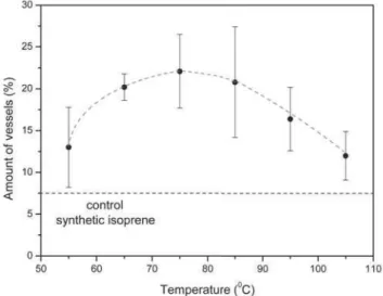
The results of Figure 5 can be quantified, as depicted in Figure 6, which again shows enhanced angiogenic activity for almost all membranes, in comparison with the synthetic substrate. Analogously to the results for the NR membranes (Figure 4), angiogenic activity is maximum for thermal treat-ments in the region of 70-85oC. The dispersion in the data, however, is now significantly less than for NR, what was ex-pected as the serum material is more homogeneous than the NR membranes.. Considering that the temperature range was essentially the same for NR and serum membranes, results indicate that the non-rubber serum fraction may be responsi-ble for angiogenesis. The trangles represent an average for data acquired in temperature points of 70oC and 80oC not evaluated for NR membranes (Figure 4).
struc-FIG. 6: Percentage of vessels vs. temperature for thermal treatment of films made with NR serum (N ranging from 5-10). The triangles represent an average for data acquired in temperature points of 70
oC and 80oC. The red dashed line is just to guide the eye. Errors
bars show standard deviation (SD).
ture of proteins, as identified by the amide bands (amide I -1620-1700 cm-1) and amide II - 1520-1580 cm-1) [32,33]. This region is represented between dashed lines in Figure 7. Because the samples investigated here comprise a mixture of non-rubber materials, and not isolated proteins, we cannot identify which type of secondary structure is being affected by the temperature in the thermal treatment. Nevertheless, one infers from Figure 7 that the conformation of the NR serum fraction does vary with the temperature.
Conclusions
The present study has shown a systematic evaluation of the natural rubber angiogenic properties using the CAM method. The results obtained shown that the natural rubber latex films produced by casting induces the vessel growth in the CAM and it can be considered as a potential biomaterial. The an-giogenic evaluation according to the heat treatment of the NR latex film revealed that the angiogenic behavior remains active and increase in temperatures ranging from 65-85oC. It is particularly interesting, since the NR films are prepared by heating the latex system. The results showed here may suggest a maximum temperature (85oC) to be used for this procedure.
The second important effect achieved in this study was the demonstration that the bioactive fraction of the NR latex is the non-rubber constituents of the latex systems and the poly-isoprene chains are not the main responsible for these
mate-rial activity. Here we proposed an easy method to isolate the angiogenic fraction from NR latex. The curve for the percentage of vessels as a function of heating for NR film and NR serum was very similar and it suggests that the ac-tive fraction extracted from the rubber could be study to be incorporated in other systems such as thin polymer films.
The FTIR experiments suggested changes in the
sec-FIG. 7: FTIR spectra of the NR serum fraction for different temper-atures of heating.
ondary structure of the NR serum fraction as a function of heating. However, the fact we did not have a completely pu-rified angiogenic fraction turned difficulty the evaluation of the structural changes as a function of heating. One of the suppositions is that the variation in the secondary structure can exhibit the angiogenic part of the NR bioactive protein and increase the potential behavior of the material.
Acknowledgement
FAPESP (03/02516-2); CNPq (420198/2005-9) and (478452/2004-7); Prof. Osvaldo Novais de Oliveira Jr. Poly-mer Group of IFSC-USP; Prof. Carlos F. de O Graeff; and Prof. Marcos Bernardes, for the latex sample donation.
[1] Folkman J, Shing Y. Angiogenesis. The Journal of biological chemistry. 1992 Jun 5;267(16):10931-4.
[2] Wilting J, Brand-Saberi B, Kurz H, Christ B. Development of the embryonic vascular system. Cellular & molecular biology
research. 1995;41(4):219-32.
[3] Folkman J. Endogenous angiogenesis inhibitors. Apmis. 2004 Jul-Aug;112(7-8):496-507.
the regulation of angiogenesis. Kidney international. 1999 Sep;56(3):794-814.
[5] Ferrara N, Gerber HP, LeCouter J. The biology of VEGF and its receptors. Nature medicine. 2003 Jun;9(6):669-76. [6] Helmlinger G, Endo M, Ferrara N, Hlatky L, Jain RK.
Formation of endothelial cell networks. Nature. 2000 May 11;405(6783):139-41.
[7] Zisch AH, Lutolf MP, Hubbell JA. Biopolymeric delivery ma-trices for angiogenic growth factors. Cardiovasc Pathol. 2003 Nov-Dec;12(6):295-310.
[8] Folkman J. Angiogenesis and apoptosis. Seminars in cancer biology. 2003 Apr;13(2):159-67.
[9] Gupta MK, Qin RY. Mechanism and its regulation of tumor-induced angiogenesis. World J Gastroenterol. 2003 Jun;9(6):1144-55.
[10] Schultz GS, Sibbald RG, Falanga V, Ayello EA, Dowsett C, Harding K, et al. Wound bed preparation: a systematic ap-proach to wound management. Wound Repair Regen. 2003 Mar;11 Suppl 1:S1-28.
[11] Shen JT, Falanga V. Innovative therapies in wound heal-ing. Journal of cutaneous medicine and surgery. 2003 May-Jun;7(3):217-24.
[12] Wissink MJ, Beernink R, Poot AA, Engbers GH, Beugeling T, van Aken WG, et al. Improved endothelialization of vascular grafts by local release of growth factor from heparinized col-lagen matrices. J Control Release. 2000 Feb 14;64(1-3):103-14.[13]
[13] Folkman J, Kalluri R. Cancer without disease. Nature. 2004 Feb 26;427(6977):787.
[14] Prestrelski SJ, Fox GM, Arakawa T. Binding of heparin to basic fibroblast growth factor induces a conformational change. Archives of biochemistry and biophysics. 1992 Mar;293(2):314-9.
[15] Mao Z, Ma L, Zhou J, Gao C, Shen J. Bioactive thin film of acidic fibroblast growth factor fabricated by layer-by-layer assembly. Bioconjugate chemistry. 2005 Sep-Oct;16(5):1316-22.
[16] Sharp WV, Falor WH. Rubber latex tubing as a vascular pros-thesis. American journal of surgery. 1963 Jun;105:802-11. [17] Sharp WV. Vascular prostheses. Transactions - American
So-ciety for Artificial Internal Organs. 1964;10:223-6.
[18] Sharp WV, Gardner DL, Andresen GJ. Adaptation of Elastic Materials for Small Vessel Replacement. Transactions - Amer-ican Society for Artificial Internal Organs. 1965;11:336-40. [19] Frade MA, Valverde RV, de Assis RV, Coutinho-Netto J,
Foss NT. Chronic phlebopathic cutaneous ulcer: a thera-peutic proposal. International journal of dermatology. 2001 Mar;40(3):238-40.
[20] Mru´e F. Neoformac¸˜ao tecidual induzida por biomembrana de l´atex natural com poli-lisina. Aplicabilidade na neoformac¸˜ao
esof´agica e da parede abdominal. Estudo experimantal em c˜aes. Ribeir˜ao Preto: Faculdade de Medicina de Ribeir˜ao Preto - USP; 2000.
[21] Neves Junior WP, Ferreira M, Alves MCO, Graeff CFO, Mulato M, Coutinho-Netto J, et al. Influence of fabrica-tion process on the final properties of Natural-Rubber La-tex tudes for vascular prothesis. Brazilian Journal of Physics. 2006;36(2B):586-91.
[22] Neves Junior WP, Graeff CFO, Ferreira M, Mulato M, Bernardes M, Coutinho-Netto J. Influence of the fabrication process on the final properties of vascular prothesis made of natural rubber latex. Journal of Applied Polymer Science. 2006;100:702-7.
[23] Mru´e F, da Silva Zborowski AC, inventors; Biomembrane suit-able for use in substituition, reconstruction of angiogenesis, neoformation or regeneration of human or animal organs or tissues. European Union. 2006.
[24] Grave H-H, Bayer AG, Leverkusen AG. Ullmanns Encyclope-dia of Industrial Chemistry 1993:221-35.
[25] Hasma H. Journal of Natural Rubber Research. 1993;12:21-32.
[26] Ferreira M, Moreno RMB, Goncalves PS, Mattoso LHC. Eval-uation of natural rubber from clones of Hevea brasiliensis. Rubber Chemistry and Technology. 2002 Mar-Apr;75(1):171-7.
[27] Yip E, Sussman G. Allergenicity of latex gloves with reference to latex protein sensitive individuals in a canadian population. Journal of Natural Rubber Research. 2000;3(3):129-41. [28] Yip E, Turjanmaa K, Ng KP, Mok KL. Residual estractable
proteins and allergenicity of natural rubber products. Journal of Natural Rubber Research. 1994;9:79-86.
[29] Mendonc¸a RJ. Caracterizac¸ ao biolgica de uma frac¸˜ao an-giogˆenica do l´atex natural da seringueira Hevea brasiliensis. Ribeiro Preto: USP; 2004.
[30] Auerbach R, Kubai L, Knighton D, Folkman J. A simple pro-cedure for the long-term cultivation of chicken embryos. De-velopmental biology. 1974 Dec;41(2):391-4.
[31] Hasma H. Lipids associated with rubber particles and their possible role in mechanical stability of latex concentrates. Journal of Natural Rubber Research. 1991;6(2):105-14. [32] Havel HA, Chao RS, Haskell RJ, Thamann TJ.
Investigations of ProteinStructure with Optical Spectroscopy -Bovine Growth-Hormone. Analytical chemistry. 1989 Apr 1;61(7):642-50.