Effects of constraint-induced movement
therapy in children with hemiplegia:
a single case experimental study
Efeitos da terapia de restrição por movimento induzido em crianças com
hemiplegia: desenho experimental de caso único
Marina B. Brandão1, Marisa C. Mancini2, Daniela V. Vaz3, Ângela M. Bueno4, Sheyla R. C. Furtado3, Zélia A. C. Coelho2
Abstract
Objective: To investigate the profile of changes in the use of the upper extremity in three children with hemiplegia submitted to an
adapted protocol of constraint-induced movement therapy (CIMT). Methods: A single-subject design (ABA) was replicated in three children aged 8 to 11 years old. Baseline phases (A1) and (A2) and the intervention phase (B) lasted 2 weeks each. During the intervention period, children wore a splint on the non-affected extremity for 10 hours a day and were submitted to 3 hours of therapy a day during 10 days. Training consisted of activities with the affected upper extremity, with gradually increasing complexity and verbal feedback. Hand function was classified according to the Manual Ability Classification System (MACS). Children were assessed four times every week with the Toddler Arm Use Test (TAUT) and three adapted tasks from the Jebsen-Taylor Hand Function test (JTHF), and once a week with the Pediatric Motor Activity Log (PMAL) and self-care scales of the Pediatric Evaluation of Disability Inventory (PEDI). Celeration Line, Two-Standard Deviation Band and visual analysis methods were used for data analyses. Results: Significant improvements in the amount and quality of upper extremity use (PMAL), TAUT quality of use for children 2 and 3, and participation for child 1, as well as decreased time to complete JTHF tasks for children 2 and 3 were observed. No changes were observed in the PEDI self-care scales. Conclusion: CIMT effects were associated with improvements in manual dexterity, amount and quality of use of the affected upper extremity in children with hemiplegia.
Key words: constraint therapy; hemiplegia; cerebral palsy; manual function.
Resumo
Objetivo: Investigar mudanças longitudinais no uso da extremidade superior em três crianças com hemiplegia submetidas a um
protocolo adaptado de terapia de movimento induzido por restrição (CIMT). Métodos: Um desenho experimental de caso único (ABA) foi replicado em três crianças entre 8 e 11 anos de idade. Fases de baseline (A1) e (A2) e fase de intervenção (B) duraram duas semanas cada. Durante a fase de intervenção, as crianças usaram um splint na extremidade não afetada por dez horas por dia e foram submetidas a três horas de terapia diária por dez dias. O treinamento consistiu em atividades para a extremidade superior acometida, com aumento gradual da complexidade da tarefa e reforço verbal. A função manual foi classificada de acordo com o Manual Ability Classification System (MACS). As crianças foram avaliadas quatro vezes por semana com o Toddler Arm Use Test (TAUT) e três provas adaptadas do Teste Jebsen-Taylor de Função Manual (JTHF) e uma vez por semana com o Pediatric Motor Activity Log (PMAL) e escalas de autocuidado do Inventário de Avaliação Pediátrica de Incapacidade (PEDI). Os métodos Celeration Line, Banda de Dois Desvios-Padrão e análise visual foram usados para análise de dados. Resultados: Resultados significativos demonstraram melhora na qualidade e frequência de uso da extremidade superior (PMAL), qualidade de uso do TAUT nas crianças 2 e 3, participação na criança 1, bem como diminuição do tempo gasto para completar as tarefas do JTHF para as crianças 2 e 3. Nenhuma mudança foi observada nas escalas de autocuidado do teste PEDI. Conclusão: Efeitos da CIMT associaram-se à melhora na destreza manual, frequência e qualidade de uso da extremidade acometida em crianças com hemiplegia.
Palavras-chave: terapia de restrição; hemiplegia; paralisia cerebral; função manual.
Received: 14/10/2008 – Revised: 13/03/2009 – Accepted: 05/05/2009
1 Universidade FUMEC, Associação Mineira de Reabilitação (AMR), Belo Horizonte (MG), Brazil
2 Occupational Therapy Department, Universidade Federal de Minas Gerais (UFMG), Belo Horizonte (MG), Brazil 3 Physical Therapy Department, UFMG
4 Occupational Therapist
Correspondence to: Marisa Cotta Mancini, Departamento de Terapia Ocupacional, Escola Educação Física, Fisioterapia e Terapia Ocupacional, Universidade Federal de Minas Gerais, Campus Pampulha, Av. Antônio Carlos, 6.627, CEP 31270-910, Belo Horizonte (MG); Brazil, e-mail: [email protected]
Introduction
Children with hemiplegia have unilateral involvement of upper and lower extremities opposite to the side of cerebral injury, often characterized as muscle weakness, spasticity and hypertonia1. hese factors may decrease movement eiciency2, especially in the use of the upper extremity, which can also limit performance in functional activities at home and school1,3,4.
Constraint-induced movement therapy (CIMT) has been used to promote functional gains in individuals with neurological dysfunctions5-11. CIMT consists of constraining movement of the non-afected upper extremity and providing intensive training to the involved upper extremity3,5,12. Intensive training is based on shapingprinciples3,5,7, which include the selection of activities suited to the client’s individual abilities, with progressive increase in diiculty and complexity. Procedures also involve providing assistance and support when the individual is unable to perform the task independently, as well as verbal rewards for observed improvements9. One of the main objectives of the intervention is to overcome the learned nonuse, deined as the diminished use of the afected extremity due to the perception of failure during the performance of manual tasks13,14.
he original CIMT protocol consists of 2 or 3 weeks of daily intensive training of the afected extremity for 6 hours in association with restriction of the non-afected extremity for 10 hours a day13. According to Gordon, Charles and Wolf3, the original CIMT protocol consisting of six hours of daily training and use of restraint for 90% of waking hours could be tiring for children. hus, modiications in speciic aspects of the origi-nal CIMT protocol have been proposed by some authors2,6,14-16. hese adaptations include decreased training time and/or de-creased use of the restriction15-18 which are often compensated for with increased protocol lengths6. Charles et al.16 investiga-ted a protocol in which the use of the restriction occurred only during the intensive training of 6 hours daily, for 12 days. hey documented signiicant improvements in children’s manual dexterity and parents reported improvements in the amount and quality of use of the afected extremity16. Eliasson et al.6 studied the efects of a two-month intervention protocol with two hours of training and restriction use every day in young
children. he authors reported positive results with signiicant improvements in children’s bimanual abilities6.
he replication of available evidence in a diferent cultural environment involves testing the efects of this intervention considering the clinical particularities of the Brazilian chil-dren, as well as our rehabilitation services. he investigation of the feasibility of adapted CIMT models to meet Brazilian children’s needs and promote improvements in manual dexte-rity and daily functioning may help broaden clinical actions of rehabilitation professionals. he objective of this study was to investigate the proile of changes in the functional use of the upper extremity in three children with hemiplegia submitted to an adapted CIMT protocol. he study’s hypotheses were: H1: there will be improvements in the quality and in the amount of use of the afected extremity in children with hemiplegia, after administration of an adapted CIMT protocol; H2: there will be improvements in the manual dexterity of children with hemiple-gia submitted to an adapted CIMT protocol; H3: there will be improvements in daily functioning of children with hemiplegia after the administration of an adapted CIMT protocol.
Method
Study design
An ABA experimental single-subject design replicated in three children was conducted. Baseline phases (A1) and (A2) consisted of repeated assessments without treatment. Phase B consisted of CIMT intervention and periodic assessments. Each phase lasted two weeks, totaling six weeks of study.
Participants
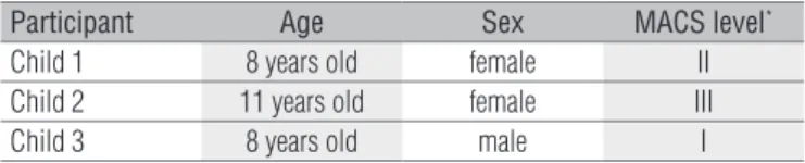
Participants of this study had a medical diagnosis of spastic hemiplegic cerebral palsy or stroke, and had cognitive abilities to follow verbal commands. hey had no associated patholo-gies or movement disorders, did not make use of hand splints or received botulinum toxin in the upper extremity within six months prior to the beginning of the study. During the study, children were not receiving other interventions to improve upper extremity function. he three children were contacted by convenience by one of the investigators. hey attended rehabilitation services in Belo Horizonte/Brazil, and agreed, with permission from their parents, to participate in this study. hey all conformed to the inclusion criteria described above. Table 1 presents descriptive information of each child.
Child 1 was a girl with right hemiplegia due to cerebral palsy. She demonstrated diminished use of the right upper extremity in daily activities, weakness of the intrinsic hand muscles and
Participant Age Sex MACS level*
Child 1 8 years old female II
Child 2 11 years old female III
Child 3 8 years old male I
Table 1. Descriptive analysis of the three children in terms of age, sex
and MACS level.
*MACS: Manual Ability Classification System which includes five levels; level I indicates no difficulty to use the hands to perform daily activities, level II indicates that use of the hands in daily activities shows lower quality and/or takes longer, level III informs that child shows difficulty in using hands and needs help to set-up for activity performance and/or requires modification of activity.
diiculty to perform tasks that required manual dexterity. his child was discharged from occupational therapy services one month before the beginning of this study.
Child 2 was a girl with right hemiplegia due to stroke at age 9. Her thumb was adducted and her wrist was lexed at rest, and wrist extensors and intrinsic hand muscles demonstrated weakness. Her occupational and physical therapy sessions were interrupted two weeks prior to the beginning of the study.
Child 3 was a boy with right hemiplegia due to cerebral palsy. He was able to use the afected upper extremity in bimanual ac-tivities, but had diiculties with inger movement dissociation in tasks demanding ine motor coordination. He had not received any therapeutic intervention since he was six years old.
Instruments and procedures
he study was approved by the Ethics Review Committee from the Universidade Federal de Minas Gerais (ETIC 147/05) and parents signed a consent form authorizing the child’s par-ticipation. Before data collection, researchers were trained in the application of all instruments. Test-retest reliability assessed with Intraclass Correlation Coeicients (ICC) varied between 0.73 and 0.99. Speciic reliability values for each assessment are indicated below. he same evaluator, who did not take part in the intervention program (an occupational therapist), performed all the assessments throughout the study.
Upper extremity functional performance was assessed four times a week during the six weeks of the study. Hand function was classiied by observation of each child performing typical daily tasks, according to the Manual Ability Classiication System (MACS)19, which provides ive categories, varying from complete dependence to perform manual activities (level V), to the use of both hands independently and without diiculty (level I).Hand dexterity was assessed with three manual tasks based on the Je-bsen Taylor Hand Function Test (JTHF)20-22: 1) picking up small objects, 2) stacking four wooden discs, 3) picking up ive round containers. he time to complete each task, as well as the total time to complete the three tasks was used for analyses. In the pre-sent study, JTHF reliability coeicients varied from 0.86 to 0.96.
Functional use of the afected upper extremity was evalua-ted with the Toddler Arm Use Test (TAUT)5 four times a week throughout the study, totaling 24 administrations of this test. he TAUT is a standardized observational test that includes 21 functional activities rated according to the child’s willingness to use the afected extremity, the amount of participation and the quality of movement of the afected extremity5. Children were videotaped during the assessments and after the end of data collection the images were scored in random order by consen-sus between two examiners blind to the study phases. TAUT ICC varied from 0.73 to 0.99.
Functioning in daily living activities was assessed for descriptive purposes with the Pediatric Motor Activity Log (PMAL)5 and the Pediatric Evaluation of Disability Inventory (PEDI)23. Frequency of administration of each test was once a week (i.e., twice in each phase), totaling six assessments throu-ghout the study. he PMAL is a semi-structured interview with the child’s caregiver. Caregiver’s perceptions about the amount and quality of use of the child’s afected upper extremity in 22 typical daily living activities were scored on a 0 to 5 scale5. A Portuguese-translated version of the PMAL was used for data collection. PMAL ICC varied from 0.75 to 0.90. he PEDI con-sists of an interview with the child’s caregivers and informs about functioning in daily living tasks23. For the present study, only self-care functional skills and caregiver assistance scales were used. his test is available in a Portuguese version adap-ted to Brazilian cultural speciicities24 and is valid and reliable25. PEDI test-retest ICC was 0.99.
During phase B, children were submitted to an adapted CIMT protocol with three hours of functional training a day, ive days a week, for two weeks. his period was arranged to happen during school holidays to allow for better organization of children’s schedules and avoid interfering with their parti-cipation at school. Many authors are investigating adapted intervention protocols that better conform to children’s par-ticularities2. In an attempt to develop a more child-friendly version of this intervention, adapted for the Brazilian clinical reality, our procedure consisted of decreasing the time spent in therapy to three hours a day in association with constant caregiver input indicating activities that would be interesting for children to perform at home. Moreover, in spite of maintai-ning the use of the restriction throughout the day, restriction devices were included progressively. In the irst week of inter-vention, children used only a ventral thermoplastic splint that immobilized wrist and inger movements. In the beginning of the second week, the elbow and shoulder were restricted with the neck sling. hus, children and caregivers had time to adapt to the use of restriction progressively.
Functional training was provided by two occupational the-rapists and a physical therapist. Each child was assisted by one therapist and performed individual and group activities. he intervention schedule included functional activities (dressing, eating, brushing teeth, preparing meals), ine motor activities, arts, board and card games and gross motor function games. Intervention aimed at stimulating the use of the afected upper extremity during performance of self-care and play activities. Speciic activities involving reaching, prehension, in-hand mani-pulation, and object release were selected based on the child’s interests and goals established by the therapist5. In accordance with the principles of shaping, complexity and diiculty of tasks were progressively increased during training9,16. Tasks were
broken down into simpler components as needed, and the level of complexity and diiculty of the tasks was gradually increased with greater demands for precision, velocity, and versatility. As children demonstrated improvements, they received positive verbal feedback and rewards5. During intervention, shaping pro-cedures (i.e., grading of task’s complexity and verbal feedback/ rewards) and the use of group activities contributed to create a motivating environment in which children were enthusiastic to use the afected extremity. he restraint was used during the ses-sions without interruption only for the intervention phase (B). Parents were instructed to keep a daily log of the time of restric-tion use at home, the activities performed during the day, as well as diiculties and improvements observed at home. his log was checked daily by the therapists in order to monitor and ensure children’s adequate use of the restriction at home.
Data analyses
he Two-Standard Deviation Band (TSDB) or the Celera-tion Line (CL) methods were used to analyze results from the TAUT and dexterity tests. he CL compares the rate of change between consecutive phases through the determination of a trend line26. Diferences in the tendency of change of perfor-mance with the introduction of a new study phase is tested according to the proportion of data points above and below the
trend line of the previous phase with the Binomial test. Level of signiicance was set to α=0.0526. he TSDB is appropriate when there are no signiicant trends, as tested with the C-Statistics, or auto-correlation in baseline data27,28. According to the TSDB, the magnitude of change is considered signiicant if at least two consecutive data points fall above or below two standard deviations from the mean of the previous phase26.
Results from PMAL and PEDI tests were visually analyzed according to the level, direction of change and slope of data points across phases26. Because of the small number of data points, statistical analysis was not performed and these data were used descriptively.
Results
he TSDB was used to analyze dexterity data, except for to-tal time for child 2, which was analyzed with CL used because of auto-correlation observed in the baseline (Table 2).
According to the daily logs, parents reported that children used the sling and the splint in their home environment du-ring the two weeks of intervention, despite initial reluctance to use the restriction device in the irst week of the intervention phase. Parents reported that the use of restriction by the parti-cipants lasted, on average, 10 hours a day.
Child 1
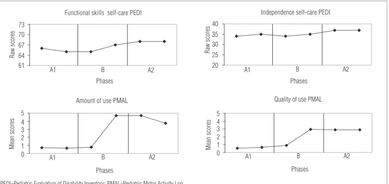
Child 1 demonstrated signiicant improvements in TAUT quality of use and participation scales between A1 and B as-sessments. No changes were observed in the JTHF test or in the TAUT willingness of use scale. Visual analysis revealed pronounced trends towards gains in PMAL amount and qua-lity of use scales. Between phases B and A2, Child 1 improved in the TAUT participation scale. No changes were observed in the JTHF test and in the TAUT quality of use and willing-ness of use scale. Visual analysis demonstrated pronounced trends towards improvements in the PMAL quality scale and maintenance of improvements in PMAL amount of use scale. No important diferences were observed in the PEDI scales (Figure 1).
After intervention, Child 1’s mother reported functional im-provements. he girl demonstrated important improvements in domestic tasks involving bimanual abilities (i.e. washing up, folding clothes).
Child 2
Child 2 demonstrated a signiicant decrease in the total time to perform the JTHF tasks and signiicant improvements in the
Phases A1-B B-A2
Child 1 Child 2 Child 3 Child 1 Child 2 Child 3
JTHF Task 1
ns ns ns ns * *
JTHF Task 2
ns ns ns ns ns ns
JTHF Task 3
ns ns ns ns ns ns
JTHF Total time
ns * ns ns * ns
TAUT Quality
* * ** ns ns **
TAUT Participation
* ns ns * ns ns
TAUT Willingness
ns ns ns ns ns ns
Table 2. Results of the Two-Standard Deviation Band Method or
Celeration Line Method to analyze JTHF test (tasks 1, 2, 3 and total) and TAUT test (quality, participation and willingness scales) of the three children, in the A1-B (baseline x intervention) and B-A2 (intervention x post-intervention) periods.
ns = non-significant result; * = significant result in the expected direction (JTHF: decrease in time to perform the test, TAUT: increase in the score); ** = significant result in the oppo-site expected direction (TAUT: decrease in the score). Note: The Two-Standard Deviation Band was used to analyze results from the JTHF tests and TAUT scales, except for the total score in JTHF from child 2, and for the scores of quality scale for children 1 and 3, which the Celeration Line Method was used.
TAUT quality of use scale,between phases A1-B. No statistically signiicant change was observed in the TAUT participation or willingness scale. he visual analysis revealed pronounced trends towards improvements in the PMAL amount of use and quality of use scales. When phase A2 was compared to phase B, Child 2 demonstrated a signiicant decrease in the time to perform JTHF Task 1 and Total Time. No changes were observed in the TAUT scales. he visual analysis demonstrated a slight trend towards decrease in the PMAL frequency of use scale, but the scores were still higher than phase A1. Child 2 maintained the previous gains in the PMAL quality of use scale. No important diferences were observed in the PEDI scales (Figure 2).
After the intervention period, parents reported that Child 2 demonstrated improved prehension of objects during recrea-tional activities, more awareness of the possibility of using the afected hand during play and school activities and less need of assistance.
Child 3
No signiicant improvements were found in the JTHF and TAUT quality and willingness of use scales when phase B was compared to A1. Visual analysis revealed a pronounced trend towards increase in the PMAL amount of use scale. No changes were observed in the PMAL quality of use scale. When phase A2 was compared to phase B, Child 3 demonstrated a signii-cant decrease in the time to perform JTHF Task 1. No changes were observed in the TAUT participation and willingness of use
scales. Visual analysis showed a slight trend towards decrease in the PMAL amount of use scale and maintenance of gains in the quality of use scale. No changes were observed in the PEDI scales because the child presented maximum scores from the beginning of the study. Child 3 demonstrated a signiicant de-crease in the TAUT quality of use scale in both A1-B and B-A2 periods. However, the visual analysis does not conform to this proile, as it presents a stable trend of data (Figure 3).
According to Child 3’s mother, he improved the ability to grasp small objects and was willing to use the afected extre-mity spontaneously during self-care and play activities.
Discussion
Results from the present study show improvements in quality and amount of use of the affected upper extremity following CIMT in two of the three children with hemiplegia. Despite a general trend towards improvement in dexterity, statistical methods detected significant gains in only 16% of the comparisons. Based on the small effects observed in two children, it is not possible to conclude that the protocol of three hours of daily training may effectively enhance ma-nual dexterity in children with hemiplegia, not confirming H2. The lack of improvements in the adapted JTHF test con-trasts with the findings of Charles et al.16. In their study, the authors observed significant improvements in the JTHF test for children submitted to the CIMT protocol. Differences in
Figure 1. Raw scores of functional skills and independence in self-care from the PEDI and mean scores of quality of use and amount of use scales
from PMAL in the A1 (baseline), B (intervention) and A2 (post-intervention) phases of Child 1.
Functional skills self-care PEDI
61 64 67 70 73
Phases
Ra
w
s
co
re
s
A1 B A2 A1 B A2
Independence self-care PEDI
20 25 30 35 40
Phases
Ra
w
s
co
re
s
Amount of use PMAL
0 1 2 3 4 5
Phases
M
ea
n
sc
or
es
B A2
A1
Phases
A1 B A2
Quality of use PMAL
0 1 2 3 4 5
M
ea
n
sc
or
es
PEDI=Pediatric Evaluation of Disability Inventory; PMAL=Pediatric Motor Activity Log.
protocol intensity between studies could explain this dis-crepancy in results. In their study, Charles et al.16 conducted training for six hours a day, while in the present study the training program lasted 3 hours. Thus, it is argued that the decreased intensity protocol used in the present study was
not sufficient to promote improvements in children’s ma-nual dexterity.
Signiicant improvements in quality of use of the afected extremity,as hypothesized in H1, were observed in two children. he gains in quality of use of the afected extremity and the lack
Functional skills self-care PEDI
61 64 67 70 73
Phases
Ra
w
s
co
re
s
A1 B A2
Phases
A1 B A2
Amount of use PMAL
0 1 2 3 4 5
M
ea
n
sc
or
es
Phases
A1 B A2
Independence self-care PEDI
20 25 30 35 40
Ra
w
s
co
re
s
Phases
A1 B A2
Quality of use PMAL
0 1 2 3 4 5
M
ea
n
sc
or
es
Figure 2. Raw scores of functional skills and independence in self-care from the PEDI and mean scores of quality of use and amount of use scales
from PMAL in the A1 (baseline), B (intervention) and A2 (post-intervention) phases of Child 2.
PEDI=Pediatric Evaluation of Disability Inventory; PMAL=Pediatric Motor Activity Log.
Figure 3. Raw scores of functional skills and independence in self-care from the PEDI and mean scores of quality of use and amount of use scales
from PMAL in the A1 (baseline), B (intervention) and A2 (post-intervention) phases of Child 3.
Phases
A1 B A2
Functional skills self-care PEDI
61 64 67 70 73
Ra
w
s
co
re
s
Phases
A1 B A2
Independence self-care PEDI
20 25 30 35 40
Raw scores
Phases
A1 B A2
Amount of use PMAL
0 1 2 3 4 5
M
ea
n
sc
or
es
Phases
A1 B A2
Quality of use PMAL
0 1 2 3 4 5
M
ea
n
sc
or
es
PEDI=Pediatric Evaluation of Disability Inventory; PMAL=Pediatric Motor Activity Log.
of signiicant improvements in manual dexterity in most tasks could be interpreted as contradictory results. However, the lack of signiicant decrease in time spent to perform tasks could be a consequence of the improvement in quality of use of the afected extremity. As children experience improved movement quality, they develop better strategies to manipulate objects. hese new abilities, however, are still emerging and performance is not yet mastered, which could have led to increased time to perform speciic tasks. Long-term follow-up is needed to document the amount of time required for stable performance following this type of training, and whether or not such stabilization is follo-wed by improvements in manual dexterity.
No changes were observed for the TAUT willingness and participation scales, except for child 1, who demonstrated improved participation scores during intervention and in the post-intervention phases. The lack of changes in the other children could be attributed to the high scores pre-sented at baseline (A1); they received the maximum score (score 2) and maintained this level throughout the study. These high scores in the TAUT willingness and participa-tion scales and the low scores in the PMAL amount of use of the affected extremity, at the baseline period, illustrate the learned nonuse phenomenon. In the current study, despite the fact that children did not present any resistance to use the affected extremity in clinical environment, parents re-ported low frequency of use of the affected upper extremity in the home environment.
According to parents’ perceptions documented with the PMAL, all children demonstrated improvements in the amount and quality of use of the afected extremity in func-tional activities during the intervention period, with mainte-nance of the quality scores and a small decrease in frequency of use in the follow-up period. his small decrease could be explained by the return of use of the non-afected extremity during performance of functional activities. he overall posi-tive results presented in PMAL scores highlight the principles of CIMT. According to Kunkel et al.29, one of the greatest bene-its of CIMT would be the decrease in the diference between current performance and real potential for the execution of daily activities, as the intervention is directed to overcome the learned nonuse.
PMAL results, however, should be interpreted with caution. Statistical analysis could not be undertaken in the current study, due to the small number of assessments. We considered that repetition of the interview four times a week could lead
parents to overestimate their child’s performance during and after intervention, and chose to perform only one interview each week in order to minimize the possible inluence of repe-ated measurements as a threat to the study’s internal validity. In the present study, the positive results documented by the TAUT quality scale corroborate the improvements in quality and the amount of use of the afected extremity reported in PMAL interviews.
The lack of functional improvements in the PEDI (H3) seems to be due to the functional characteristics of the children included in the study, as they obtained high sco-res from the beginning of baseline. In spite of the lack of significant results in PEDI, parents reported improvements in the performance of daily living activities, documented in the daily logs. Future studies should investigate the effects of CIMT on functional abilities and independence, among younger children or children with different levels of manual function.
According to the results of this study, the use of CIMT was efective to promote improvements in the amount and qua-lity of use of the afected upper extremity during functional activitiesin two of the three children with hemiplegia. Future studies analyzing children’s and parents’ perceptions regarding the training program and the use of restraint would help to further elucidate the beneits and applicability of traditional and adapted CIMT protocols in children. his has been the irst Brazilian study to investigate the use of an adapted CIMT model for children with CP, however, it is important and ne-cessary to continue testing this intervention in our clinical population in order to validate our results. Such information is of great relevance for clinical practice, considering the parti-cularities of health services which call for an adaptation of the technique in order to favor parent and therapist adhesion to the intervention.
Acknowledgments
his study was supported by grants from two Brazilian government agencies: Conselho Nacional de Desenvolvi-mento Cientíico e Tecnológico (CNPq), and Fundação de Apoio à Pesquisa do Estado de Minas Gerais (FAPEMIG). We also thank the collaboration of Luciana Braga and Rafa-ela Alvarenga, who helped during the administration of the intervention.
1. World Health Organization. International classification of functioning, disability and health (ICF). Geneva: World Health Organization; 2001.
2. Charles J, Gordon AM. A critical review of constraint-induced movement therapy and forced use in children with hemiplegia. Neural Plast. 2005;12(2-3):245-61.
3. Gordon AM, Charles J, Wolf SL. Methods of constraint-induced movement therapy for children with hemiplegic cerebral palsy: development of a child-friendly intervention for improving upper-extremity function. Arch Phys Med Rehabil. 2005;86(4):837-44.
4. Eliasson AC, Bonnier B, Krumlinde-Sundholm L. Clinical experience of constraint induced movement therapy in adolescents with hemiplegic cerebral palsy - a day camp model. Dev Med Child Neurol. 2003;45(5):357-9.
5. Taub E, Ramey SL, DeLuca S, Echols K. Efficacy of constraint-induced movement therapy for children with cerebral palsy with asymmetric motor impairment. Pediatrics. 2004;113(2):305-12.
6. Eliasson AC, Krumlinde-Sundholm L, Shaw K, Wang C. Effects of constraint-induced movement therapy in young children with hemiplegic cerebral palsy: an adapted model. Dev Med Child Neurol. 2005;47(4):266-75.
7. Liepert J, Bauder H, Wolfgang HR, Miltner WH, Taub E, Weiller C. Treatment-induced cortical reorganization after stroke in humans. Stroke. 2000;31(6):1210-6.
8. Sterr A, Elbert T, Berthold I, Kolbel S, Rockstroh B, Taub E. Longer versus shorter daily constraint-induced movement therapy of chronic hemiparesis: an exploratory study. Arch Phys Med Rehabil. 2002;83(10):1374-7.
9. Taub E, Uswatte G, Pidikiti R. Constraint-induced movement therapy: a new family of techniques with broad application to physical rehabilitation - a clinical review. J Rehabil Res Dev. 1999;36(3):237-51.
10. Pierce SR, Daly K, Gallagher KG, Gershkoff AM, Schaumburg SW. Constraint-induced therapy for a child with hemiplegic cerebral palsy: a case report. Arch Phys Med Rehabil. 2002;83(10):1462-3.
11. DeLuca SC, Echols K, Ramey SL, Taub E. Pediatric constraint-induced movement therapy for a young child with cerebral palsy: two episodes of care. Phys Ther. 2003;83(11):1003-13.
12. Miltner WH, Bauder H, Sommer M, Dettmers C, Taub E. Effects of constraint-induced movement therapy on patients with chronic motor deficits after stroke: a replication. Stroke. 1999;30(3):586-92.
13. DeLuca SC, Echols K, Law CR, Ramey SL. Intensive pediatric constraint-induced therapy for children with cerebral palsy: randomized controlled, crossover trial. J Child Neurol. 2006;21(11):931-8.
14. Sterr A, Freivogel S, Schmalohr D. Neurobehavioral aspects of recovery: assessment of the learned nonuse phenomenon in hemiparetic adolescents. Arc Phys Med Rehabil. 2002;83(12):1726-31.
15. Charles J, Lavinder G, Gordon AM. Effects of constraint-induced therapy on hand function in children with hemiplegic cerebral palsy. Pediatr Phys Ther. 2001;13(2):68-76.
16. Charles JR, Wolf SL, Schneider JA, Gordon AM. Efficacy of a child-friendly form of constraint-induced movement therapy in hemiplegic cerebral palsy: a randomized control trial. Dev Med Child Neurol. 2006;48(8):635-42.
17. Naylor CE, Bower E. Modified constraint-induced movement therapy for young children with hemiplegic cerebral palsy: a pilot study. Dev Med Child Neurol. 2005;47(6):365-9.
18. Gordon A, Connelly A, Neville B, Vargha-Khadem F, Jessop N, Murphy T, et al. Modified constraint-induced movement therapy after childhood stroke. Dev Med Child Neurol. 2007;49(1):23-7.
19. Eliasson AC, Krumlinde Sundholm L, Rösblad B, Beckung E, Arner M, Öhrvall AM, et al. The manual ability classification system (MACS) for children with cerebral palsy: scale development and evidence of validity and reliability. Dev Med Child Neurol. 2006;48(7):549-54.
20. Jebsen RH, Taylor N, Trieschmann RB, Trotter MJ, Howard LA. An objective and standardized test of hand function. Arch Phys Med Rehabil. 1969;50(6):311-9.
21. Taylor N, Sand PL, Jebsen RH. Evaluation of hand function in children. Arch Phys Med Rehabil. 1973;54(3):129-35.
22. Vaz DV, Mancini MC, Fonseca ST, Vieira DS, de Melo Pertence AE. Muscle stiffness and strength and their relation to hand function in children with hemiplegic cerebral palsy. Dev Med Child Neurol. 2006;48(9):728-33.
23. Haley SM, Coster WJ, Ludlow LH, Haltiwanger JT, Andrellow PJ. Pediatric evaluation of disability inventory: development, standardization and administration manual. Boston: New England Medical Center; 1992.
24. Mancini MC. Inventário de avaliação pediátrica de incapacidade (PEDI) - manual da versão brasileira adaptada. Belo Horizonte: UFMG; 2004.
25. Feldman AB, Haley SM, Coryell J. Concurrent and construct validity of the pediatric evaluation of disability inventory. Phys Ther. 1990;70(10): 602-10.
26. Portney LG, Watkins MP. Foundations of clinical research: applications to practice. 2ª ed. New Jersey: Prentice Hall Health; 2000.
27. Ottenbacher KJ. Evaluating clinical change: strategies for occupational and physical therapists. Baltimore: Williams & Wilkins; 1986.
28. Nourbakhsh MR, Ottenbacher KJ. The statistical analysis of single-subject data: a comparative examination. Phys Ther. 1994;74(8):768-76.
29. Kunkel A, Kopp B, Muller G, Villringer K, Villringer A, Taub E, et al. Constraint-induced movement therapy for motor recovery in chronic stroke patients. Arch Physical Med Rehabil. 1999;80(6):624-8.
534