Quim. Nova, Vol. 35, No. 11, 2222-2225, 2012
Artigo
*e-mail: [email protected]
#Artigo em homenagem ao Prof. Otto R. Gottlieb (31/8/1920-19/6/2011)
PHAEOPHYTINS FROM Thyrsacanthus ramosissimus Moric. WITH INHIBITORY ACTIVITY ON HUMAN DNA TOPOISOMERASE II-α#
Analúcia Guedes Silveira Cabral, Fábio Henrique Tenório-Souza, Marcelo Dantas Moura, Sabrina Gondim Ribeiro Mota, Antônio Cláudio da Silva Lins, Celidarque da Silva Dias e José Maria Barbosa-Filho∗
Departamento de Ciências Farmacêuticas, Universidade Federal da Paraíba, CP 5009, 58051-970 João Pessoa – PB, Brasil Ana Maria Giulietti
Departamento de Ciências Biológicas, Universidade Estadual de Feira de Santana, 44036-900 Feira de Santana – BA, Brasil / Royal Botanic Gardens, Kew, TW9 3AB, UK
Tania Maria Sarmento da Silva
Departamento de Ciências Moleculares, Universidade Federal Rural de Pernambuco, 52171-900 Recife – PE , Brasil Creusioni Figueredo dos Santos
Departamento de Biologia Molecular, Universidade Federal da Paraíba, 58051-900 João Pessoa – PB, Brasil
Recebido em 22/5/12; aceito em 21/7/12; publicado na web em 15/10/12
Our study reports the extraction and isolation of a new phaeophytin derivative 151-hydroxy-(151-S)-porphyrinolactone, designated anamariaine (1) herein, isolated from the chloroform fraction of aerial parts of Thyrsacanthus ramosissimus Moric. along with the known 151-ethoxy-(151-S)-porphyrinolactone (2). These compounds were identified by usual spectroscopic methods. Both compounds were subjected to in vitro (inhibitory activity) tests by means of supercoiled DNA relaxation techniques and were shown to display inhibitory activity against human DNA topoisomerase II-α at 50 µM. Interconversion of these two pigments under the mild conditions of the isolation techniques should be highly unlikely but cannot be entirely ruled out.
Keywords: Thyrsacanthus ramosissimus; phaeophytins; topoisomerase activity.
INTRODUCTION
The family Acanthaceae comprises around 250 genera and ap-proximately 2500 species,1 which occur mainly in the Atlantic Forest and in the mesophilic forest formations of the Central-Western and Southeastern regions of Brazil, as well as in other types of vegetation.2 Previous studies on the isolation of natural products from the family Acanthaceae have revealed the presence of alkaloids,3 flavonoids,4 terpenes,5 coumarins,6 lignans,7 and quinoids.8 These constituents have been demonstrated to have biological effects such as cytotoxicity,7 vasorelaxant,5 anti-inflammatory,9 antifungal,5 and antiviral actions,10 as well as CNS depression and immunosuppressive activities.9,11
Thyrsacanthus Moric. is a genus with five species whose presen-ce is restricted to South America and that occurs mainly in Brazil. Recently, the genus was restored to include the South American species traditionally assigned to Anisacanthus Nees. Thyrsacanthus ramosissimus Moric. (= Anisacanthus brasiliensis Lindau) is po-pularly known as “canudo” and is endemic to Brazil, in the states of Alagoas, Bahia, Minas Gerais, Pernambuco, and Rio Grande do Norte. It can be found principally in seasonally dry vegetation and in semideciduous and riparian forests.12 No biological studies have been previously performed with this species and chemical and biological studies with species of this genus are also scarce.
Interest in compounds with inhibitory activity against the enzyme DNA-topoisomerase II-α has increased,13-20 because intracellular lev-els of topoisomerase II-α (topo II-α) are elevated in a number of hu-man tumors as compared to the respective normal tissues. Moreover, the development of drugs that can affect the DNA replication process by selectively interfering with the function of topo II-α continues to draw researchers’ attention.21 Topo II-α is an essential enzyme that
plays a key role in DNA replication, but is also important in repair, transcription, and chromosome segregation. Topo II-α changes DNA topology by passing an intact DNA double helix through a transient double-stranded break, thereby producing another helix.22,23 Thus, as part of our ongoing chemical investigations of this plant species,24 in this work we evaluate the inhibitory effect of this plant on human DNA-topoisomerase II-α. The isolation of a new phaeophytin (1), as well as its structural elucidation by means of ESI-MS and 1D- and 2D-NMR experiments, will be reported here.
EXPERIMENTAL
General experimental procedures
Melting points were determined on a Koefler hot stage and are uncorrected. The infrared absorption spectra were recorded in KBr pellets, using a Bomem/MB-102 spectrophotometer operating in the 4000-400 cm-1 range. The LC-MS spectra were obtained in the positive electrospray mode using a Quattro LC-Micromass device (Waters). Silica gel 60 was used for column chromatography, and Kieselgel 60F254 (E. Merck) was employed for preparative TLC as precoated plates. Sephadex LH-20 (Sigma) was utilized for gel permeation chromatography. 1H and 13C NMR spectra were acquired on a Bruker AC 200 spectrometer (200 MHz for 1H and 50 MHz for 13C), in CDCl
3. Plant material
Phaeophytins from Thyrsacanthus ramosissimus Moric. 2223 Vol. 35, No. 11
Extraction and isolation
The powdered plant material (5.0 kg) was extracted successively with EtOH, to give 310.0 g dry extract. This extract was dissolved in MeOH/H2O (3:7) and successively fractionated with hexane, CHCl3, and AcOEt, yielding the hexane (20.0 g), CHCl3 (10.0 g), and EtOAc (5.0 g) fractions. The CHCl3 fraction was successively submitted to column chromatography using silica gel and Sephadex LH-20, followed by preparative TLC on silica, to yield 1 (29.2 mg) and 2 (27.8 mg) as dark blue amorphous solids. Analysis of the spectroscopic data (IR, LC-MS and NMR spectra, including 2D NMR) led to identification of the following compounds: anamariaine (151-hydroxy-(151-S)-porphyrinolactone, (1) and 151-ethoxy-(151 -R)--porphyrinolactone (2).
Anamariaine (1)
Dark blue amorphous solid; mp 171.0 ºC; IR (KBr) νmax 3549,
3476, 3414, 1737, 1638, 1615 cm-1. 1H NMR (CDCl
3, 200 MHz) (Table 1), 13C NMR (CDCl
3, 50 MHz) (Table 1), ESI-MS (pos) m/z 903.99 [M+H]+ (C
55H74N4O7).
151-Ethoxy-(151-R)-porphyrinolactone (2)
Dark blue amorphous solid; mp 114.0 ºC; IR (KBr) νmax 3447, 2927, 1734, 1636, 1104 cm-1, ESI-MS (pos) m/z 931.59 [M+H]+ (C57H79N4O7).
In vitro assay for DNA topoisomerase II-α
The conversion of pBR322 supercoiled plasmid DNA to the relaxed form by topo II-α was examined in the presence of phaeo-phytins 1 and 2. DNA topoisomerases are enzymes that modulate the topological state of DNA and are targets for many active drugs in cancer treatment.15,23
Enzymatic activity was analyzed by the DNA relaxation assay
Table 1. 1H (200 MHz) and 13C (50 MHz) spectral data for anamariaine (1) obtained by heteronuclear 2D shift-correlated HSQC, HMQC, and HMBC spectra, in CDCl3. Chemical shifts (δ, ppm) and coupling constants (J in Hz, in parenthesis)
a
Porphyrin moiety Phytol moiety
Position δC δH Position δC δH
1 141.18 P1 61.45 4.52 (m)
2 131.45 P2 117.70 5.20 (t, 7.0)
21 12.13 3.42 (s) P3 142.86
3 135.98 P31 16.27 1.60 (s)
31 128.93 8.05 (dd, 18.0; 12.0) P4 39.77 1.93 (m)
32 122.71 (E) 6.37 (dl, 18.0)
(Z) 6.24 (dl, 7.8)
P5 24.95 1.30 (m)
4 135.98 P6 29.69 1.30 (m)
5 99.59 9.68 (s) P7 32.73 1.55 (m)
6 155.71 P71 19.71 0.84 (d, 6.6)
7 136.46 P8 37.36 1.04 (m)
71 11.24 3.22 (s) P9 24.39 1.30 (m)
8 145.51 P10 36.59 1.21 (m)
81 19.52 3.82 (m) P11 32.58 1.35 (m)
82 17.58 1.68 (t, 8.0 Hz) P111 19.71 0.81(d, 6.6)
9 149.94 P12 37.28 1.24 (m)
10 104.10 9.91 (s) P13 24.76 1.30 (m)
11 138.70 P14 39.32 1.48 (m)
12 134.75 P15 27.94 1.52 (m)
121 12.43 3.87 (s) P151 22.70 0.88 (d, 6.6)
13 111.29 P16 22.70 0.88 (d, 6.6)
131 166.30
14 135.98
15 100.43
151 101.95
152 170.87
153 54.15 3.75 (s)
16 161.06
17 53.68 4.16 (d, 8.7)
171 32.13 171 B 1.81 (m)
172 A 2.63 (m)
172 31.29 172 B 2.22 (m)
172 A 2.49 (m)
173 173.30
18 50.12 4.51 (m)
181 22.60 1.62 (d, 8.0 Hz)
19 171.12
20 93.87 8.86 (s)
a 2D homonuclear 1H-1H-COSY and heteronuclear HMBC spectra were also used for these assignments. Hydrogen atoms chemical shifts obtained from 1D 1H NMR spectra. Carbon signals corresponding to C, CH, CH2, and CH3 as deduced by comparative analysis of the APT and HSQC spectra. Superimposed
Cabral et al.
2224 Quim. Nova
according to the protocol described by USB Corporation (USB Corporation, Cleveland, OH, USA). One unit of topo II-α (USB Corporation, Cleveland, OH, USA) was incubated with 0.152 µg pBR322 DNA (human recombinant from E. coli, Invitrogen) and with 100 or 50 µM of compound 1 or 2, or without the test compounds, in 10 µL reaction mixture containing 10 mM Tris, pH 7.9, 50 mM NaCl, 50 mM KCl, 5 mM MgCl2, 0.1 mM EDTA, 15 µg/mL BSA, 1 mM ATP, 10 mM Na2HPO4, and 0.2 mM DTT for 40 min, at 37 °C. The reaction was terminated by addition of 1µL stop solution consisting of 50% glycerol, 10% sodium dodecyl sulfate (SDS), and 25 % bromophenol blue. Electrophoresis was carried out on 1% agarose gel (Sigma-Aldrich) equilibrated with TAE buffer (4.84 g L-1 Tris-base, pH 8.5, 1.14 g L-1 glacial acetic acid and 100 mL of 0.74 g L-1 EDTA) for 120 min at 40 V. Etoposide was used as the positive control. The gels were stained with ethidium bromide solution (5 g L-1) after electrophoresis for 30 min, washed with water, and photo-graphed under UV light with a digital camera.
RESULTS AND DISCUSSION
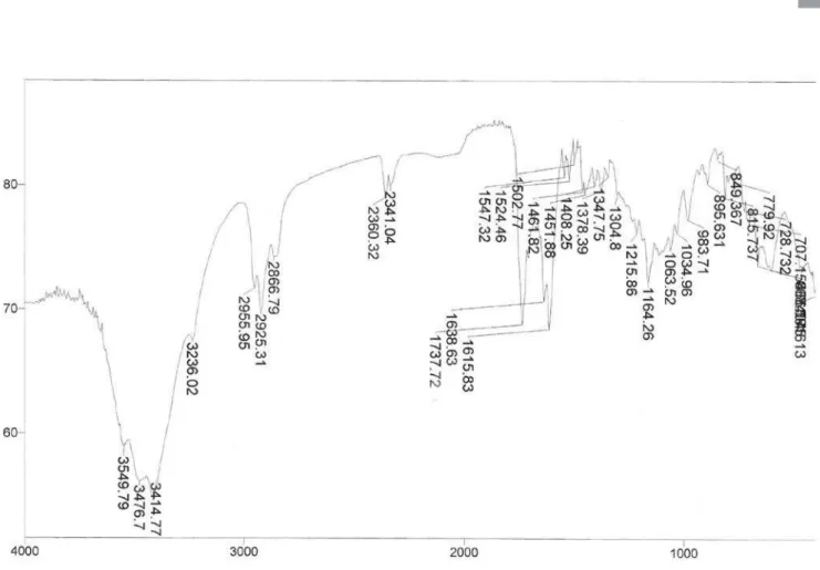
Extensive chromatography of the CHCl3 fraction of Thyrsacanthus ramosissimus resulted in the isolation and characterization of two pha-eophytins 1 and 2 (Figure 1). Compound 1 was isolated as a dark blue amorphous solid. ESI-MS analysis in the positive ionization mode resulted in [M+H]+m/z 903.99, indicating 21 degrees of insaturation for a molecular formula of C55H74N4O7. The IR absorptions at 3549, 3476, 3414, 1737, and 1615 cm-1 and UV data, and overall color characteristics suggested the presence of a large chromophore related to the chlorophyll derivative containing a hydroxyl group.
The 200 MHz 1HNMR spectrum displayed all the resonances of phaeophytin A and its derivatives:25 three aromatic methyl groups (δ
H 3.95, 3.48, and 3.33), a methoxyl group (δH 3.79), three singlet olefinic protons (δH 9.91, 9.68, and 8.86), vinyl substitution (δH 6.37, 6.24, and 8.05) with a characteristic exomethylene coupling pattern, and two singlets at δH 1.50 and 1.41 indicative of two pyrrole NH functiona-lities within the shielding ring current.26 The attached phytol moiety was recognized by a large number of overlapping proton signals of aliphatic methylene and methyl functions. Proton resonances sugges-ting the phytol moiety were the carbinol resonances CH2-P1 (δH 4.51), the olefinic proton H-P2 (δH 5.20) and CH3-P3
1 (δ
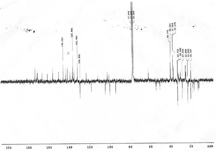
H 1.6) (Figure 1). Thirty-five carbon signals were detected in the 13C NMR spectrum for the porphyrin system of compound 1, while the aliphatic phytol
moiety afforded a partial overlapping set of signals in the aliphatic region. Overall, the 1H and 13C-NMR spectra were in accordance with the literature data.27,28
The presence of the hydroxyl group in ring E was deduced from the carbinol resonance C-151 at δ
C 101.95, with a quaternary nature confirmed by the APT and HSQS experiment. The NMR and ESI-MS data, together with the degrees of insaturation, allowed us to esta-blish a conjugated d-lactone structure, which appeared to be formed between the C-131-C-132 bond at ring E of 132-hydroxyphaeophytin A. There was evidence of a carbonyl carbon at δC 166.30 instead of the resonances at dC 192.2 for the corresponding carbon.
29
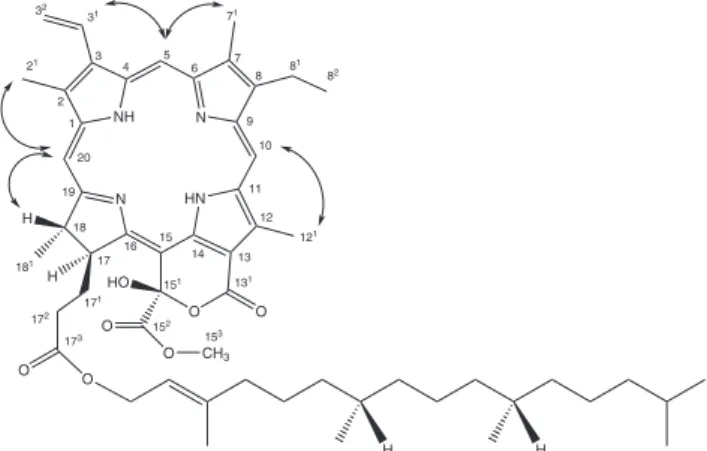
The complete analysis of the porphyrin structure was conducted on the basis of the correlations detected in the 2,3J
CH long-range of the olefinic protons H-5, H-10, H-20, which provide the connectivities between the four pyrrole rings (Figure 2). The signal corresponding to H-5 (δH 9.68) correlated with C-7 in ring B. Similarly, H-10 (δH 9.91) presented connectivity with C-8 (ring B) and C-11 in ring C, while H-20 (dH 8.86) correlated with C-1 and C-2 (ring A). The signal relative to 3H-181 (δ
H 1.62) in ring D showed long-range connectivity with C-19, and H-18 (δH 4.51) correlated with C-16. The 3J
CH correlation of OCH3-15 3 (d
H 3.75) with the carboxyl group C-152 confirmed the position of the methyl-ester. The presence of the phytol-moiety attachment to the porphyrin system was indicated by the 2J
CH correlation of 17 2-B (δ
H 2.22) and 17
2-A (2.49) to the carboxyl function C-173 (δ
C 173.30).
The configuration of C-151 was determined as S due up-field of H-17 (dH 4.16),
30 since the Nuclear Overhauser enhancements in the NOESY spectrum did not show signals referring to this chiral cen-ter. Other significant signals were observed in the porphyrin system (Figure 3). Therefore, the structure of compound 1 was identified as 151-hydroxy-(151-S)-porphyrinolactone (anamariaine). This is its first isolation as a natural compound.
Compound 2 displays identical signals as compared to compound 1, except for an additional ethoxy group identified from 1H resonances at dH 4.36 (q, J = 7.1 Hz, H-2-1’’) and 1.50 (t, J = 7.10 Hz, H3-2”) and the respective carbons at dC 62.44 and 15.62. The ethoxy group was deduced as being positioned at C-151 on the basis of the HMBC correlation between H2-1” and C-151 (δ
C 106.31). The spectral data of compound 2, including the chiral center at C-151 (δ
H 4.82), agrees with those described in the literature and allows for its identification as 151-ethoxy-(151-R)-porphyrinolactone.31 This compound has been previously isolated from the green alga Cladophora fascicularis.31
The isolation of phaeophytins from Thyrsacanthus ramosissimus raises the question as to whether this material occurs naturally or is
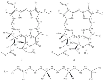
N NH N HN O O HO O
O CH3
O O H 1 2 3 31 32 5 4 6 7 71 8 81 82 9 10 11 12 121 13 131 14 15 16 17 18 181 171 172 173 21 151 152 153 20 19 R N NH N HN O O EtO O
O CH3
O O H 1 2 3 31 32 5 4 6 7 71 8 81 82 9 10 11 12 121 13 131 14 15 16 17 18 181 171 172 173 21 151 152 153 20 19 R H H P1
H P111 H
P71 P2 P31 P3 P4 P5 P6 P8 P7 P10 P9 P11 P12 P13 P15 P14 P16 P151 R = 1 2
Figure 1. Structures of anamariaine (1) and 151-ethoxy-(151
-R)-porphyri-nolactone (2)
N NH N HN O O HO O
O CH3
O O H 1 2 3 31 32 5 4 6 7 71 8 81 82 9 10 11 12 121 13 131 14 15 16 17 18 181 171 172 173 21 151 152 153 20 19 H H H
Phaeophytins from Thyrsacanthus ramosissimus Moric. 2225 Vol. 35, No. 11
a possible artifact produced during chromatographic separation. Our experience suggests that this rare pigment is not commonly observed in plant species.32,33 Comparison between the structures of compounds 1 and 2 shows that an interconversion of these two pigments under the mild conditions of the isolation techniques should be highly unlikely. The effects of the compounds on the catalytic activity of the DNA topo II-a enzyme were observed in the relaxation assays using pBR322 in the presence of ATP. Compounds 1 and 2 were evaluated at 50 µM (lanes 4 and 5, respectively) (Figure 4). The two compounds promoted significant inhibition of the catalytic activity of topo II-α
as compared to etoposide, which was used as the positive control.
SUPPLEMENTARY MATERIAL
1H and 13C NMR, HMQC, COSY, HMBC, NOESY, IR, and ESI-MS spectra of compound 1 are available at http://quimicanova.sbq. org.br, in PDF file, with free access.
ACKNOWLEDGEMENTS
This work was financially supported by CNPq/CAPES/FAPESQ/ PRONEX. We are also extremely grateful to CENAUREM/UFC for conducting the 200 MHz spectra. The authors are also thankful to the technicians R. N. da Silva Filho (UFPB) and D. E. de A. Uchoa (UFC) for the technical support. Dr. A. Leyva helped with editing of the English language.
REFERENCES
1. Barroso, G. M.; Peixoto, A. L.; Costa, C. G.; Ichaso, C. L. F.; Guimarães, E. F.; Lima, H. C.; Sistemática das Angiospermas do Brasil,
N NH N HN O O HO O
O CH3
O O H 1 2 3 31 32 5 4 6 7 71 8 81 82 9 10 11 12 121 13 131 14 15 16 17 18 181 171 172 173 21 151 152 153 20 19 H H H
Figure 3. Structure corresponding to NOESY correlation signals observed in compound 1 showing proton-proton through-space interactions in the porphyrin-ring system
Figure 4. All lanes contain 0.152 µg DNA (pBR322) and 1.0 unit of topo II-a, with the exception of Lane 1. Lane 1: negative control (pBR322 only). Lane 2: positive control (pBR322 and topo II-a). Lane 3: etoposide (100 mM). Lane 4: compound 1 (50 µM). Lane 5: compound 2 (50 µM)
Imprensa Universitária: Viçosa, 1991, vol. 3.
2. Braz, D. M.; Carvalho-Okano, R. M.; Kameyama, C.; Rev. Bras. Bot. 2002, 25, 495.
3. Amer, M. E.; Abou-Shoer, M. I.; Abdel-Kader, M. S.; El-Shaibany, A. M. S.; Abdel-Salama, N. A.; J. Braz. Chem. Soc. 2004, 15, 262. 4. Rao, Y. K.; Vimalamma, G.; Rao, C. V.; Tzeng, Y.; Phytochemistry 2004,
65, 2317.
5. Rasoamiaranjanahary, L.; Marston, A.; Guilet, D.; Schenk, K.; Randim-bivololona, F.; Hostettmann, K.; Phytochemistry 2003, 62, 333. 6. Leal, L. K. A. M.; Ferreira, A. A. G.; Bezerra, G. A. A.; Matos, F. J. A.;
Viana, G. S. B.; J. Ethnopharmacol. 2000, 70, 151.
7. Venkataraman, R.; Gopalakrishnan, S.; Phytochemistry 2002, 61, 963. 8. Siripong, P.; Yahuafai, J.; Shimizu, K.; Ichikawa, K.; Yonezawa, S.;
Asai, T.; Kanokmedakul, K.; Ruchirawat, S.; Oku, N.; Biol. Pharm. Bull. 2006, 29, 2279.
9. Kanchanapoom, T.; Noiarsa, P.; Ruchirawat, S.; Kasai, R.; Otsuka, H.; Phytochemistry 2004, 65, 2613.
10. Asano, J.; Chiba, K.; Tada, M.; Yosmil, T.; Phytochemistry 1996, 42, 713.
11. Luo, Y.; Feng, C.; Tian, Y.; Zhang, G.; Phytochemistry 2002, 61, 449. 12. Côrtes, A. L. A.; Borges, R. L. B.; Rapini, A.; Taxon 2010, 59, 965. 13. Silva, M. N.; Ferreira, V. F.; Souza, M. C. B. V.; Quim. Nova 2003, 26,
407.
14. Oliveira, M. C. C.; Carvalho, M. G.; Grynberg, N. F.; Brioso, P. S. T.; Planta Med. 2005, 71, 561.
15. Cunha, A. S.; Lima, E. L. S.; Pinto, A. C.; Esteves-Souza, A.; Echevarria, A.; Câmara, C. A.; Vargas, M. D.; Torres J. C.; J. Braz. Chem. Soc. 2006, 17, 439.
16. Vega, M. R. G.; Esteves-Souza, A.; Vieira, I. J. C.; Mathias, L.; Braz-Filho, R.; Echevarria, A.; J. Braz. Chem. Soc. 2007, 18, 1554. 17. Esteves-Souza, A.; Figueiredo, D. V.; Esteves, A.; Câmara, C. A.; Vargas,
M. D.; Pinto, A. C.; Echevarria, A.; Braz. J. Med. Biol. Res. 2007, 40, 1399.
18. Branco, A.; Pinto, A. C.; Braz-Filho, R.; Silva, E. F.; Grynberg, N. F.; Echevarria, A.; Rev. Bras. Farmacogn. 2008, 18, 703.
19. Cotrim, C. A.; Garrido, S. S.; Trovatti, E.; Marchetto, R.; Quim. Nova 2010, 33, 841.
20. Bruxel, F.; Guterres, S. S.; Teixeira, H. F.; Quim. Nova 2011, 34, 1643. 21. Corbett, A. H.; Osheroff, N.; Chem. Res. Toxicol. 1993, 6, 585. 22. Nitiss, J. L.; Biochim. Biophys. Acta 1998, 1400, 63. 23. Wang, J. C.; Annu. Rev. Biochem. 1996, 65, 635.
24. Dias, C. S.; Moura, M. D.; Cabral, A. G. S.; Mota, S. G. R.; Cunha, E. V. L.; Silva, T. M. S.; Harley, A. M. G.; Barbosa-Filho, J. M.; Labciencia 2007, 1, 14.
25. Buchanan, M. S.; Hashimoto, T.; Asakawa, Y.; Phytochemistry 1996, 41, 1373.
26. Helaja, J.; Stapelbroek-Möllmann, M.; Hynninen, P. H.; J. Org. Chem. 2000, 65, 3700.
27. Schwikkard, S. L.; Mulholland, D. A.; Hutchings, A.; Phytochemistry 1998, 49, 2391.
28. Tomaz, A. C. A.; Nogueira, R. B. S. S.; Pinto, D. S.; Agra, M. F.; Souza, M. F. V.; Cunha, E. V. L.; Rev. Bras. Farmacogn. 2008, 18, 47. 29. Jerz, G.; Arrey, T. N.; Wray, V.; Du, Q.; Winterhalter, P.; Innov. Food Sci.
Emerging Technol. 2007, 8, 413.
30. Nakatani, Y.; Ourisson, G.; Beck, J. P.; Chem. Pharm. Bull. 1981, 29, 2261.
31. Huang, X.; Li, M.; Xu, B.; Zhu, X.; Deng, Z.; Lin, W.; Molecules 2007, 12, 582.
32. Silva, T. M. S.; Câmara, C. A.; Medeiros, F. D.; Oliveira, E. J.; Agra, M. F.; Harley, R. M.; Giulietti, A. M.; Biochem. Syst. Ecol. 2006, 34, 263. 33. Silva, T. M. S.; Câmara, C. A.; Barbosa-Filho, J. M.; Giulietti, A. M.;
Quim. Nova, Vol. 35, No. 11, S1-S10, 2012
Supplementary Material
*e-mail: [email protected]
#Artigo em homenagem ao Prof. Otto R. Gottlieb (31/8/1920-19/6/2011)
PHAEOPHYTINS FROM Thyrsacanthus ramosissimus Moric. WITH INHIBITORY ACTIVITY ON HUMAN DNA
TOPOISOMERASE II-α#
Analúcia Guedes Silveira Cabral, Fábio Henrique Tenório-Souza, Marcelo Dantas Moura, Sabrina Gondim Ribeiro Mota,
Antônio Cláudio da Silva Lins, Celidarque da Silva Dias e José Maria Barbosa-Filho∗
Departamento de Ciências Farmacêuticas, Universidade Federal da Paraíba, CP 5009, 58051-970 João Pessoa – PB, Brasil
Ana Maria Giulietti
Departamento de Ciências Biológicas, Universidade Estadual de Feira de Santana, 44036-900 Feira de Santana – BA, Brasil / Royal Botanic Gardens, Kew, TW9 3AB, UK
Tania Maria Sarmento da Silva
Departamento de Ciências Moleculares, Universidade Federal Rural de Pernambuco, 52171-900 Recife – PE , Brasil
Creusioni Figueredo dos Santos
Departamento de Biologia Molecular, Universidade Federal da Paraíba, 58051-900 João Pessoa – PB, Brasil
Cabral et al.
S2 Quim. Nova
Figure 2S. ESI-MS spectrum of compound 1
Figure 3S. NMR 13C-APT spectrum of compound 1 (CDCl
Phaeophytins from Thyrsacanthus ramosissimus Moric. S3
Vol. 35, No. 11
Figure 5S. Expansion of the NMR 13C-APT spectrum in the region of 65.0 – 8.0 of compound 1 (CDCl
3, 50 MHz)
Figure 4S. Expansion of NMR 13C-APT spectrum in the region of 178.0 – 90.0 of compound 1 (CDCl
Cabral et al.
S4 Quim. Nova
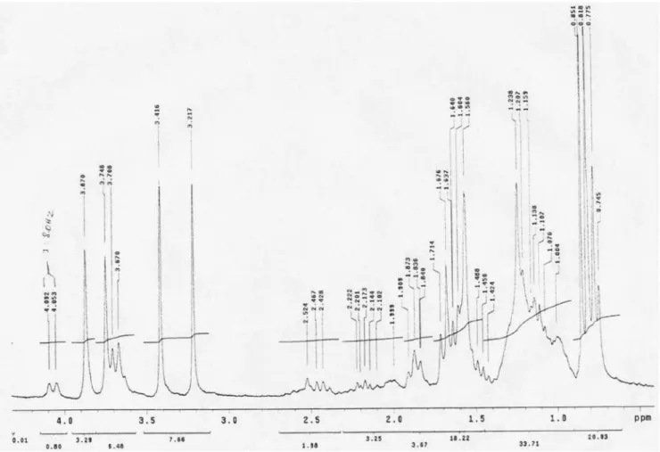
Figure 6S. NMR 1H spectrum of compound 1 (CDCl
3, 200 MHz)
Figure 7S. Expansion of the NMR 1H spectrum in the region of 10.0 – 4.5 of compound 1 (CDCl
Phaeophytins from Thyrsacanthus ramosissimus Moric. S5
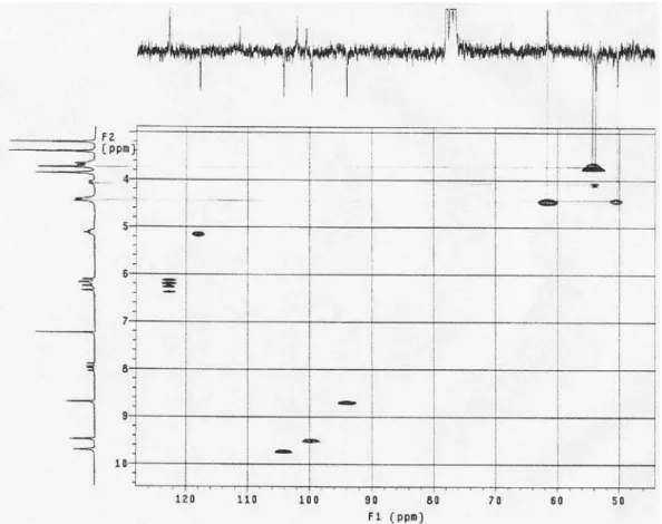
Vol. 35, No. 11
Figure 9S. 1H x 13C-HMQC correlation spectrum of compound 1 (CDCl
3, 200 and 50 MHz respectively)
Figure 8S. Expansion of the NMR 1H spectrum in the region of 4.5 – 0.5 of compound 1 (CDCl
Cabral et al.
S6 Quim. Nova
Figure 10S. Expansion of the1H x 13C-HMQC correlation spectrum of compound 1 (CDCl
3, 200 and 50 MHz respectively)
Figure 11S. Expansion of the1H x 13C-HMQC correlation spectrum of compound 1 (CDCl
Phaeophytins from Thyrsacanthus ramosissimus Moric. S7
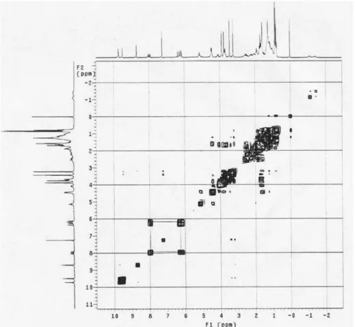
Vol. 35, No. 11
Figure 13S. Expansion of the1H x 1H-COSY correlation spectrum of compound 1 (CDCl
3, 200 MHz)
Figure 12S. 1H x 1H-COSY correlation spectrum of compound 1 (CDCl
Cabral et al.
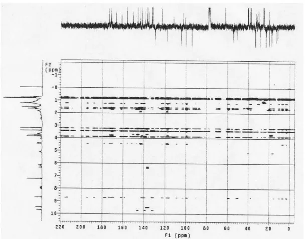
S8 Quim. Nova
Figure 14S. 1H x 13C-HMBC correlation spectrum of compound 1 (CDCl
3, 200 and 50 MHz respectively)
Figure 15S. Expansion of the1H x 13C-HMBC correlation spectrum of compound 1 (CDCl
Phaeophytins from Thyrsacanthus ramosissimus Moric. S9
Vol. 35, No. 11
Figure 17S. 1H x 1H-NOESY spatial correlation spectrum of compound 1 (CDCl
3, 200 MHz)
Figure 16S. Expansion of the1H x 13C-HMBC correlation spectrum of compound 1 (CDCl
Cabral et al.
S10 Quim. Nova
Figure 18S. Expansion of the1H x 1H-NOESY spatial correlation spectrum of compound 1 (CDCl