Streamlining DNA Barcoding Protocols:
Automated DNA Extraction and a New
cox1
Primer in Arachnid Systematics
Nina Vidergar1,5, Natasˇa Toplak4, Matjazˇ Kuntner1,2,3*
1.Institute of Biology, Scientific Research Centre of the Slovenian Academy of Sciences and Arts, Ljubljana, Slovenia,2.Centre for Behavioural Ecology & Evolution, College of Life Sciences, Hubei University, Wuhan, China,3.National Museum of Natural History, Smithsonian Institution, Washington, DC, United States of America,4.Omega d.o.o., Ljubljana, Slovenia,5.Molecular Virology lab, International Centre for Genetic Engineering and Biotechnology–ICGEB, Trieste, Italy
Abstract
Background: DNA barcoding is a popular tool in taxonomic and phylogenetic
studies, but for most animal lineages protocols for obtaining the barcoding
sequences—mitochondrial cytochrome C oxidase subunit I (cox1AKA CO1)—are not standardized. Our aim was to explore an optimal strategy for arachnids, focusing on the species-richest lineage, spiders by (1) improving an automated DNA extraction protocol, (2) testing the performance of commonly used primer combinations, and (3) developing a new cox1primer suitable for more efficient alignment and phylogenetic analyses.
Methodology: We used exemplars of 15 species from all major spider clades,
processed a range of spider tissues of varying size and quality, optimized genomic DNA extraction using the MagMAX Express magnetic particle processor—an automated high throughput DNA extraction system—and testedcox1amplification protocols emphasizing the standard barcoding region using ten routinely employed primer pairs.
Results: The best results were obtained with the commonly used Folmer primers
(LCO1490/HCO2198) that capture the standard barcode region, and with the C1-J-2183/C1-N-2776 primer pair that amplifies its extension. However, C1-J-2183 is designed too close to HCO2198 for well-interpreted, continuous sequence data, and in practice the resulting sequences from the two primer pairs rarely overlap. We therefore designed a new forward primer C1-J-2123 60 base pairs upstream of the C1-J-2183 binding site. The success rate of this new primer (93%) matched that of C1-J-2183.
Conclusions: The use of C1-J-2123 allows full, indel-free overlap of sequences
obtained with the standard Folmer primers and with C1-J-2123 primer pair. Our
OPEN ACCESS
Citation:Vidergar N, Toplak N, Kuntner M (2014) Streamlining DNA Barcoding Protocols: Automated DNA Extraction and a Newcox1Primer in Arachnid Systematics. PLoS ONE 9(11): e113030. doi:10.1371/journal.pone.0113030
Editor:Damon P. Little, The New York Botanical Garden, United States of America
Received:June 11, 2014
Accepted:October 17, 2014
Published:November 21, 2014
Copyright:ß2014 Vidergar et al. This is an open-access article distributed under the terms of theCreative Commons Attribution License, which permits unrestricted use, distribution, and repro-duction in any medium, provided the original author and source are credited.
Data Availability:The authors confirm that all data underlying the findings are fully available without restriction. All relevant data are within the paper and its Supporting Information files.
Funding:This research was supported by the Slovenian Research Agency (grants P1-0236 and MR-2013) and a Swiss Contribution to the enlarged EU grant (C1536-1 1T440013). The funders had no role in study design, data collection and analysis, decision to publish, or preparation of the manu-script.
preliminary tests suggest that in addition to spiders, C1-J-2123 will also perform in other arachnids and several other invertebrates. We provide optimal PCR protocols for these primer sets, and recommend using them for systematic efforts beyond DNA barcoding.
Background and Objectives
DNA barcoding in animals routinely uses the mitochondrial gene cytochrome C
oxidase subunit I (cox1, also CO1) [1–8] and the same gene is also among the
usual markers employed in phylogenetic, genetic and genomic analyses [9–27]. In spiders and other arachnids, the standard barcoding region — 650 base pair long fragment ofcox1 —is usually targeted with the use of a few selected primer pairs
(Table 1). For phylogenetic and phylogenomic analyses, however, a longer stretch of cox1 is targeted [9,13–14,28–29], but the primer pairs, or combinations of
them yielding these nucleotide data may provide only limited amplification success [12,30–32] whose outcome are data deficient alignments between two targeted cox1 regions such as between those targeted by the Folmer region [33]
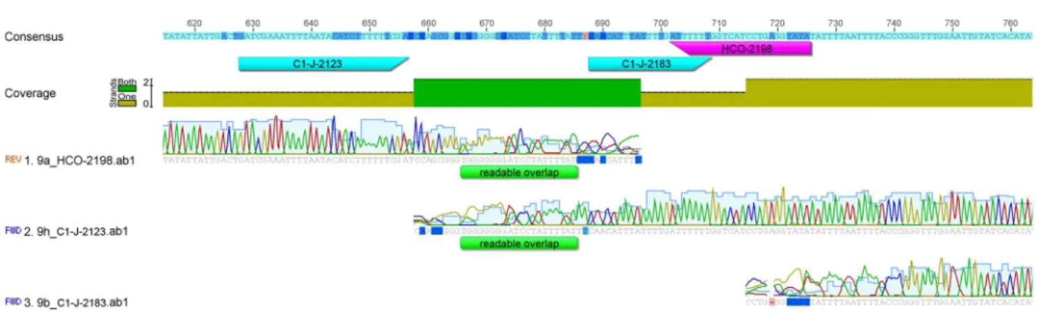
and the C1-J-2183/C1-N-2776 extension [28,30] (Fig. 1). Such indel region, arising through incomplete or poor reads, is artificial due to simple lack of data, and may reduce the accuracy of phylogenetic analyses.
The objective of our study was to explore an optimal strategy for extracting and analyzing arachnid DNA focusing on the barcoding and adjacent cox1regions.
Our work focused on the species richest arachnid lineage, spiders.
Our first goal was to improve an automated DNA extraction protocol. Compared with manual extraction procedures using kits, robotic DNA extraction methods often yield lower quantity of extracted DNA [34]. To maximize its efficiency, we experimentally adjusted an internal robotic DNA extraction program and improved it for acquisition of high concentration of genomic DNA from different quality tissues. Our second goal was to test the performance of commonly used cox1primer combinations and to identify the optimal primer set
over the major phylogenetic lineages of spiders. We screened and tested the high throughput utility with a single PCR program of ten cox1primer pairs. Our third
goal was to develop a new cox1 primer that would produce an indel-free
alignment resulting in more accurate phylogenetic analyses.
Materials and Methods
Specimens and taxonomic coverage
Table 1.Commoncox1primers used in arachnid systematics, and tested in this study.
Name Primer Sequence Reference
LCO1490 GGTCAACAAATCATAAAGATATTGG [33]
HCO2198 TAAACTTCAGGGTGACCAAAAAAT [33]
C1-J-2183 CAACATTTATTTTGATTTTTT [30]
CO1-J-1718 GGAGGATTTGGAAATTGATTAGTTCC [30]
C1-N-2776 GGATAATCAGAATATCGTCGAGG [28]
dgLCO1490 GGTCAACAAATCATAAAGAYATYGG [40]
dgHCO2198 TAAACTTCAGGGTGACCAAARAAYCA [40]
CO1-N-2735 AAAATGTTGAGGGAAAAAATGTTA [41]
Chelicerate_R2 GGATGGCCAAAAAATCAAAATAAATG [42]
CO1-RCF1 GTYTCTTCWATAGTWGAAATRGG [43]
CO1-RCR1 ACAGAAAAYATATGATGRGCYCAYAC [43]
C1-J-2123 GATCGAAATTTTAATACTTCTTTTTTTGA This study
doi:10.1371/journal.pone.0113030.t001
Figure 1. An artificial indel region in the alignment ofcox1sequences between the Folmer region and the C1-J-2183/C1-N-2776 extension.Such indel region arising due to incomplete or poor reads, commonly hampers the accuracy of phylogenetic analyses.
ezlab.zrc-sazu.si/) for every numbered species and the size of tissue samples varied from 0.3 to 3.0 mm3volume of spider’s leg.
Automated DNA extraction
Robotic DNA extraction was done with MagMAX Express Magnetic Particle Processor (Life Technologies). DNA from muscle cells was extracted from fresh tissue or tissue frozen at -80
˚
C after collection and species identification. DNA was extracted using MagMAX DNA Multi-Sample Kit (Life Technologies) bymodifying the manufacturer protocol for manual extraction.
The MagMAX plate was loaded as follows; row A: 80mL of Multisample DNA
Lysis Buffer, 96 mL of Isopropanol, 80 mL of tissue sample in phosphate buffered
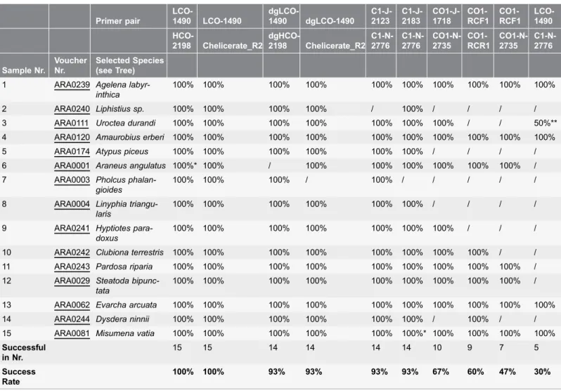
saline (PBS: 137 mM NaCl, 2.7 mM KCl, 8 mM Na2HPO4, and 2 mM KH2PO4, Table 2.The success rate of different primer combinations for the fifteen selected spider species varies from 100% to 30%.
Primer pair LCO-1490 LCO-1490 dgLCO-1490 dgLCO-1490 C1-J-2123 C1-J-2183 CO1-J-1718 CO1-RCF1 CO1-RCF1 LCO-1490 HCO-2198 Chelicerate_R2 dgHCO-2198 Chelicerate_R2 C1-N-2776 C1-N-2776 CO1-N-2735 CO1-RCR1 CO1-N-2735 C1-N-2776 Sample Nr. Voucher Nr. Selected Species (see Tree)
1 ARA0239 Agelena labyr-inthica
100% 100% 100% 100% 100% 100% 100% 100% 100% 100%
2 ARA0240 Liphistius sp. 100% 100% 100% 100% / 100% / / / /
3 ARA0111 Uroctea durandi 100% 100% 100% 100% 100% 100% 100% / / 50%**
4 ARA0120 Amaurobius erberi 100% 100% 100% 100% 100% 100% 100% 100% 100% 100%
5 ARA0174 Atypus piceus 100% 100% 100% 100% 100% 100% / / / /
6 ARA0001 Araneus angulatus 100%* 100% / 100% 100% 100% 100% 100% 100% / 7 ARA0003 Pholcus
phalan-gioides
100% 100% 100% / 100% / / / / /
8 ARA0004 Linyphia triangu-laris
100% 100% 100% 100% 100% 100% / / / /
9 ARA0241 Hyptiotes para-doxus
100% 100% 100% 100% 100% 100% 100% / / /
10 ARA0242 Clubiona terrestris 100% 100% 100% 100% 100% 100% 100% 100% / / 11 ARA0243 Pardosa riparia 100% 100% 100% 100% 100% 100% 100% 100% 100% / 12 ARA0029 Steatoda
bipunc-tata
100% 100% 100% 100% 100% 100% 100% 100% 100% /
13 ARA0062 Evarcha arcuata 100% 100% 100% 100% 100% 100% 100% 100% 100% 100%
14 ARA0244 Dysdera ninnii 100% 100% 100% 100% 100% 100% / 100% / /
15 ARA0081 Misumena vatia 100% 100% 100% 100% 100% 100%* 100% 100% 100% 100%
Successful in Nr.
15 15 14 14 14 14 10 9 7 5
Success Rate
100% 100% 93% 93% 93% 93% 67% 60% 47% 30%
Voucher numbers refer to EZ Lab (http://ezlab.zrc-sazu.si/) cryo-collection.
*excised gel band used for 2nd PCR; **only C1-N-2776 binds, one way sequence obtained.
Figure 2. Fifteen selected spider species (seeTable 2) representing the major phylogenetic lineages on a simplified phylogeny[35].
pH 7.4); into row B: 120 mL of Wash Solution I, into row C, E, F: 120 mL of Wash
Solution II, into row D: 38 mL of nuclease-free water, into row G: 40 mL of Elution
Buffer I. During the run in the MagMAX Express Magnetic Particle Processor the magnetic beads solution (6.4 mL DNA binding beads with 9.6 mL nuclease-free
water) into row A and 2 mL of RNase A into row D were added. In second pause
40 mL of Multisample DNA Lysis Buffer and 48 mL of Isopropanol were added
into row D. During the third pause a step of incubation in thermoblock at 70
˚
C for 5 min was made. After the incubation, 40 mL of Elution Buffer II was addedinto row G (from 70 mL down to 30 mL minimum is allowed) and the run
continued in the instrument. Samples of purified DNA were transferred to cryovials for storage from row G (see Appendix S1).
The additional step of overnight incubation of starting material with Proteinase K was added to the protocol improving extraction efficiency. Differently sized tissue was cut and thoroughly homogenized with a pestle in a tube with 73.6 mL
PK Buffer and 6.4 mL Proteinase K (100 mg/mL), shortly centrifuged and
incubated over night at 55
˚
C on a shaker. All reagents and buffers (with exception of PBS) used for DNA extraction are components of MagMAX DNA Multi-Sample Kit (Life Technologies).During the optimization step with MagMAX DNA Multi-Sample Kit also the comparison with MagMAX Total Nucleic Acid Isolation Kit (Life Technologies) was done (data not shown). After the quantification of extracted nucleic acids with NanoDrop Lite (Thermo Scientific) the amount of extracted DNA was up to 5-fold higher in comparison to sample concentration prepared with MagMAX DNA Multi-Sample Kit. However, better extraction of nucleic acids could also be related to the MagMAX Total Nucleic Acid Isolation Kit specifications where the isolation of all the nucleic acids (ssDNA, dsDNA and RNA) is performed at once. The comparison of material costs showed substantial differences between the kits used in this study, with the MagMAX DNA Multi-Sample Kit being the most economic.
PCR optimization strategy
For PCR amplification reaction we achieved the best results using GoTaq Flexi Polymerase Kit (Promega) and the cycling program forcox1gene. All of the PCR
reaction mixtures had a total volume of 25 mL and 1 mL of DNA template (in the
range between 3.0 to 50 ng) per reaction was generally used. Each reaction included 5.1 mL of Promega’s GoTaq Flexi Buffer, 0.14 mL of GoTaq Flexi
Polymerase, 2.5 mL dNTP’s (2 mM each), 2.3 mL MgCl2 (25 nM), 0.5mL of each
primer (forward and reverse, 20 mM), 0.14mL BSA (10 mg/mL) and 12.8 mL of
sterile distilled water. The thermocycle program for cox1 amplification consisted
PCR amplification verification and sequencing
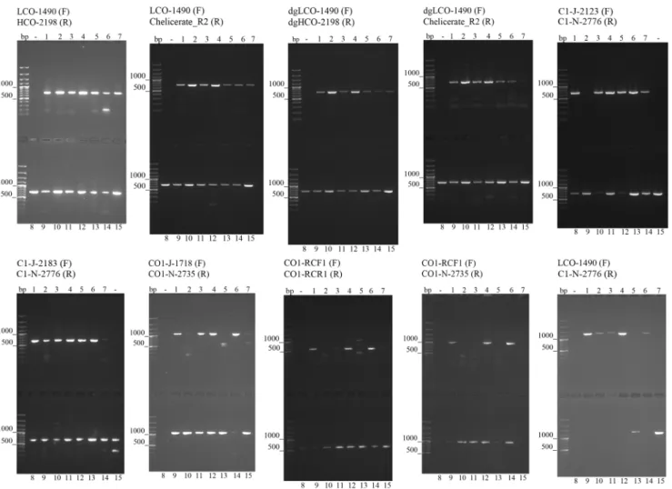
PCR products were stained with Sybr Safe (Invitrogen), separated by standard 1.5% agarose gel using Owl B2 EasyCast Mini Gel Electrophoresis System and visualized by UV gel imager E-box VX2/20LM (Vilber Lourmat). All of the PCRs were repeated three times to verify strong signals obtained in every reaction where specific PCR fragment was generated. Primers (Table 1) were used in PCRs in 10 different combinations: LCO-1490/HCO-2198, LCO-1490/Chelicerate_R2, dgLCO-1490/dgHCO-2198, dgLCO-1490/Chelicerate_R2, C1-J-2123/C1-N-2776, C1-J-2183/C1-N-2776, J-1718/N-2735, LCO-1490/C1-N-2776, CO1-RCF1/CO1-N-2735, CO1-RCF1/CO1-RCR1. Samples were sequenced bidirec-tionally using Standard-Seq method (Macrogen). Sequence data were edited and assembled using Geneious Pro version 5.4 [36] and further handled in Mesquite version 2.74 [37].
Primer design
In order to construct a new primer that would bind upstream of the C1-J-2183 primer binding site, we searched for the most conserved region in that area. We used the NCBI nucleotide search facility within Geneious Pro to gather all cox1
sequences of the order Araneaethat contained the keyword ‘‘BARCODE’’. This
search tagged sequences meeting all CBOL criteria [38]. All sequences longer or shorter than 658 bp were deleted and the remaining 1672 sequences, already aligned, were used for primer design (see Appendix S2). Potential primers were evaluated using the program Primer3 [39].
Results
We optimized and improved the manufacturer’s protocol for extraction of genomic DNA using the MagMAX Express Magnetic Particle Processor, an automated high throughput DNA extraction system. We processed a wide range of spider tissue of different taxonomic affiliation, size and quality, to improve the protocol and increase the efficiency of the procedure. The manufacturer’s protocol only specifies the use of the kit with MagMAXExpress-96 Standard Magnetic Particle Processor or manually. Our procedure describes the use of the MagMAX Express Magnetic Particle Processor in combination with the MagMAX DNA Multi-Sample Kit. Additionally, we optimized the workflow for smaller quantities of starting material and accordingly adjusted and modified the internal program. Changes were made to the volume of reagents used and timing of specific steps (see Appendix S1).
To assess the amplification success of ten primer pairs routinely employed for barcoding cox1gene, we tested fifteen target species throughout the spider
success rate of different primer pairs varied from 30 to 100% (Fig. 3;Table 2). To target solely the short barcoding cox1 region, we recommend using the
combination of Folmer primers (LCO1490/HCO2198) [33].
To maximize sequence data for genetic, genomic, and phylogenetic analyses, however, a longer stretch ofcox1is desired. Existing primers can fail to provide a
continuous stretch if used in a single pair or in combinations of pairs [12,31–32]. The forward primer C1-J-2183, for example, is designed too close to the reverse
Figure 3. Gel images showing different success rates incox1amplification using the ten tested primer pairs. doi:10.1371/journal.pone.0113030.g003
Figure 4. The new primer C1-J-2123 binding site is 60 bp upstream of the C1-J-2183 binding site.
HCO2198 for accurate chromatogram reads, and in practice the resulting interpretations of base pairs result in two partial cox1 sequences (Fig. 1). We
therefore designed, via analysis of the consensus alignment of 1672 arachnidcox1
sequences a new forward DNA primer situated 60 base pairs upstream of the C1-J-2183 binding site. The sequence GATCGAAATTTTAATACTTCTTTTTTTGA was chosen as the most conserved and appropriate for the binding of a new primer, named C1-J-2123 (Fig. 4). Our preliminary tests (data not shown) suggest that this primer will work not only in spiders, but also other arachnids (scorpions, mites and ticks) and other invertebrates (bivalves, gastropods, tunicates and others).
C1-J-2123 performed with the same success rate (93%) as the alternative primer C1-J-2183, amplifying in 14 out of 15 spider species (Fig. 3;Table 2). We recommend using the C1-J-2123/C1-N-2776 primer pair extended in upstream direction, which will allow for full overlap of this extended sequence with that obtained with the standard Folmer primers LCO1490 and HCO2198. Cox1
sequences obtained with the C1-J-2123/C1-N-2776 primer pair can fully sequence both regions (Fig. 5).
Conclusions
This study assessed the usefulness, measured as amplification success, of ten primer pairs routinely employed for targeting the barcoding and other cox1gene
regions in arachnids. Aiming to optimize the efforts in pursuing a longer stretch of cox1 that would maximize the data versus effort ratio for phylogenetic use of
the barcode data, we sought an ideal protocol for automatic, reliable and fast extraction of genomic DNA, and developed a new cox1primer for routine spider
systematic work. Our newly designed cox1 primer C1-J-2123 replaces C1-J-2183
to avoid creating an indel region after the Folmer region. This may be especially useful to obtain more complete cox1 data for phylogenetic analyses.
Figure 5. The newly amplified and elongated C1-J-2123/C1-N-2776 sequence overlaps with the Folmer (LCO1490/HCO2198) sequence.
We also improved the robotic DNA extraction protocol from the manufac-turer’s version, adapting it for spider tissue. We are the first to convey usage and set protocol for DNA extraction with MagMAX Express Magnetic Particle Processor in combination with MagMAX DNA Multi-Sample Kit. Our protocol allows for higher DNA concentration output compared with manual DNA extraction using commercial kits. It is thus suitable for semi-high throughput preparation of arachnid DNA. With the 1.5 hour DNA extraction run, the system can be loaded about 8 times per day providing DNA isolated from 192 samples. Following our protocol, PCR amplification of 96 samples is possible in only two hours using a single PCR program. Our protocol is fast and effective and able to provide up to 1000 amplifications per week. Using an even higher throughput system such as the MagMAX Express-96 Magnetic Particle Processor that processes 96 samples at a time, this time could be further cut in half.
Supporting Information
Appendix S1. Internal Program of MagMAX Express DNA Extraction Robot (Life Technologies) Protocol, modified. See separate file.
doi:10.1371/journal.pone.0113030.s001 (DOCX)
Appendix S2. Final DNA sequence assembly accession information. See separate file.
doi:10.1371/journal.pone.0113030.s002 (DOCX)
Acknowledgments
We thank Ren-Chung Cheng, Gregor Guncˇar, Matjazˇ Gregoricˇ, Simona Kralj-Fisˇer, Klemen Cˇ andek and Tjasˇa Lokovsˇek for their field and lab help, Miquel Arnedo for technical advice, and Ingi Agnarsson and Cor Vink for constructive reviews.
Author Contributions
Conceived and designed the experiments: NV NT MK. Performed the
experiments: NV. Analyzed the data: NV MK. Contributed reagents/materials/ analysis tools: NV NT MK. Wrote the paper: NV NT MK.
References
1. Hebert PDN, Cywinska A, Ball SL, DeWaard JR (2003) Biological identifications through DNA barcodes. Proceedings of the Royal Society of London Series B-Biological Sciences 270: 313–321.
2. Hebert PDN, deWaard JR, Landry JF(2010) DNA barcodes for 1/1000 of the animal kingdom. Biology Letters 6: 359–362.
4. Hebert PDN, Gregory TR(2005) The promise of DNA barcoding for taxonomy. Systematic Biology 54: 852–859.
5. Hebert PDN, Penton EH, Burns JM, Janzen DH, Hallwachs W(2004) Ten species in one: DNA barcoding reveals cryptic species in the neotropical skipper butterfly Astraptes fulgerator. Proceedings of the National Academy of Sciences of the United States of America 101: 14812–14817.
6. Hebert PDN, Ratnasingham S, deWaard JR (2003) Barcoding animal life: cytochrome c oxidase subunit 1 divergences among closely related species. Proceedings of the Royal Society of London Series B-Biological Sciences 270: S96–S99.
7. Hebert PDN, Stoeckle MY, Zemlak TS, Francis CM (2004) Identification of birds through DNA barcodes. Plos Biology 2: 1657–1663.
8. Blagoev GA, Nikolova NI, Sobel CN, Hebert PDN, Adamowicz SJ (2013) Spiders (Araneae) of Churchill, Manitoba: DNA barcodes and morphology reveal high species diversity and new Canadian records. Bmc Ecology 13.
9. Agnarsson I, Gregoric M, Blackledge TA, Kuntner M (2013) The phylogenetic placement of Psechridae within Entelegynae and the convergent origin of orb-like spider webs. Journal of Zoological Systematics and Evolutionary Research 51: 100–106.
10. Arnedo MA, Coddington IA, Agnarsson I, Gillespie RG(2004) From a comb to a tree: phylogenetic relationships of the comb-footed spiders (Araneae, Theridiidae) inferred from nuclear and mitochondrial genes. Molecular Phylogenetics and Evolution 31: 225–245.
11. Bidegaray-Batista L, Arnedo MA (2011) Gone with the plate: the opening of the Western Mediterranean basin drove the diversification of ground-dweller spiders. Bmc Evolutionary Biology 11: 317.
12. Kuntner M, Arnedo MA, Trontelj P, Lokovsˇek T, Agnarsson I (2013) A molecular phylogeny of nephilid spiders: Evolutionary history of a model lineage. Molecular Phylogenetics and Evolution 69: 961–979.
13. Agnarsson I, Maddison WP, Aviles L(2007) The phylogeny of the socialAnelosimusspiders (Araneae: Theridiidae) inferred from six molecular loci and morphology. Molecular Phylogenetics and Evolution 43: 833–851.
14. Agnarsson I, Coddington JA, Kuntner M(2013) Systematics: Progress in the Study of Spider Diversity and Evolution. In:, Penney D, , editor. Spider Research in the 21st Century: Trends and Perspectives. Manchester: Siri Scientific Press. pp. 58–111.
15. Havird JC, Santos SR(2014) Performance of Single and Concatenated Sets of Mitochondrial Genes at Inferring Metazoan Relationships Relative to Full Mitogenome Data. Plos One 9.
16. Gomez-Zurita J, Cardoso A(2014) Systematics of the New Caledonian endemic genus Taophila Heller (Coleoptera: Chrysomelidae, Eumolpinae) combining morphological, molecular and ecological data, with description of two new species. Systematic Entomology 39: 111–126.
17. Brugler MR, Opresko DM, France SC (2013) The evolutionary history of the order Antipatharia (Cnidaria: Anthozoa: Hexacorallia) as inferred from mitochondrial and nuclear DNA: implications for black coral taxonomy and systematics. Zoological Journal of the Linnean Society 169: 312–361.
18. Ahrens D, Fabrizi S, Sipek P, Lago PK (2013) Integrative analysis of DNA phylogeography and morphology of the European rose chafer (Cetonia aurata) to infer species taxonomy and patterns of postglacial colonisation in Europe. Molecular Phylogenetics and Evolution 69: 83–94.
19. Lloyd RE, Foster PG, Guille M, Littlewood DTJ(2012) Next generation sequencing and comparative analyses of Xenopus mitogenomes. Bmc Genomics 13.
20. Horak M, Day MF, Barlow C, Edwards ED, Su YN, et al.(2012) Systematics and biology of the iconic Australian scribbly gum moths Ogmograptis Meyrick (Lepidoptera: Bucculatricidae) and their unique insect-plant interaction. Invertebrate Systematics 26: 357–398.
21. McDonagh LM, Stevens JR (2011) The molecular systematics of blowflies and screwworm flies (Diptera: Calliphoridae) using 28S rRNA, COX1 and EF-1 alpha: insights into the evolution of dipteran parasitism. Parasitology 138: 1760–1777.
23. Cohen BL, Bitner MA, Harper EM, Lee DE, Mutschke E, et al.(2011) Vicariance and convergence in Magellanic and New Zealand long-looped brachiopod clades (Pan-Brachiopoda: Terebratelloidea). Zoological Journal of the Linnean Society 162: 631–645.
24. Gower DJ, San Mauro D, Giri V, Bhatta G, Govindappa V, et al.(2011) Molecular systematics of caeciliid caecilians (Amphibia: Gymnophiona) of the Western Ghats, India. Molecular Phylogenetics and Evolution 59: 698–707.
25. Borrero-Perez GH, Gomez-Zurita J, Gonzalez-Wanguemert M, Marcos C, Perez-Ruzafa A(2010) Molecular systematics of the genus Holothuria in the Mediterranean and Northeastern Atlantic and a molecular clock for the diversification of the Holothuriidae (Echinodermata: Holothuroidea). Molecular Phylogenetics and Evolution 57: 899–906.
26. Timmermans M, Dodsworth S, Culverwell CL, Bocak L, Ahrens D, et al.(2010) Why barcode? High-throughput multiplex sequencing of mitochondrial genomes for molecular systematics. Nucleic Acids Research 38.
27. Mladineo I, Bott NJ, Nowak BF, Block BA(2010) Multilocus phylogenetic analyses reveal that habitat selection drives the speciation of Didymozoidae (Digenea) parasitizing Pacific and Atlantic bluefin tunas. Parasitology 137: 1013–1025.
28. Hedin MC, Maddison WP(2001) A combined molecular approach to phylogeny of the jumping spider subfamily Dendryphantinae (Araneae: Salticidae). Molecular Phylogenetics and Evolution 18: 386–403.
29. Arnedo MA, Coddington J, Agnarsson I, Gillespie RG(2004) From a comb to a tree: phylogenetic relationships of the comb-footed spiders (Araneae, Theridiidae) inferred from nuclear and mitochondrial genes. Molecular Phylogenetics and Evolution 31: 225–245.
30. Simon C, Frati F, Beckenbach A, Crespi B, Liu H, et al.(1994) Evolution, weighting, and phylogenetic utility of mitochondrial gene sequences and a compilation of conserved polymerase chain reaction primers. Annals of the Entomological Society of America 87: 651–701.
31. Kuntner M, Agnarsson I(2011) Phylogeography of a successful aerial disperser: the golden orb spider Nephila on Indian Ocean islands. Bmc Evolutionary Biology 11.
32. Kuntner M, Agnarsson I(2011) Biogeography and diversification of hermit spiders on Indian Ocean islands (Nephilidae: Nephilengys). Molecular Phylogenetics and Evolution 59: 477–488.
33. Folmer O, Black M, Hoeh W, Lutz R, Vrijenhoek R (1994) DNA primers for amplification of mitochondrial cytochrome c oxidase subunit I from diverse metazoan invertebrates. Molecular Marine Biology and Biotechnology 3: 294–299.
34. Francesconi A, Kasai M, Harrington SM, Beveridge MG, Petraitiene R, et al.(2008) Automated and manual methods of DNA extraction for Aspergillus fumigatus and Rhizopus oryzae analyzed by quantitative real-time PCR. Journal of Clinical Microbiology 46: 1978–1984.
35. Coddington JA(2005) Phylogeny and classification of spiders. In:, Ubick D, Paquin P, Cushing PE, Roth V, , editors. Spiders of North America: An Identification Manual: American Arachnological Society. pp. 18–24.
36. Drummond A, Ashton B, Buxton S, Cheung M, Cooper A, et al.(2011) Geneious v 5.4.
37. Maddison WP, Maddison DR(2014) Mesquite: a modular system for evolutionary analysis, version 2.74. Available:http://mesquiteproject.org. Accessed 2014 May 31.
38. Hanner R(2009) Data standards for BARCODE records in INSDC (BRIs), version 2.3. Washington, DC.
39. Koressaar T, Remm M(2007) Enhancements and modifications of primer design program Primer3. Bioinformatics 23: 1289–1291.
40. Meyer CP, Geller JB, Paulay G(2005) Fine scale endemism on coral reefs: Archipelagic differentiation in turbinid gastropods. Evolution 59: 113–125.
41. Lunt DH, Zhang DX, Szymura JM, Hewitt GM (1996) The insect cytochrome oxidase I gene: Evolutionary patterns and conserved primers for phylogenetic studies. Insect Molecular Biology 5: 153– 165.
42. Barrett RDH, Hebert PDN (2005) Identifying spiders through DNA barcodes. Canadian Journal of Zoology-Revue Canadienne De Zoologie 83: 481–491.
![Figure 2. Fifteen selected spider species (see Table 2) representing the major phylogenetic lineages on a simplified phylogeny [35].](https://thumb-eu.123doks.com/thumbv2/123dok_br/18458140.364851/5.918.59.807.104.1004/figure-selected-species-representing-phylogenetic-lineages-simplified-phylogeny.webp)