American Journal of Infectious Diseases 3 (4): 225-229, 2007 ISSN 1553-6203
© 2007 Science Publications
Corresponding Author: Dr. C.N. Ramchand, Kemin Nutritional Technologies (India) Pvt. Ltd, 39, The Trapezium, Nelson Manickam Road, Chennai 600 029, India Tel: +91-44-4220 2842
Extraction of Genomic DNA Using Magnetic Nanoparticles (Fe
3O
4) as a Solid-Phase
Support
1, 2
Z.M. Saiyed and
2C.N. Ramchand
1
Department of Biochemistry, The M.S. University of Baroda, Vadodara, India.
2Kemin Nutritional Technologies (India) Pvt. Ltd., Chennai, India
Abstract: Magnetic separation technology, using magnetic particles, is quick and easy method for sensitive and reliable captures of specific proteins, genetic material and other biomolecules. The current paper describes a universal genomic DNA extraction method optimized in our laboratory using magnetic nanoparticles as a solid phase adsorbent. The yields of the isolated DNA with magnetic method were higher or equivalent to the conventional procedures in all the samples tested. Additionally, the magnetic method takes less than 15 minutes to extract DNA as against several hours taken by conventional protocols. Furthermore, the isolated DNA was found to function satisfactorily in PCR amplification and restriction endonuclease digestion. The developed procedure is simple, quick, cheap, robust and does not require the use of organic solvents or sophisticated equipments; thereby making it more amenable to automation.
Key word: magnetic nanoparticles, mammalian cells, PCR, agarose gel
INTRODUCTION
The sequencing of human genome marked the end of the first phase of genomics revolution. The second phase that has already begun, involves evaluating genetic sequences for genotyping (single-nucleotide polymorphism analysis) and allelic polymorphism analysis. This requires unprecedented capabilities for DNA purification and sequencing. In addition to DNA analysis, the rapidly growing field of molecular diagnostics and molecular phylogeny requires a need for quick, simple, robust, and high throughput procedures for extraction of DNA from diverse organisms and tissues. The process of genomic DNA isolation and purification has evolved considerably within the last decade. The new demands of high throughput facilities have resulted in the development of new technologies for easier and faster DNA processing than ever before.
During recent years, magnetic techniques employing magnetic particles coated with different polymers (e.g., agarose, silica) have been used increasingly for molecular biology applications[1,2].The purification of genomic and plasmid DNA using magnetizable support (beads or matrix) has already been attempted from different biological sources[3-7]. Furthermore, carboxyl coated magnetic particles
(BioMag) have been used as adsorbent for DNA purification under high-salt conditions[8].All the above-mentioned extraction procedures have used coated magnetic particles, which means only the magnetic property of the particles was used to achieve quick separation. However, the use of naked (uncoated) magnetic nanoparticles (Fe3O4) permits to exploit also
its property to reversibly bind DNA under specific conditions. Additionally, there are several inherent advantages of the use of naked particles; where molecules are directly linked to the magnetic support. Due to the absence of polymer coating the particle size is small (≤100 nm), which provides higher surface area (on a weight basis) for the binding of the biomolecules; as well as this allows the particles to a higher magnetic susceptibility to the external magnetic field. Moreover, magnetic nanoparticles can exist as stable colloidal suspension that will not aggregate, allowing for uniform distribution in a reaction mixture. Therefore, we have recently reported the use of naked magnetic nanoparticles as a support for the isolation of genomic DNA from mammalian cells[9]. The applicability of these magnetic nanoparticles for elution of DNA from agarose gels was also successfully demonstrated[10]. The aim of the current work was to produce a universal approach for extraction of genomic DNA using magnetic nanoparticles as a purification medium.
SCI-PUBLICATION
Author Manuscrip
t
SCI-PUBLICATIONS
Author Manuscrip
t
Open
Access
MATERIAL AND METHODS
Preparation of magnetic nanoparticles: Magnetic nanoparticles were prepared by chemical co-precipitation of Fe+2 and Fe+3 ions in an alkaline solution and followed by a treatment under hydrothermal conditions[11]. 100 ml solution of 1M FeSO4.7H2O and 2M FeCl3 (Qualigens Fine chemicals,
Mumbai, India) were thoroughly mixed and added to 8M ammonium hydroxide (Sisco Research Laboratories Limited, Mumbai, India) with constant stirring at 25°C. The particles thus obtained exhibited a strong magnetic response. Impurity ions such as chlorides and sulphates were removed by washing the particles several times with hot distilled water. The yield of precipitated magnetic nanoparticles was determined by removing known aliquots of the suspension and drying to a constant mass in an oven at 60°C. Finally, the magnetic particles were dispersed in TE buffer (10 mM Tris-HCl and 1mM EDTA, pH 8.0) and stored at a stock concentration of 10 mg/ml. The magnetic nanoparticles prepared were stable at room temperature (25 - 30°C) without getting agglomerated. The particles were characterized for size using transmission electron microscopy. The mean particle size determined from transmission electron microscopy was about 40 nm.
Isolation of DNA from mammalian cells using magnetic nanoparticles: In the current study, method for isolation of genomic DNA was initially optimized using whole blood and then investigated for its suitability in cultured cells (HCT116) and tissue (rat liver and brain) homogenates. The sample preparation for DNA extraction involved collection of whole blood in a tube containing ethylenediaminetetraacetic acid (EDTA) as anticoagulant. Cultured cells used in this study were of colon carcinoma cell lines (HCT116), trypsinized, and adjusted to a cell density of 7 x 10⁶ cells/ml with phosphate buffered saline (PBS, pH 7.4). For tissue DNA extraction, a 10% homogenate of rat liver and brain was prepared in 0.32 M buffered sucrose (pH 7.5).
In a typical extraction, a 30 µl of sample (whole blood, cultured cell suspension, or tissue homogenate), 30 µl of 1% (w/v) sodium dodecyl sulphate solution was added. The tube was mixed by gentle inversion for two or three times and incubated at room temperature for a minute. After incubation, to the cell lysate 10 µl of
magnetic nanoparticles were added, followed by addition of 75 µl of binding buffer (1.25 M sodium chloride and 10% PEG 6000). The suspension was mixed by inversion and allowed to stand at room temperature for 3 minutes. The magnetic pellet was immobilized by application of an external magnet and supernatant removed. The magnetic pellet was washed with 70 % ethanol and dried. The pellet was then completely resuspended in 50 µl of TE buffer (10 mM Tris-HCl, 1 mM EDTA, pH 8.0) and magnetic particle bound DNA eluted by incubation at 65 °C with continuous agitation. Finally, the supernatant containing the DNA was transferred to a fresh tube and analyzed with agarose gel electrophoresis.
Elution of DNA from agarose gel using magnetic nanoparticles: For a standardized procedure, DNA samples isolated from human blood were electrophoresed on a 0.8 % agarose gel (Fig. 3a). The separated DNA was visualized on a UV transilluminator and the band of interest was excised with a sterile blade and transferred to a microcentrifuge tube. Four volumes of SSC (0.75 M sodium chloride, 0.0075 M sodium citrate, pH 7.0) buffer was added to the agarose plug and the tubes were incubated at 80°C for 5 min to allow agarose to melt. After incubation, immediately 20 µl of magnetic nanoparticles were added from the stock, followed by addition of 200 µl of binding buffer (1.25 M sodium chloride and 10% polyethylene glycol 6000 [PEG 6000]). The suspension was mixed by gentle inversion and the tube was allowed to stand at room temperature for 5 min. The magnetic pellet was then immobilized by application of an external magnet and the supernatant was discarded. The magnetic pellet was then washed twice with 70 % ethanol and the pellet was dried. The pellet was then resuspended in 30 µl of TE buffer (10 mM Tris-HCl, 1 mM EDTA, pH 8.0) and magnetic particle bound DNA was eluted by incubation at 65 °C with continuous agitation. Finally, the particles were separated magnetically and supernatant containing the DNA was transferred to a fresh tube. The current magnetic method was compared with conventional phenol extraction[12] and glass wool spin column procedures (www.protocol-online.org) that are used for extraction of DNA from agarose gel.
PCR amplification: All PCRs were performed in a 50 µl reaction volume; 25 µl of PCR 2X master mix
Author Manuscrip
(Genetix, USA) was added. Five picomoles each of
primers GAPDH-forward
(5-ACAGTCCATGCCATCACTGCC-3) and GAPDH-reverse (5-GCCTGCTTCACCACCTTCTTG-3) were added per reaction for amplification of an amplicon in the glyceraldehyde 3-phosphate dehydrogenase (GAPDH) gene[13]. 100 ng of template DNA was used in the reaction mixture; PCR was performed on Techne thermal cycler PCR system (Roche, USA). PCR conditions were 4 min at 94°C; 34 cycles of 30 seconds at 94°C, 30 seconds at 61°C and 1 min at 72°C; and 10 min at 72°C. The PCR products were analyzed on 1.5% agarose gel stained with ethidium bromide.
Restriction endonuclease digestion of extraction DNA: A 10-µl volume of the eluted DNA solution was mixed with the manufacturer’s reaction (Bangalore Genei, Bangalore, India) buffer (1 µl), sterile water (quantity sufficient) and incubated with the restriction endonuclease Eco R1/Hind III (1 µl, 10 units) at 37°C for 4 hours. The digestion mixture was analyzed directly after electrophoresis on 0.8% agarose gel.
RESULTS AND DISCUSSION
Isolation of DNA from mammalian cells: We found that using magnetic nanoparticles as solid phase support, yields of recovered genomic DNA was up to 1.2 µg per 30 µl of whole blood or 2.0 x 10⁵
cultured cells, while it was about 1.8 µg and 2 µg per 30 µl of liver and brain tissue homogenate, respectively. The DNA yield in each case was estimated fluorimetrically by Hoechst 33258, or preferably, comparison of intensity of DNA bands in ethidium bromide stained agarose gel. The molecular mass of the isolated DNA was more than 20 kb, as the band migrated at a slower rate than the 23.13-kb band of the λ phage/Hind III molecular mass marker (Fig. 1). Further to this the isolated DNA from all the samples were successfully amplified for a 226-bp glyceraldehyde 3-phosphate dehydrogenase amplicon (Fig. 2a). Also, the restriction digestion of isolated DNA showed no inhibitors of the enzymes present in the sample and the DNA can be used for downstream applications (Fig. 2b). To check the robustness and reproducibility of the method, genomic DNA isolation from blood and cultured cells were performed in ten sets. The recovered genomic DNA from all ten extractions was pooled and
spectrophotometric assessment was performed. The yield of DNA was in accordance to previously estimated and it was about 12 – 15 µg per 0.3 ml of blood or cultured cells. The average OD 260/ OD 280 ratio was 1.8 indicating that the DNA was of good quality with negligible protein contamination. Also, as seen in the gel picture (Fig. 1), no low molecular weight bands or smear was detected, indicating the absence of RNA contamination. This is in agreement with the previous reports; which mention that in presence of high-salt conditions or chaotropes, the adsorption of double stranded DNA onto silica support and magnetite (Fe3O4) is thermodynamically favored, while the
adsorption of proteins and single-stranded RNA is not[14,15]. The current DNA extraction method was tested for its efficiency of DNA extraction and ease of use compared with a commercially available kit. The yield of DNA extracted using magnetic nanoparticles was on average 1.3-fold greater than that using the Qiagen method. Moreover, the magnetic DNA isolation procedure can be carried out in a single microcentrifuge tube per sample, whereas the Qiagen procedure requires a number of tube transfers.
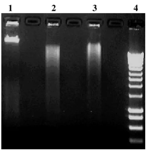
Elution of DNA from agarose gel: The yield of DNA was quantified after electrophoresis in 0.8% agarose gel containing 0.5 µg/ml ethidium bromide, visualized by UV transilluminator using a gel documentation system (UVP Bioimaging Systems, Cambridge, UK). As observed from Fig. 3b, the yield of recovered DNA from agarose gel using magnetic nanoparticles as solid- support was on average 80% (80 ± 5%), whereas that obtained with conventional phenol-extraction and spin-column method was in the range of 50 – 60%. The DNA yield obtained with the current magnetic method (≥ 80%) is similar to that reported by Hawkins and coworkers; where an excess of carboxyl coated magnetic microparticles were used to elute DNA from agarose gels[16]. In order to check the robustness and reproducibility of the current method, DNA fragments were eluted in triplicate and the results were found to be consistent. The yield of recovered DNA of size 23 kb was more than 80% indicating consistency and applicability of this method. The higher yield with the current method is probably attributed to the nano-size of the magnetic particles and optimum conditions for DNA binding[9].
Author Manuscrip
1 2 3 4 5
Fig. 1: Agarose gel electrophoresis of genomic DNA isolated using magnetic nanoparticles. Lanes: 1 = DNA molecular weight marker (λ phage DNA/Hind III digest); 2 = genomic DNA isolated from rat liver homogenate; 3 = genomic DNA isolated from rat brain homogenate; 4 = genomic DNA isolated from cultured cells (HCT116); 5 = genomic DNA isolated from human blood.
The isolated DNA was also checked for its compatibility for restriction endonuclease digestion. The results showed a successful restriction digestion (Fig. 4) indicating absence of enzyme inhibitors and that the purified DNA can be used for downstream applications.
In summary, the developed procedure for DNA extraction has several advantages. It is simple, quick, cheap, robust and does not require use of organic solvents. Also, the method needs only a magnet and a heating block and can be performed in any laboratory without the requirement of sophisticated equipments. The procedure yields enough DNA for 30 PCR reactions from small (30 µl) quantity of biological specimens in less than 15 minutes. Furthermore the whole procedure can be accomplished in a single tube, thereby making it more amenable to automation.
1 2 3 4 5 6 7 8 9 10
(a)
1 2 3 4 5 6
(b)
Fig 2: (a) Agarose gel electrophoresis of 226-bp amplicon of GAPDH gene. Lanes: 1 = DNA molecular weight marker(100 bp ladder); 2-4 = PCR product from genomic DNA isolated from human blood using magnetic particles; 5-6 = PCR product from genomic DNA isolated from cultured cells (HCT116) using magnetic particles; 7-8 = PCR product from genomic DNA isolated from human blood using Qiagen kit; 9-10 = PCR product from genomic DNA isolated from cultured cells (HCT 116) using Qiagen kit.
(b) Restriction endonuclease digestion of extracted DNA. Lanes: 1,2= undigested genomic DNA isolated using magnetic nanoparticles from human blood and cultured cells (HCT116), respectively; 3,5 = HindIII digested genomic DNA isolated from human blood using magnetic nanoparticles; 4,6 = EcoR1 digested genomic DNA isolated from cultured cells using magnetic nanoparticles
Fig 3: (a) Agarose gel electrophoresis of genomic DNA isolated from human blood cells using magnetic nanoparticles. Lanes: 1= DNA molecular weight marker (λ phage DNA/HindIII digest); 2-4= genomic DNA (23 kb) isolated from human blood cells (equal volume was loaded from the tube containing the isolated DNA).
(b) Agarose gel electrophoresis of extracted DNA eluted from agarose gel. Lanes: 1 = DNA molecular weight marker (λ phage DNA/HindIII digest); 2 = DNA eluted with magnetic nanoparticles as solid-phase adsorbent; 3 = DNA eluted using phenol extraction method; Lane 4: DNA eluted by glass wool spin-column method.
Author Manuscrip
1 2 3 4
Fig 4: Restriction analysis of DNA extracted from agarose gel. Lanes: 1 = undigested genomic DNA; 2 = BamH1 digested genomic DNA extracted from agarose gel using magnetic nanoparticles; 3 = Hind III digested genomic DNA extracted from agarose gel using magnetic nanoparticles; 4 = DNA molecular weight marker (1kb DNA ladder).
ACKNOWLEDGEMENT
The financial support to ZMS [F.NO. 9/114(131)/2K2/EMR-I] from Council of Scientific and Industrial Research (CSIR), New Delhi, India is gratefully acknowledged.
REFERENCES
1. Saiyed, Z.M., S.D. Telang and C.N. Ramchand, 2003. Application of magnetic techniques in the field of drug discovery and biomedicine. Biomagn. Res. Technol., 1: e2. 2. Safarik, I. and M. Safarikova, 2002. Magnetic
nanoparticles and biosciences. Monatshefte für Chemie, 133: 737-759.
3. Davies, M.J., D.E. Smethurst, K. Howard, M. Todd, L.M. Higgins and I.J. Bruce, 1997. Improved manufacture and application of an agarose magnetizable solid-phase support. Appl. Biochem. Biotechnol., 68: 95-112. 4. Prodelalova, J., B. Rittich, A. Spanova, K.
Petrova and M.J. Benes, 2004. Isolation of genomic DNA using magnetic cobalt ferrite and silica particles. J. Chromatogr. A, 1056: 43-48.
5. Xie, X., X. Zhang, H. Zhang, D. Chen and W. Fei, 2004. Preparation and application of surface-coated superparamagnetic nanobeads in the isolation of genomic DNA. J. Magn. Magn. Mater., 277: 16-23.
6. Nagy, M., P. Otremba, C. Kruger, S. Bergner-Greiner, P. Anders, B. Henske, M. Prinz and L. Roewer, 2005. Optimization and validation of a fully automated silica-coated magnetic beads purification technology in forensics. Forensic Sci. Int., 152: 13-22.
7. Chiang, C.L., C.S. Sung, T.F. Wu, C.Y. Chen and C.Y. Hsu, 2005. Application of superparamagnetic nanoparticles in purification of plasmid DNA from bacterial cells. J. Chromatogr. B Analyt. Technol. Biomed. Life Sci., 822: 54-60.
8. Hawkins, T.L., T. O’Connor-Morin, A. Roy and C. Santillan, 1994. DNA purification and isolation using a solid-phase. Nucleic Acids Res., 22: 4543-4544.
9. Saiyed, Z.M., C. Bochiwal, H Gorasia, S.D. Telang and C.N. Ramchand, 2006. Application of magnetic particles (Fe3O4) for isolation of
genomic DNA from mammalian cells. Anal. Biochem., 356: 306-308.
10. Saiyed, Z.M., M. Parasramka, S.D. Telang and C.N. Ramchand, 2007. Extraction of DNA from agarose gel using magnetic nanoparticles (magnetite or Fe3O4). Anal. Biochem., 363:
288-290.
11. Mehta, R.V., R.V. Upadhyay, S.W. Charles and C.N. Ramchand, 1997. Direct binding of protein to magnetic particles. Biotechnol. Tech., 11: 493-496.
12. Sambrook, J., E.F. Fritsch and T. Maniatis, 1989. Molecular Cloning: A Laboratory Manual, 2nd ed. Cold Spring Harbor Laboratory Press, Cold Spring Harbor, NY. 13. Deggerdal, A. and F. Larsen, 1997. Rapid
isolation of PCR-ready DNA from blood, bone marrow, and cultured cells, based on paramagnetic beads. BioTechniques, 22: 554-557.
14. Davies, M.J., J.I. Taylor, N. Sachsinger and I.J. Bruce, 1998. Isolation of plasmid DNA using magnetite as a solid phase adsorbent. Anal. Biochem., 262: 92-94.
15. Taylor, J.I., C.D. Hurst, M.J. Davies, N. Sachsinger and I.J. Bruce, 2000. Application of magnetite and silica-magnetite composites to the isolation of genomic DNA. J. Chromatogr. A, 890: 159-166.
16. Hawkins, T., 1998. DNA purification and isolation using magnetic particles, United States Patent Application No. 5705628.