Emerging Novel ECSA Genotype of Chikungunya Virus in
Aedes aegypti
Ankita Agarwal1, Paban Kumar Dash1*, Anil Kumar Singh2, Shashi Sharma1, Natarajan Gopalan2¤, Putcha Venkata Lakshmana Rao1¤, Man Mohan Parida1, Paul Reiter3*
1Division of Virology, Defence R and D Establishment, Gwalior, Madhya Pradesh, India,2Division of Vector Management, Defence R and D Establishment, Gwalior, Madhya Pradesh, India,3Insects and Infectious Disease Unit, Institut Pasteur, Paris, France
Abstract
Background: Chikungunya virus (CHIKV) has emerged as one of the most important arboviruses of public health significance in the past decade. The virus is mainly maintained through human-mosquito-human cycle. Other routes of transmission and the mechanism of maintenance of the virus in nature are not clearly known. Vertical transmission may be a mechanism of sustaining the virus during inter-epidemic periods. Laboratory experiments were conducted to determine whetherAedes aegypti, a principal vector, is capable of vertically transmitting CHIKV or not.
Methodology/Principal Findings:FemaleAe. aegyptiwere orally infected with a novel ECSA genotype of CHIKV in the 2nd gonotrophic cycle. On day 10 post infection, a non-infectious blood meal was provided to obtain another cycle of eggs. Larvae and adults developed from the eggs obtained following both infectious and non-infectious blood meal were tested for the presence of CHIKV specific RNA through real time RT-PCR. The results revealed that the larvae and adults developed from eggs derived from the infectious blood meal (2ndgonotrophic cycle) were negative for CHIKV RNA. However, the larvae and adults developed after subsequent non-infectious blood meal (3rd gonotrophic cycle) were positive with minimum filial infection rates of 28.2 (1:35.5) and 20.2 (1:49.5) respectively.
Conclusion/Significance: This study is the first to confirm experimental vertical transmission of emerging novel ECSA genotype of CHIKV inAe. aegyptifrom India, indicating the possibilities of occurrence of this phenomenon in nature. This evidence may have important consequence for survival of CHIKV during adverse climatic conditions and inter-epidemic periods.
Citation:Agarwal A, Dash PK, Singh AK, Sharma S, Gopalan N, et al. (2014) Evidence of Experimental Vertical Transmission of Emerging Novel ECSA Genotype of Chikungunya Virus inAedes aegypti. PLoS Negl Trop Dis 8(7): e2990. doi:10.1371/journal.pntd.0002990
Editor:Michael J. Turell, United States Army Medical Research Institute of Infectious Diseases, United States of America
ReceivedDecember 30, 2013;AcceptedMay 19, 2014;PublishedJuly 31, 2014
Copyright:ß2014 Agarwal et al. This is an open-access article distributed under the terms of the Creative Commons Attribution License, which permits unrestricted use, distribution, and reproduction in any medium, provided the original author and source are credited.
Funding:This work was funded by Defence Research Development Organization, Ministry of Defence, Govt of India. The authors are thankful to the Director, Defence Research Development Establishment (DRDE), Gwalior for his keen interest and support in this study. AA thanks Department of Biotechnology (DBT), Govt. of India for providing Fellowship and Contingency grants. The funders had no role in study design, data collection and analysis, decision to publish, or preparation of the manuscript.
Competing Interests:The authors have declared that no competing interests exist.
* Email: pabandash@drde.drdo.in (PKD); paul.reiter@pasteur.fr (PR)
¤ Current address: DRDO-Bharathiar University Center for Life Sciences (DRDO – BU CLS), Bharathiar University Campus, Coimbatore, India
Introduction
Chikungunya virus (CHIKV) (genusAlphavirus, family Toga-viridae) is a mosquito-borne pathogen, native to Africa that is transmitted between non human primates, mainly by forest dwellingAedesspecies. The virus is also widespread as an urban infection throughout the old world tropics and subtropics, transmitted by two species of mosquito- Aedes aegypti and Ae. albopictus, both closely associated with the human peridomestic environment [1]. In Asia, theAe. aegyptimosquitoes are primarily responsible for the maintenance of urban cycle, while in Africa, CHIKV transmission involves a sylvatic cycle, primarily withAe. furcifer and Ae. africanusmosquitoes [2]. Autochthonous cases have also occurred in Europe, most notably in 2007 in an epidemic in northeast Italy that affected nearly 300 people [3]. In
this case the vector wasAe. albopictus, an invasive species that is rapidly expanding its distribution in Europe and is already present in at least 27 countries.
encodes the polyprotein precursor of the structural proteins (C, E1, E2) [5].
Vertical transmission is the passage of virus between generations via the egg stage. Virus that infects the ovaries must persist through the larval instars, survive histolysis in the pupal instar and continue through to the adult stage [6]. Vertical transmission is considered to be a primary means by which some arboviruses are maintained during adverse environmental conditions. During this period, the arthropod hosts are either inactive or unable to survive, thus acting as a mechanism for virus persistence in environments where amplifying hosts are temporarily absent or immune. Because theAedeseggs are desiccation resistant, these can survive for longer durations, leading to the possibility of persistence of CHIKV in the eggs [7]. The mechanisms responsible for prevalence of CHIKV during unfavourable periods, especially during winter seasons are unknown. So, vertical transmission is considered as an unresolved issue that has important bearing on the persistence of virus in periods when horizontal transmission is low or non-existent.
Low rates of vertical transmission of the three main groups of mosquito-borne arboviruses- flaviviruses, alphaviruses, and bunyaviruses have been demonstrated in the field [8–10]. The existence of vertical transmission has also been demonstrated experimentally in these three groups. [11–14]. Among the alphaviruses, Ross River virus, Sindbis virus, western equine encephalomyelitis virus, and CHIKV have been isolated from adult Aedes species reared from larvae collected from natural habitats, confirming existence of natural vertical transmission [15– 20]. Lindsay and coworkers isolated Ross River virus from wild-caught male Aedes mosquitoes, further providing evidence of natural vertical transmission [21]. However, to the best of our knowledge, there is no evidence of experimental vertical trans-mission among alphaviruses other than some conflicting reports in CHIKV [13,22,23].
In reviewing the literature on laboratory infections we noted that in nearly all studies, the infective blood meal was given to nulliparous mosquitoes [11,12] and detection of virus was limited to the offspring of the first gonotrophic cycle, whereas, when
studies continued through subsequent cycles, rates of vertical transmission were found much higher [11,24,25]. We speculated that the difference could be attributable to two factors:(i) the first batch of eggs is laid several days before virus has begun rapid replication after passing via the gut wall into the hemolymph, and/ or (ii) the enormous increase in the volume of the ovaries during oogenesis might increase its permeability to virus. We explored these possibilities by investigating the occurrence of vertical transmission in experimentally infectedAe. aegypti, the principal vector of CHIKV.
Materials and Methods
Viruses and mosquitoes
CHIKV belonging to novel Indian Ocean lineage (IOL) of ECSA genotype, obtained from an epidemic in India in 2006 (DRDE06) (GenBank Accession No. EF210157). Initially it was isolated in BHK-21 cells and subsequently passaged in C6/36 cells. The virus is maintained at Virology Division, DRDE, Gwalior. In the present study, it was used at passage level 10 in C6/36 cells. Titre was found to be 108PFU/ml through plaque assay [26] in Vero cells (African green monkey kidney cells). The virus was aliquoted and stored in280uC until use.
Ae. aegypti used in this study, were collected from Gwalior district, Madhya Pradesh, India in 2005 and maintained as laboratory colony in Vector Management Division, Defence Research and Development Establishment (DRDE), Gwalior at 2762uC with 70% relative humidity and 14:10 light:dark photo period. Adult mosquitoes were provided with 10% sucrose solution soaked in cotton pads and larvae were provided with 2–3 yeast tablets per day in a pan containing tap water.
Oral infection of mosquitoes
The female Ae. aegypti mosquitoes were provided with three serial blood meals during this experiment to investigate vertical transmission. Schematic representation of the experimental design is shown in Fig. 1.
I Non infectious blood meal. First, a non infectious blood meal (only rabbit blood) was given to 4–5 days old female Ae. aegypti. Fully engorged females were separated and allowed to lay eggs. The eggs thus obtained were not utilized for this experiment. II Infectious blood meal. Following oviposition from the 1st non infectious blood meal, these Ae. aegypti were starved for 24 hours before providing infectious blood meal in a BSL-3 laboratory. Around 50 of these mosquitoes kept in plastic boxes with mesh covers, were allowed to feed for 45 minutes through a hemotek feeding membrane (Discovery workshops, Accrinton, UK) covering the base of a glass feeder containing the blood-virus mixture. The temperature of the blood meal was maintained at 37uC through a water circulator. The infectious blood meal was prepared by adding 1 ml of viral suspension (108PFU/ml) in 2 ml of washed rabbit erythrocytes to a final concentration of 3.36107PFU/ml. ATP was added to a final concentration of 5 mM as a phagostimulant. After 45 minutes of feeding, mosquitoes were cold anaesthetized and sorted on ice. 20 fully engorged females were transferred to each small cardboard container and maintained with 10% sucrose for 9 days. The eggs (obtained within 3–4 days) following this infectious blood meal were further processed.
III Non infectious blood meal. On day 10 post infection, the mosquitoes were provided with a non-infectious blood meal (only rabbit blood). Fully engorged females were separated and kept individually in conical tubes (50 ml Falcon tubes) containing moistened filter paper and cotton at the bottom. The eggs laid were then further processed.
Author Summary
Although vertical transmission of arboviruses has been recognized for nearly a century, rates of transmission in laboratory experiments are low and their significance in terms of survival of virus during periods of low transmis-sion appears debatable. Recently, major urban outbreaks of chikungunya have been recorded in many parts of Asia, Africa, and Europe. The occurrence of random sporadic cases of the disease in years following a major outbreak prompted us to investigate whether these might be attributable to survival of the virus by vertical transmission. Our experiments were designed to test two hypotheses: (1) The development of an egg-batch derived from an infectious blood meal is too rapid for the infection to reach ovaries; (2) The enormous distension of the membrane enveloping ovaries and ovarioles following oviposition, might facilitate virus penetration. We conclude that after the infected blood meal, oogenesis and oviposition were complete before virus had disseminated to infect the ovaries. Because similar experiments with infection in first gonotrophic cycle did not lead to infected progenies, it is presumed that expanded parous ovaries might support efficient infection. Therefore, it may be concluded that vertical transmission is a more common phenomena in mosquitoes during subsequent gonotrophic cycles follow-ing arboviral infection.
Testing of parental females for disseminated infection At day 14 post infection, 10Ae. aegyptiparental females (after laying eggs from 3rdgonotrophic cycle) were randomly selected and their midgut, legs & wings were tested for the presence of CHIKV to analyze infection and dissemination status.
Processing of larvae and adults
The eggs obtained after infectious blood meal and second non-infectious blood meal were reared under standard laboratory conditions i.e. 2762uC at 70% relative humidity. The 4th instar larvae and 4–5 days old adults were screened for the presence
of virus. The larvae and adults were processed in pools (#20/ pool).
Extraction of RNA
Each pool of adults and larvae were homogenized in 2 ml tubes with 800ml of Eagles Minimum Essential Medium (EMEM) (Sigma, St. Louis, USA) and stainless steel beads in Tissuelyser LT (Qiagen, Hilden, Germany). The homogenate was clarified by centrifugation at 45006gfor 10 minutes. 140ml of supernatant was used to extract RNA using QIAamp viral RNA mini kit (Qiagen, Hilden, Germany) according to the manufacturer’s
protocol. The RNA was finally eluted in 50mL elution buffer and stored at280uC until use.
CHIKV specific SYBR Green-I based Real time RT-PCR CHIKV specific SYBR Green I based one step real time quantitative RT-PCR targeting to E1 gene was performed to screen the presence of CHIKV RNA in test samples [27]. Briefly, the quantitative RT-PCR was carried out using SS III Platinum one step qRT-PCR kit (Invitrogen, Carlsbad, USA) in Mx3005P system (Stratagene, La Jolla, USA). Samples were assayed in a 25mL reaction volume containing 12.5mL of 26master mix, 0.125mL (0.25mmol) each of forward and reverse primer, 0.25mL of enzyme mix comprising of Taq DNA polymerase and MMLV Reverse transcriptase, 9.5mL of nuclease free water and 2.5mL of RNA. The thermal profile comprised of 30 min of reverse transcription at 50uC, 10 min of polymerase activation at 95uC, followed by 40 cycles of PCR at 95uC for 30 s, 55uC for 60 s, and 72uC for 30 s. Following amplification, a melting curve analysis was performed with the melting curve analysis software of the Mx3005P according to the instructions of manufacturer. Positive and negative template control was also included along side in all experiments.
Statistical analysis
CHIKV RNA mean titre in larvae and adults were analyzed. Comparison between two groups was performed by 2 tailed unpaired t-test using GraphPad Prism software (Version 6.04).
Results
Detection of CHIKV in parental females
CHIKV RNA was detected in body, legs & wings of all the 10 randomly selected female Ae. aegypti that were provided with an infectious blood meal. This indicates 100% infection and dissemination of virus at day 14 post infection. The mean titre of CHIKV RNA in midgut, legs & wings of femaleAe. aegyptiwas found to be 106.46101.4and 106.06101.0per reaction respectively.
Vertical transmission rates in larvae and adults
The total number of larvae and adults obtained after infectious blood meal were 230 and 485 respectively. All these larvae and adults were found to be negative for CHIKV. The total number of larvae and adults obtained after second non infectious blood meal were 284 and 693 respectively. These were divided and processed in pools (#20/pool). Out of a total 30 pools of larvae, 8 pools were found positive. Out of 33 pools of adults, 14 were found positive. Minimum infection Rate (MIR) was calculated by the following formula: No. of positive pools/No. of individuals tested X 1000 [25]. The minimum infection rates achieved for larvae and adults were 28.2 and 20.2 respectively. MIR is also expressed as ratio i.e. Proportion of positive mosquitoes by uninfected mosquitoes. Thus
the ratio was found to be 1:35.5 for larvae and 1:49.5 for adults [25] (Table 1).
The results indicated above are cumulative of three experiments. Because the feeding efficiency was low and fewer mosquitoes underwent oviposition with successive gonotrophic cycles, fewer larvae and adults were obtained.
Quantification of CHIKV in larvae and adults
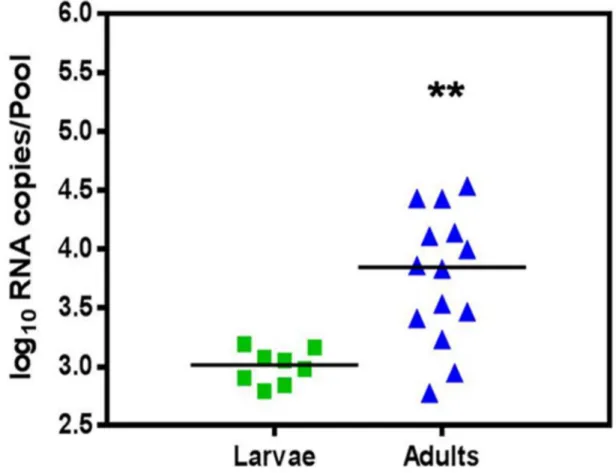
The CHIKV RNA titre as determined by real time RT-PCR in larvae was 102.8to 103.2(103.06100.1). The CHIKV RNA titre in adults was 102.8 to 104.5 (103.86100.6) (Fig. 2). Significant difference was observed between the two groups (p = 0.0012) with t = 3.788 and df = 20. The result indicated the amplification of virus following transition from larvae to adult stage.
Discussion
Since the emergence of the novel ECSA genotype of CHIKV in 2005, widespread outbreaks have been reported in many parts of Africa, Asia and Europe. Intense speculation has been generated regarding different transmission pattern of this emerging virus. So far horizontal transmission is the only method clearly linked to transmission of CHIKV. However, natural vertical transmission of CHIKV has been recently reported from India [17], Reunion Island [18], Thailand [19] and Madagascar [20]. This mode of transmission was recorded in bothAe. albopictusand Ae. aegypti
mosquitoes. In contrast, the vertical transmission experiments in laboratory have generated conflicting reports [13,22,23]. Though a very high rate of vertical transmission (18–62%) was recorded among different species ofAedes mosquito in one experimental study [13], two other studies failed to document existence of vertical transmission [22,23]. A number of variable factors including the viral assay methods, mosquito species, viral isolate, day of sampling and blood meal feeding might have contributed to these conflicting results. Thus, the present study was carried out to investigate the existence of vertical transmission of novel IOL lineage of ECSA genotype of CHIKV inAe. aegyptifrom India, because it is widely reported to be the major Aedes species in northern India [28].
Our earlier experiment using the same CHIKV isolate andAe. aegypti, revealed that the viral dissemination started from day 3 post infection onwards and reached its peak on day 10 post infection [27]. Other replication kinetics experiments of CHIKV inAe. aegypticlearly revealed high viral dissemination from day 8–12 post infection [29]. These observations guided us to provide a non infectious blood meal at day 10 post infection to maximize infected eggs. The infectivity status of the parental female Ae. aegypti on day 14 post infection was confirmed through the detection of CHIKV RNA in the midgut, indicating successful CHIKV replication following oral infection. Further, the viral RNA titers in legs and wings of parental females also suggested efficient dissemination of virus within the mosquito. This was
Table 1.Rate of vertical transmission of Chikungunya virus in 3rdgonotrophic cycle.
Stage Examined Number Analyzed Positive pools/tested pools MIR Ratio
Larvae 284 8/30 28.2 1:35.5
Adults 693 14/33 20.2 1:49.5
Total 977 22/63 22.5 1:44.4
Ratio = Proportion of positive mosquitoes by uninfected mosquitoes. MIR (Minimum infection rate) expressed as Ratio.
doi:10.1371/journal.pntd.0002990.t001
considered as an evidence of dissemination of CHIKV to various secondary organs, presumably ovaries also. In our experiment, larvae and adults developed directly from the eggs after infectious blood meal were found negative for CHIKV RNA. This was primarily due to shorter gonotrophic cycle, where the eggs were laid within 2–3 days of ingestion of infectious blood meal, prior to the adequate dissemination of the virus to ovaries and oviduct. However, larvae and adults developed after the subsequent non infectious blood meal (3rdgonotrophic cycle) were found positive for CHIKV RNA, suggesting an adequate incubation period is required for viral dissemination to the ovaries. Considerable amount of viral particles was earlier demonstrated in the ovaries on 6th day post infection, indicating possibility of vertical transmission of CHIKV [30]. The result obtained by Anderson et al. also suggested the importance of extrinsic incubation period on vertical transmission. In their experiment, F1 progeny developed from eggs laid after 1st infectious blood meal were negative for West Nile virus, whereas progeny obtained after subsequent blood meals were positive [31]. Similar observations were also reported for LaCrosse virus [32], Kunjin virus [33] and yellow fever virus [34].
Comparison with recent laboratory vertical transmission results, where progenies were tested during 1stand 2ndgonotrophic cycle following infectious blood meal of ECSA genotype of CHIKV revealed negative [23] to very low positive results (0.43%) [35]. Similar results were also reported with Asian genotype CHIKV and Indian Aedes mosquitoes in consecutive gonotrophic cycles [22]. Due to these negative results, we tried to evaluate the effect of
infection in 2ndgonotrophic cycle and continued the experiments till 3rdgonotrophic cycle, which resulted in high positivity. The infection of expanded parous ovaries with massive stretching of peritrophic membrane might have facilitated better viral infection of the oocytes. In contrast, Vazeille and co-authors [23] offered a non infectious blood meal prior to an infected one, however, the evaluation of progenies were only restricted to 2nd gonotrophic cycle, that failed to document evidence of vertical transmission. The follow up study to the next gonotrophic cycle, which was not performed, might have contributed to the negative result in their study.
The minimum infection rates achieved for larvae and adults were 28.2 (1:35.5) and 20.2 (1:49.5) respectively. Such high MIR in vertical transmission experiments was also described earlier in Japanese encephalitis and West Nile viruses [11,12,36]. High MIR for West Nile virus (up to 31.1) was observed among male
Culexmosquitoes collected in Connecticut, New Haven, USA [9]. These high MIR rates are likely to be indicator of potential outbreaks.
The demonstration of high viral titer of CHIKV in midgut, legs and wings of IndianAe. aegypti, implicate this species as a major vector of CHIKV (IOL of ECSA genotype). Further, vertical transmission is a plausible survival mechanism for the virus, enabling the virus to be endemic during inter-epidemic periods. In view of the increasing reports regarding identification of natural vertical transmission of CHIKV from different continents, the conflicting results from artificial vertical transmission experiments may be laboratory artifacts.
Figure 2. Vertical transmission of Chikungunya virus inAe. aegyptias determined by measuring log10RNA copies/pool of larvae and adults by real time RT-PCR.Horizontal line represent mean value for each group. Asterisks are indicating significant differences between the two groups (**p,0.01).
Conclusion
The identification of vertical transmission of CHIKV in both natural and experimental settings, confirms the existence of this transmission pattern. The present study indicated that vertical transmission is a more common phenomena in mosquitoes during subsequent gonotrophic cycles following an arboviral infection. In view of the desiccation resistance nature of Aedes eggs, vertical transmission is likely to facilitate the persistent survival of virus during unfavourable inter-epidemic periods. This survival of virus
has immense epidemiological implication further enhancing the risk of potential future outbreaks.
Author Contributions
Conceived and designed the experiments: PKD PR. Performed the experiments: AA PKD AKS SS. Analyzed the data: PKD AA NG PVLR MMP PR. Contributed reagents/materials/analysis tools: NG PVLR MMP. Wrote the paper: AA PKD PR.
References
1. Jupp PG, McIntosh BM, (1988) Chikungunya virus disease, in: Monath, T.P., (Ed.), The arboviruses: epidemiology and ecology vol. II. CRC Press, Boca Raton, FL, pp. 137–157.
2. Powers AM, Brault AC, Tesh RB, Weaver SC (2000) Re-emergence of Chikungunya and O’nyong-nyong viruses: Evidence for distinct geographical lineages and distant evolutionary relationships. J Gen Virol 81: 471–479. 3. Rezza G, Nicoletti L, Angelini R, Romi R, Finarelli AC, et al. (2007) Infection
with Chikungunya virus in Italy: An outbreak a temperate region. Lancet 370: 1840–1846.
4. Lakshmi V, Neeraja M, Subbalaxmi MV, Parida MM, Dash PK, et al. (2008) Clinical features and molecular diagnosis of Chikungunya fever from South India. Clin Infect Dis 46: 1436–1442.
5. Chevillon C, Briant L, Renaud F, Devaux C (2008) The Chikungunya threat: an ecological and evolutionary perspective. Trends Microbiol 16: 80–88. 6. Hardy JL, Houk EJ, Kramer LD, and Reeves WC (1983) Intrinsic factors
affecting vector competence of mosquitoes for Arboviruses. Annu Rev Entomol 28: 229–262.
7. Miller BR, Nasci RS, Godsey MS, Savage HM, Lutwama JJ, et al. (2000) First field evidence for natural vertical transmission of West Nile virus inCulex univittatuscomplex mosquitoes from Rift Valley Province, Kenya. Am J Trop Med Hyg 62: 240–246.
8. Martins VE, Alencar CH, Kamimura MT, de Carvalho Araujo FM, De Simone SG, et al. (2012) Occurrence of natural vertical transmission of Dengue-2 and Dengue-3 Viruses inAedes aegyptiandAedes albopictusin Fortaleza, Ceara, Brazil. PLoS One 7: e41386.
9. Anderson JF, Andreadis TG, Main AJ, Ferrandino FJ, Vossbrinck CR (2006) West Nile virus from female and male mosquitoes (Diptera: Culicidae) in subterranean, ground, and canopy habitats in Connecticut. J Med Entomol 43: 1010–1019.
10. Fontenille D, Diallo M, Mondo M, Ndiaye M, Thonnon J (1997) First evidence of natural vertical transmission of yellow fever virus inAedes aegypti, its epidemic vector. Trans R Soc Trop Med Hyg 91: 533–535.
11. Rosen L, Lien JC, Shroyer DA, Baker RH, Lu LC (1989) Experimental vertical transmission of Japanese encephalitis virus byCulex tritaeniorhynchusand other mosquitoes. Am J Trop Med Hyg 40: 548–556.
12. Baqar S, Hayes CG, Murphy JR, Watts DM (1993) Vertical transmission of West Nile virus byCulexandAedesspecies of mosquitoes. Am J Trop Med Hyg 48: 757–762.
13. Hailin Z, Yunzhi Z, ZhuqingM (1993) Transovarial transmission of Chikungu-nya virus inAedes albopictusandAedes aegyptimosquitoes. Chin J Virol 9: 222– 227.
14. Tesh RB, Gubler DJ (1975) Laboratory studies of transovarial transmission of La Crosse and other arboviruses byAedes albopictusandCulex fatigans. Am J Trop Med Hyg 24: 876–880.
15. Dhileepan K, Azuolas JK, Gibson CA (1996) Evidence of vertical transmission of Ross River and Sindbis viruses (Togaviridae: Alphavirus) by mosquitoes (Diptera: Culicidae) in southeastern Australia. J Med Entomol 33: 180–182. 16. Fulhorst CF, Hardy JL, Eldridge BF, Presser SB, Reeves WC (1994) Natural
vertical transmission of western equine encephalomyelitis virus in mosquitoes. Science 263: 676–678.
17. Niyas KP, Abraham R, Unnikrishnan RN, Mathew T, Nair S, et al. (2010) Molecular characterization of Chikungunya virus isolates from clinical samples and adultAedes albopictusmosquitoes emerged from larvae from Kerala, South India. Virol J 7: 189.
18. Delatte H, Paupy C, Dehecq JS, Thiria J, Failloux AB, et al. (2008)Aedes albopictus, vector of chikungunya and dengue viruses in Reunion Island: biology and control. Parasite 15: 3–13.
19. Thavara U, Tawatsin A, Pengsakul T, Bhakdeenuan P, Chanama S, et al. (2009) Outbreak of Chikungunya fever in Thailand and virus detection in field population of vector mosquitoes, Aedes aegypti (L.) and Aedes albopictus Skuse (Diptera: Culicidae). Southeast Asian J Trop Med Public Health 40: 951– 962.
20. Ratsitorahina M, Harisoa J, Ratovonjato J, Biacabe S, Reynes JM, et al. (2008) Outbreak of dengue and Chikungunya fevers, Toamasina, Madagascar, 2006. Emerg Infect Dis 14: 1135–1137.
21. Lindsay MD, Broom AK, Wright AE, Johansen CA, Mackenzie JS (1993) Ross River virus isolations from mosquitoes in arid regions of Western Australia: implication of vertical transmission as a means of persistence of the virus. Am J Trop Med Hyg 49: 686–696.
22. Mourya DT (1987) Absence of transovarial transmission of Chikungunya virus inAedes aegypti&Ae. albopictusmosquitoes. Indian J Med Res 85: 593–595. 23. Vazeille M, Mousson L, Failloux AB (2009) Failure to demonstrate experimental
vertical transmission of the epidemic strain of Chikungunya virus in Aedes albopictusfrom La Reunion Island, Indian Ocean. Mem Inst Oswaldo Cruz 104: 632–635.
24. Flores FS, Diaz LA, Batallan GP, Almiron WR, Contigiani MS (2010) Vertical transmission of St. Louis encephalitis virus inCulex quinquefasciatus(Diptera: Culicidae) in Cordoba, Argentina. Vector Borne Zoonotic Dis 10: 999–1002. 25. Rosen L (1988) Further observations on the mechanism of vertical transmission
of flaviviruses byAedesmosquitoes. Am J Trop Med Hyg 39: 123–126. 26. Flint SJ, Enquist LW, Racaniello VR, Skalka AM (2004) Principles of Virology:
Molecular Biology, Pathogenesis and Control of Animal viruses. 2nd edition.. Washington, D.C: ASM press. 31–32 p.
27. Agarwal A, Singh AK, Sharma S, Soni M, Thakur AK, et al. (2013) Application of Real-time RT-PCR in vector surveillance and assessment of replication kinetics of an emerging Novel ECSA genotype of Chikungunya virus inAedes aegypti. J Virol Methods 193: 419–425.
28. Angel B, Joshi V (2009) Distribution of dengue virus types inAedes aegyptiin dengue endemic districts of Rajasthan, India. Indian J Med Res 129: 665–668. 29. Dubrulle M, Mousson L, Moutailler S, Vazeille M, Failloux AB (2009) Chikungunya virus andAedesmosquitoes: Saliva is infectious as soon as two days after oral infection. PLoS One 4: e5895.
30. Vazeille M, Moutailler S, Coudrier D, Rousseaux C, Khun H, et al. (2007) Two Chikungunya isolates from the outbreak of La Reunion (Indian Ocean) exhibit different patterns of infection in the mosquito,Aedes albopictus. PLoS One 2: e1168.
31. Anderson JF, Main AJ, Delroux K, Fikrig E (2008) Extrinsic incubation periods for horizontal and vertical transmission of West Nile virus byCulex pipiens pipiens(Diptera: Culicidae). J Med Entomol 45: 445–451.
32. Miller BR, DeFoliart GR, Yuill TM (1979)Aedes triseriatusand La Crosse virus: lack of infection in eggs of the first ovarian cycle following oral infection of females. Am J Trop Med Hyg 28: 897–901.
33. Tesh RB (1980) Experimental studies on the transovarial transmission of Kunjin and San Angelo viruses in mosquitoes. Am J Trop Med Hyg 29: 657–666. 34. Diallo M1, Thonnon J, Fontenille D (2000) Vertical transmission of the yellow
fever virus byAedes aegypti(Diptera, Culicidae): dynamics of infection in F1 adult progeny of orally infected females. Am J Trop Med Hyg 62: 151–156. 35. Bellini R, Medici A, Calzolari M, Bonilauri P, Cavrini F, et al. (2012) Impact of
Chikungunya virus on Aedes albopictus females and possibility of vertical transmission using the actors of the 2007 outbreak in Italy. PLoS One 7: e28360 36. Goddard LB, Roth AE, Reisen WK, Scott TW (2003) Vertical transmission of West Nile virus by three CaliforniaCulex(Diptera: Culicidae) species. J Med Entomol 40: 743–746.