Prevalence of hepatitis C virus infection and HCV genotypes
among hemophiliacs in the State of Bahia, Northeastern Brazil:
analysis of serological and virological parameters
Prevalência da infecção pelo vírus da hepatite C e genótipos
entre hemofílicos no Estado da Bahia, nordeste do Brasil:
análise de parâmetros sorológicos e virológicos
Luciano Kalabric Silva
1, Maria Betânia Souza da Silva
1, Gisele Barreto Lopes
1,
Itatiana Ferreira Rodart
1, Fernando Quadros Costa
1, Nelma P. Santana
2, Raymundo Paraná
3,
Aurelino Santana
4and Mitermayer Galvão dos Reis
1, 3, 4ABSTRACT
The objective of the present study was to analyze HCV serological and virological parameters from hemophiliacs in the State of
Bahia. Anti-HCV was investigated by ELISA in a cohort of 268 hemophiliacs A/B who were followed-up in a reference unit for
hemotherapy in the State of Bahia. HCV viremia and genotypes were also determined from a subset of 66 anti-HCV seropositive
hemophiliacs. Seroprevalence among hemophiliacs was 42.2% (95% CI 36.5-48.1) and was significantly higher (p<0.05)
according to age >10 years, presence of factor VIII/IX inhibitory antibodies and other infection markers. None of the
hemophiliacs less than 5 years of age were anti-HCV seropositive. Viremia was detectable in 77.3% (51/66). HCV genotype 1
(74%) was the most prevalent followed by genotype 3 (22%) and genotype 2 (4%). Our results indicate that HCV prevalence
is still high among hemophiliacs, although HCV transmission was not observed in young hemophiliacs.
Key-words:
Hepatitis C virus. Hemophilia. Prevalence. Genotype. Bahia.
RESUMO
O objetivo deste estudo foi analisar parâmetros sorológicos e virológicos em hemofílicos no Estado da Bahia. O anti-VHC foi
investigado por ELISA em uma coorte de 268 hemofílicos A/B sob acompanhamento em uma unidade de referência do Estado
da Bahia. A viremia do VHC e genótipos foram determinados em um subgrupo de 66 hemofílicos soropositivos para o
anti-VHC. A soroprevalência do anti-VHC entre os hemofílicos foi de 42,2% (IC 95% 36,5-48,1) e foi associada significativamente
(p<0,05) a idade >10 anos, presença de anticorpos antifator VIII/IX e outros marcadores sorológicos de infecção. Nenhum
dos hemofílicos com idade inferior a 5 anos foram anti-VHC positivos. A viremia foi detectada em 77,3% (51/66), sendo o
genótipo 1 do VHC (74%) o mais prevalente, seguido pelos genótipos 3 (22%) e 2 (4%). Nossos resultados indicam que a
prevalência do VHC é ainda alta entre os hemofílicos, muito embora a transmissão não tenha sido observada entre os menores
de 5 anos.
Palavras-chaves:
Vírus da hepatite C. Hemofilia. Prevalência. Genótipos. Bahia.
1. Laboratory of Pathology and Molecular Biology, Gonçalo Moniz Research Center, Fundação Oswaldo Cruz, Salvador, BA, Brazil. 2. Hemotransfusion and Hemotherapy Foundation (HEMOBA), Secretaria de Saúde do Estado da Bahia, Salvador, BA, Brazil. 3. School of Medicine, Universidade Federal da Bahia, Salvador, BA, Brazil. 4. School of Medicine and Public Health, FDC, Salvador, BA, Brazil.
Informed consent was obtained from all subjects who participated in this study. Guidelines of the Ethical Committee of the Gonçalo Moniz Research Center, FIOCRUZ, were followed in the conduct of this research.
Financial support: This work was partially supported by research grants from CNPq, FIOCRUZ, FAPESB, and CAPES/FIOCRUZ (doctorate scholarship, between March 1999 to March 2003).
Address to: Prof. Mitermayer G. Reis. Centro de Pesquisas Gonçalo Moniz/FIOCRUZ. R. Waldemar Falcão 121, Brotas, 40295-001 Salvador-BA, Brasil. Tel: 55 71 3356-4320; ext: 205; Fax: 55 71 3356-4292.
Hepatitis C virus (HCV), a positive-stranded RNA virus,
has been identified as the major causative agent of non-A
non-B post transfusion hepatitis
8 17. Prior to the introduction
of screening candidate blood donors for antibody to hepatitis
C virus (anti-HCV) and inactivation methods for pooled
plasma products, nearly all hemophiliacs became infected
with HCV soon after their first exposure
23. In the early 1990s,
data from around the world showed that the seroprevalence
of anti-HCV antibodies in hemophiliacs could reach up
to 90%
11 21 26 31 47 48, because virtually all clotting factor
concentrates manufactured before the late 1980s were
contaminated
22. As a consequence, HCV is by far the most
common cause of infection among hemophiliacs.
Since the mid 1970s, with evidence of the existence of non-A
non-B hepatitis viruses and the early 1980s with the epidemic of
human immunodeficiency virus (HIV), physicians involved in
hemophiliac care and manufacturers of clotting factors have been
aware of the risk of transmission of unknown viruses
12 13 15.
Consequently, methods have been developed to inactivate these
viruses, which became accessible in the developed countries in
the mid 1980s. It is now known that some of these methods such
as pasteurization in liquid state (10 hr at 60°C), viral inactivation
by the so-called solvent detergent (SD) and dry heat treatment up
to 80°C were effective, while others were inappropriate
9 14 15 30 32 41.
Other risk factors for HCV infection in hemophiliacs include age,
severity of disease, previous blood transfusion, annual quantity
of clotting factor and infection with human immunodeficiency
virus (HIV)
6 34.
In the majority of the developing countries, these inactivated
products were introduced only in the mid 1990s, substituting
locally produced cryoprecipitate and fresh-frozen plasma. In
Brazil, seroprevalence of HCV infection in hemophiliacs can range
from 44.6% to 60% according to studies conducted in
Southeastern and Central areas of Brazil
6 24 34. Carmo et al
6published a follow-up study from the State of Minas Gerais,
Southeastern Brazil, revealing a tendency towards a decrease in
the general seroprevalence of HCV infection among hemophiliacs.
However, the majority of seropositive hemophiliacs identified
were viremic, indicating the necessity of specialized health care
and treatment.
The study of the genetic variability of HCV strains have
led to a consensus classification of six major genotypes, many
of which include a number of closely related subtypes
7 44.
Studies suggest that the clinical features of liver disease
depend on HCV genotypes
20 33 42 49. It is also noteworthy that
the success of interferon treatment seems to be genotype
related
16. These observations make the identification of
infecting HCV genotypes from different geographical regions
and groups under risk of great interest. Furthermore,
genotypes can be a very useful tool for molecular
epidemiology purposes.
Thus, the primary objective of the present study was to
determine the hepatitis C virus (HCV) seroprevalence, viremia
and genotypes isolated from a cohort of hemophiliacs in the
State of Bahia, Northeastern Brazil.
PATIENTS AND METHODS
Patients.
Between November 1999 and August 2000, the
Hemotransfusion and Hemotherapy Foundation of Bahia
(HEMOBA) attended a total of 339 patients with hemophilia
A or B. However only 268 were tested in HEMOBA for the
presence of anti-HCV by ELISA (3
rdGeneration, Abbott Murex,
IL, USA) and became eligible for the study.
Laboratory data.
Clinical and laboratory data were
collected from patients’ records and are summarized in Table 1.
Serological tests for anti-HCV (Abbott Murex, IL, USA), HBsAg/
HBc (Biorad, CA, USA), HIV (Biorad, CA, USA),
anti-HTLV I/II (Abbott Murex, IL, USA), Chagas’ disease (Embrabio,
SP, Brazil) and Syphilis (Weiner Lab, Argentine) were based on
ELISA according to each manufacturer’s instructions. To test the
coagulation factors and the presence of inhibitor antibody activity,
the functional method based on the comparative activity assays
against a factor deficient plasma was used according to the kit
instructions (Biomérieux, NC, USA).
Samples for molecular assays.
Of the total of anti-HCV
positive hemophiliacs, only 66 patients returned to HEMOBA to
receive treatment during the period of this study and were
interviewed and had their serum collected for molecular assays.
The Institution Ethical Committee approved this study and
informed consent was obtained from all subjects enrolled in the
study. Within 2 hours after venopuncture all samples were
aliquoted and stored immediately at -70ºC until use. Aliquots
were not thawed more than once prior to analysis.
Extraction of HCV RNA and cDNA synthesis.
Two
hundred microliters of serum were used for HCV RNA
extraction using Trizol LS reagent (Invitrogen Life
Technologies, CA, USA) following the manufacturer’s
instructions and were precipitated with ethanol and then
dried
36. HCV RNA was immediately transcribed into cDNA
using random primers (Amersham Biosciences, NJ, USA).
Samples with HCV RNA undetectable by nested-PCR described
below were extracted twice in different experiments. Even if
they were confirmed negative, all these patients were recalled
to repeat the blood collection within six months after the
first examination in order to avoid false negative results.
HCV RNA detection and genotyping.
cDNA was targeted
by a nested-PCR directed to the 5’UTR using specific primers 939,
209, 940 and 211 as described previously
7. The size of the
nested-PCR product was 251 bp. Positivity was confirmed by identification
of this fragment after electrophoresis on a 1.5% routine agarose
gel and ethidium bromide staining under UV light. Positive samples
were genotyped by RFLP
10 28. Briefly, restriction digests were carried
out on the 251 bp PCR products for 4-16h after adjustment with
10x enzyme reaction buffer as appropriate. Reactions were at 37°C
in the presence of 10 units each of (a)
Rsa
I and
Hae
III, and (b)
Statistical analysis.
Data were entered into two
statistical database packages: Epi Info 6.04d (Center for
Disease Control, GA, USA) and SPSS v 10 (IL, USA). Estimates
for 95% confidence intervals (95% CI) of prevalence were
calculated using the Mid-p algorithmic. Fisher’s exact test
and
χ
2test (Yates corrected) were used to compare
frequencies between groups when appropriate. ANOVA was
used to compare means or Kruskal-Wallis test when the
variances were heterogeneous. In all tests, p values < 0.05
were considered statistically significant.
RESULTS
Baseline characteristics of the 268 hemophiliacs
participating in this study are shown in Table 1. The mean
age for all hemophiliacs was 19.5 ± 12.1 years old, age range
from <1 to 66 years old, 238 (88.8%) were hemophilia A
and 30 (11.2%) were hemophilia B, most live in the interior
of the State (66.8%), the severity of disease was predominately
mild to moderate (89.6%) and there was a high prevalence
of factor VIII/IX inhibitory antibodies (35.7%).
Of the 268 hemophiliacs, 113 were found to be anti-HCV
antibody positive resulting in an overall seroprevalence of
42.2% (95% CI 36.5-48.1). Other serological markers were
also determined: HBV in 22.5%, HIV in 10.4%, HTLV I/II in
3.7%, Chagas’ disease in 4.1% and syphilis in 1.1%. Almost
half of the patients (128) presented at least one marker and
50% (64/128) of these presented multiple markers.
Seroprevalence for anti-HCV according to age is presented
in Figure 2. Seroprevalence decreased significantly during
the last decade. There were no seropositive cases among
hemophiliacs younger than 5 years old. By bivariate analysis,
anti-HCV antibody positivity was associated with age older
than 10 years, residence in Salvador, the capital of the State,
1 2 3 4 5 6 7 8 9
(A)
198/251 (origin) 114/115 hp
44/56 26
(B)
142/143 94 63 53/56 48 251 (origin)
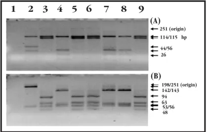
Figure 1- Electrophoresis through a 4% Metaphor agarose of restriction digests carried out on the 251 bp PCR fragment. Reactions were at 37°C in the presence of 10 units each of (A) RsaI and HaeIII,
(B) HinfI and MvaI as described by McOmish et al28 and Davidson et al10 28. Lane 1 - blank control;
lane 2 genotype 2 control; lane 3 genotype 1 control; lane 4 genotype 3 control; lanes 5, 6, 9 -genotype 1 samples and lanes 7, 8 - -genotype 3 samples. Genotype was deduced from the banding patterns produced by the two restriction enzyme combinations.
Table 1 - Baseline characteristics of hemophiliacs participating in this study.
Type of disease
hemophilia A hemophilia B Total Characteristics no = 238 (88.8%) no = 30 (11.2%) no (%)§
Age (yr)
mean ± σ 19.6 ± 12.3 18.5 ± 11.1 19.5 ± 12.1 range <1 - 66 2 - 40 <1 - 66 Residence
capital 79 (33.2) 10 (33.3) 89 (33.2) other cities 159 (66.8) 20 (66.7) 179 (66.8) Severity of disease
severe (<1%) 18 (10.5) 2 (9.1) 20 (10.4) mild/moderate (≥ 1%) 153 (89.5) 20 (90.9) 173 (89.6) Factor VIII/IX inhibitory antibodies
present 15 (41.7) 0 (0) 15 (35.7) absent 21 (58.3) 6 (100) 27 (64.3) Seropositivity
anti-HCV 105 (44.1) 8 (26.7) 113 (42.2) AgHBs/anti-HBc 53 (22.3) 7 (23.3) 60 (22.5) anti-HIV 24 (10.1) 4 (13.3) 28 (10.4) anti-HTLV I/II 10 (4.2) 0 (0) 10 (3.7) Chagas’ disease 9 (3.8) 2 (6.7) 11 (4.1) syphilis 2 (0.8) 1 (3.3) 3 (1.1) § Totals vary according to the availability of data.
Figure 2 - Seroprevalence of anti-HCV antibody according to age in hemophiliacs in the State of Bahia, northeastern Brazil, 2000.
-10 20 30 40 50 60 70 80
0-4 5-9 10-14 15-19 20-29 30-39 40-49 >=50 Age group (Years )
P
rev
al
en
ce
(%
)
Introduction of blood screening (1992-3)
presence of inhibitory antibodies, and positivity to HBV, HIV,
HTLV-I/II and Chagas’ disease (
T.
cruzi
infection) but not with
the type of hemophilia, the severity of the disease or positivity
to syphilis (Table 2).
From a subset of 66 hemophiliacs, HCV-RNA could be
detected in 77.3% (51/66). The prevalence of HCV infection
confirmed by the detection of HCV-RNA in hemophiliacs in
Salvador is therefore estimated to be 32.6% (95% CI
24.3-42.6). However, this sample may not be representative, since
the presence of HCV-RNA could not confirm any of the
associations with potential risk factors found for anti-HCV+
hemophiliacs (Table 2).
Specimens from 50 hemophiliacs could be genotyped.
Despite several attempts, one sample from a hemophilia patient
could not be genotyped successfully. The distribution of HCV
genotypes is shown in Table 3. HCV genotype distribution was
similar to the distribution from local candidate blood donors
(data not shown), with predominance of genotype 1 (74%),
followed by genotype 3 (22%) and the rare genotype 2 (4%). As
for viremia, the genotype could not be associated with any of the
characteristics analyzed previously (Table 4).
Table 2 - Potential risk factors associated with the prevalence of anti-HCV and viremia of hemophiliacs in Salvador, BA, Brazil.
Characteristics anti-HCV antibodies positivity HCV-RNA detectable
no tested§ % positive OR (95% CI) p values no tested § % detectable OR (95% CI) p values
All subjects 268 42.2 - - 66 77.3 -
-Age, years
>10 198 56.1 43.48 (9.94-266.79) <0.01 65 78.5 7.43 (0.47-224.72) ¥ NS¶
< 10† 70 2.9 1.00 1 0.0 1.00
Residence
capital 89 59.6 2.92 (1.73-4.94) <0.01 38 76.3 0.88 (0.23-3.30) NS¶ other cities† 179 33.5 1.00 28 78.6 1.00
Type of disease
hemophilia A 238 44.1 2.17 (0.93-5.07) NS 60 78.3 1.81 (0.20-13.90) NS¶
hemophilia B† 30 26.7 1.00 6 66.7 1.00
Severity of hemophilia
severe (<1%) 20 55.0 2.36 (0.85-6.62) NS 6 66.7 0.47 (0.05-4.60) NS¶ mild/moderate (>1%)† 173 34.1 1.00 37 81.1 1.00
Inhibitory antibodies
present 15 60.0 4.29 (0.93-20.88) 0.06‡ 7 85.7 4.00 (0.15-186.31) NS¶
absent† 27 25.9 1.00 5 60.0 1.00
Other viral markers
AgHBs/anti-HBc positive 60 81.7 9.95 (4.60-21.97) 0.01 28 85.7 2.44 (0.59-10.75 NS¶
negative† 207 30.9 1.00 38 71.1 1.00
anti-HIV positive 28 85.7 10.18 (3.18-36.24) <0.01¶ 15 80.0 0.81 (0.15-3.95) NS¶
negative† 240 37.1 1.00 51 76.5 1.00
anti-HTLV I/II positive 10 100.0 16.50 (2.16-347.00)¥ <0.01¶ 7 71.4 0.71 (0.10-6.08) NS¶
negative† 258 39.9 1.00 59 78.0 1.00
OR = odds ratio and CI confidence interval. †Subjects in this category served as reference group. § Totals vary according to the availability of data. ‡Inhibitory antibodies were significantly associated with anti-HCV positivity by Mantel-Haenszel. ¥OR was estimated by adding 1 in each cell.¶Fisher e-test.
Table 3 - Prevalence of HCV genotypes in hemophiliacs in State of Bahia, Northeastern Brazil.
No (%) of isolates of the genotype
Exposure category All Type 1 Type 2 Type 3 Hemophiliacs 50 (100.0) 37 (74.0) 2 (4.0) 11 (22.0) Note: Genotyping by RFLP analysis of the PCR product from the 5’UTR as described previously10 28
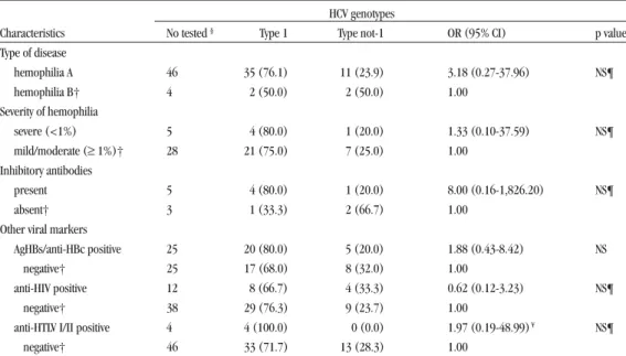
Table 4 - HCV genotype and potential risk factors in hemophiliacs in Salvador-BA, Brazil.
HCV genotypes
Characteristics No tested § Type 1 Type not-1 OR (95% CI) p values
All subjects 50 37 (74.0) 13 (26.0) -
-Age, years
>10 50 37 (74.0) 13 (26.0) undefined NS < 10† 0
Residence
capital 28 20 (71.4) 8 (28.6) 0.74 (0.16-3.20) NS other cities† 22 17 (77.3) 5 (22.7) 1.00
DISCUSSION
The overall prevalence of anti-HCV in our population of
hemophiliacs (42.2%) was similar to other studies performed
in different regions of Brazil
4 6 34. However, because most of
our patients were also infected with HIV and HTLV I/II, it
cannot be excluded that the detection rate of anti-HCV in this
group may underestimate the real seroprevalence, as has been
demonstrated previously
18.
With the use of HCV-safe clotting products and the
introduction of screening tests in the blood banks, there is a
very important tendency towards decrease of seropositivity for
anti-HCV antibodies among hemophiliac patients in Brazil and
other countries
4 6 32. In our study, HCV seroprevalence among
hemophiliacs younger than 10 years old was quite similar to
that found among candidate blood donors screened at HEMOBA
(1.5%, personal communication) during the same period of time.
However, it will need another five to ten years to confirm that
young hemophiliacs are really protected from HCV infection.
Residence in Salvador, the state capital, was shown to be an
independent risk factor for acquiring HCV but not for developing
chronic infection. This association may be particularly influenced
by age, access to the service, time of residence in the city and
other confounding factors (data not shown), and more
exploratory analysis is necessary. Although the risk of HCV
infection from environmental exposure (not related to transfusion
of blood or derivates) is low, it is not altogether absent. In Bahia,
Silva et al (1995) studied the seroprevalence of anti-HCV in urban
and rural populations and demonstrated that HCV was 1.5%
prevalent in Salvador and absent in a city of the interior. This
data suggests that HCV can be an endemic urban disease.
Even though the detection of anti-HCV could be used as a
part of this study, these results should be interpreted with
some care. Anti-HCV antibodies were associated with
serological markers for other blood-borne infections (HBV,
HIV and HTLV-I/II). These findings surely reflect the similar
modes of transmission. However, it is still unclear whether
HIV infection can interfere negatively by immune-suppression
of some individuals, which could increase the uncertainty of
our results and lead to underestimation of the prevalence
6.
Some investigators have reported HCV seronegative candidate
blood donors or patients that were HCV-RNA positive by PCR,
specially when associated with HIV and low CD4 cells counts
(<200x10
9/l)
3 43 45. On the other hand, the possibility of false
positive results caused by unspecific reactions by the kits
cannot be excluded. Therefore, HCV viremia and genotyping
were also investigated in hemophiliacs.
HCV-RNA could be detected in the majority of anti-HCV positive
hemophiliacs (77.3%) which is compatible to the chronicity rates
reported for this infection
2. Hence, about 20% of HCV-infected
hemophiliacs seem to have resolved HCV infection. It cannot be
ruled out that some hemophiliacs have viral replication below the
detection limit of polymerase chain reaction (PCR). To increase
the sensitivity of PCR some authors have suggested the application
of the PCR to detect HCV viremia in whole blood instead of in
serum
27 39 40. Furthermore, a new promising methodology base on
transcription-mediated amplification has become available for
more accurate HCV-RNA detection
37 38.
While certain risk factors could be identified for anti-HCV
hemophiliacs in Salvador, no significant association could be linked
to the presence of HCV-RNA in serum or the HCV genotypes. Some
studies have reported that HCV viremia was associated with older
age and abnormal ALT levels, while the presence of inhibitory
antibodies and HBsAg were protective factors against the detection
of HCV-RNA. The mechanisms for this have been not explained
6,
and we have not found significant evidence in favor.
In our study, HCV genotype 1 was most frequent (74%),
followed by genotype 3 (22%) and genotype 2 (4%). Although
heterotypic superinfection and mixed infections of hepatitis
Table 4 - Continuation.
HCV genotypes
Characteristics No tested § Type 1 Type not-1 OR (95% CI) p values
Type of disease
hemophilia A 46 35 (76.1) 11 (23.9) 3.18 (0.27-37.96) NS¶ hemophilia B† 4 2 (50.0) 2 (50.0) 1.00
Severity of hemophilia
severe (<1%) 5 4 (80.0) 1 (20.0) 1.33 (0.10-37.59) NS¶ mild/moderate (≥ 1%)† 28 21 (75.0) 7 (25.0) 1.00
Inhibitory antibodies
present 5 4 (80.0) 1 (20.0) 8.00 (0.16-1,826.20) NS¶ absent† 3 1 (33.3) 2 (66.7) 1.00
Other viral markers
AgHBs/anti-HBc positive 25 20 (80.0) 5 (20.0) 1.88 (0.43-8.42) NS negative† 25 17 (68.0) 8 (32.0) 1.00
anti-HIV positive 12 8 (66.7) 4 (33.3) 0.62 (0.12-3.23) NS¶ negative† 38 29 (76.3) 9 (23.7) 1.00
anti-HTLV I/II positive 4 4 (100.0) 0 (0.0) 1.97 (0.19-48.99) ¥ NS¶
negative† 46 33 (71.7) 13 (28.3) 1.00
C virus may be possible
46, the distribution of HCV genotypes
among the hemophiliacs was similar to the candidate blood
donors (data not shown). Similar results have already been
reported for other regions in Brazil in the same exposure
group
4 5 25. Considering the dynamic behavior of HCV infection,
a study has shown that a phenomenon of HCV superinfection
and overgrowth can occur in chronically infected patients and
suggests that HCV genotypes 1a and 1b may possess replicative
advantages over other genotypes
19. However, this has not been
confirmed experimentally
29. It is therefore probable that the high
prevalence of HCV genotype 1 in this population reflects the origin
of the blood and derivates.
It was not possible to study all the hemophiliacs in this region,
which would have enabled us to draw firm conclusions. However,
these findings clarify the status of HCV infection in hemophiliacs
from a northeastern area of Brazil, and highlight the importance
of studying the HCV genotypes due to their relevance in the
management of patients under interferon therapy. Special
consideration has to be taken since most of our patients are
infected by HCV genotype 1 and are co-infected with HIV, which
can lead to more rapidly developing progressive liver disease in
infected hemophiliacs
1 35. Therefore, this study demonstrates that
hemophiliacs are a group at high risk for severe chronic hepatitis
C disease in Bahia and will require hepatological assistance and
longer antiviral therapy.
ACKNOWLEDGMENTS
We thank the patients for compliance in participating in this
study, the Associação Bahiana dos Hemofílicos, and the staff from
Hemotransfusion and Hemotherapy Foundation (HEMOBA/
SESAB) for efficient assistance during the field surveys.
REFERENCES
1. Allain JP, Dailey SH, Laurian Y, Vallari DS, Rafowicz A, Desai SM, Devare SG. Evidence for persistent hepatitis C virus (HCV) infection in hemophiliacs. Journal of Clinical Investigation 88: 1672-1679, 1991. 2. Alric L, Fort M, Izopet J, Vinel JP, Bureau C, Sandre K, Charlet JP, Beraud M,
Abbal M, Duffaut M. Study of host- and virus-related factors associated with spontaneous hepatitis C virus clearance. Tissue Antigens 56: 154-158, 2000. 3. Alter MJ, Margolis HS, Krawczynski K, Judson FN, Mares A, Alexander WJ, Hu PY, Miller JK, Gerber MA, Sampliner RE, et al. The natural history of community-acquired hepatitis C in the United States. The Sentinel Counties Chronic non-A, non-B Hepatitis Study Team. New England Journal of Medicine 327: 1899-1905, 1992.
4. Barbosa AP, Martins RM, Teles SA, Silva SA, Oliveira JM, Yoshida CF. Prevalence of hepatitis C Virus infection among hemophiliacs in Central Brazil. Memórias do Instituto Oswaldo Cruz 97: 643-644, 2002. 5. Bassit L, Ribeiro-Dos-Santos G, Da Silva LC, Takei K, Villaca P, David-Neto E,
Chamone D, Saez-Alquezar A. Genotype distributions of hepatitis C virus in Sao Paulo, Brazil: rare subtype found [letter]. Hepatology 29: 994-995, 1999. 6. Carmo RA, Oliveira GC, Guimaraes MD, Oliveira MS, Lima AA, Buzek SC,
Correa-Oliveira R, Rocha MO. Hepatitis C virus infection among Brazilian hemophiliacs: a virological, clinical and epidemiological study. Brazilian Journal of Medical and Biological Research 35: 589-598, 2002. 7. Chan SW, McOmish F, Holmes EC, Dow B, Peutherer JF, Follett E, Yap PL,
Simmonds P. Analysis of a new hepatitis C virus type and its phylogenetic
relationship to existing variants. Journal of General Virology 73: 1131-1141, 1992.
8. Choo QL, Kuo G, Weiner AJ, Overby LR, Bradley DW, Houghton M. Isolation of a cDNA clone derived from a blood-borne non-A, non-B viral hepatitis genome. Science 244: 359-362, 1989.
9. Colombo M, Mannucci PM, Carnelli V, Savidge GF, Gazengel C, Schimpf K. Transmission of non-A, non-B hepatitis by heat-treated factor VIII concentrate. Lancet 2: 1-4, 1985.
10. Davidson F, Simmonds P, Ferguson JC, Jarvis LM, Dow BC, Follett EA, Seed CR, Krusius T, Lin C, Medgyesi GA, et al. Survey of major genotypes and subtypes of hepatitis C virus using RFLP of sequences amplified from the 5' non-coding region. Journal of General Virology 76: 1197-1204, 1995. 11. Esteban JI, Esteban R, Viladomiu L, Lopez-Talavera JC, Gonzalez A, Hernandez JM, Roget M, Vargas V, Genesca J, Buti M, et al. Hepatitis C virus antibodies among risk groups in Spain. Lancet 2: 294-297, 1989. 12. Feinstone SM, Kapikian AZ, Purcell RH, Alter HJ, Holland PV.
Transfusion-associated hepatitis not due to viral hepatitis type A or B. New England Journal of Medicine 292: 767-770, 1975.
13. Fletcher ML, Trowell JM, Craske J, Pavier K, Rizza CR. Non-A non-B hepatitis after transfusion of factor VIII in infrequently treated patients. British Medical Journal 287: 1754-1757, 1983.
14. Horowitz MS, Rooks C, Horowitz B, Hilgartner MW. Virus safety of solvent/ detergent-treated antihaemophilic factor concentrate. Lancet 2: 186-189, 1988.
15. Kernoff PB, Lee CA, Karayiannis P, Thomas HC. High risk of non-A non-B hepatitis after a first exposure to volunteer or commercial clotting factor concentrates: effects of prophylactic immune serum globulin. British Journal of Haematology 60: 469-479, 1985.
16. Kohara M, Tanaka T, Tsukiyama-Kohara K, Tanaka S, Mizokami M, Lau JY, Hattori N. Hepatitis C virus genotypes 1 and 2 respond to interferon-alpha with different virologic kinetics. Journal of Infectious Diseases 172: 934-938, 1995.
17. Kuo G, Choo QL, Alter HJ, Gitnick GL, Redeker AG, Purcell RH, Miyamura T, Dienstag JL, Alter MJ, Stevens CE, An assay for circulating antibodies to a major etiologic virus of human non-A, non-B hepatitis. Science 244: 362-364, 1989.
18. Lanotte P, Dubois F, Le Pogam S, Guerois C, Fimbel B, Bacq Y, Gruel Y, Goudeau A, Barin F. The kinetics of antibodies against hepatitis C virus may predict viral clearance in exposed hemophiliacs. Journal of Infectious Diseases 178: 556-559, 1998.
19. Laskus T, Wang LF, Rakela J, Vargas H, Pinna AD, Tsamandas AC, Demetris AJ, Fung J. Dynamic behavior of hepatitis C virus in chronically infected patients receiving liver graft from infected donors. Virology 220: 171-176, 1996.
20. Le Guen B, Squadrito G, Nalpas B, Berthelot P, Pol S, Brechot C. Hepatitis C virus genome complexity correlates with response to interferon therapy: a study in French patients with chronic hepatitis C. Hepatology 25: 1250-1254, 1997.
21. Maisonneuve P, Laurian Y, Guerois C, Verroust F, Ferrer Le Coeur F, Courouce AM, Noel L. Antibody to hepatitis C (anti C 100-3) in French hemophiliacs. Nouvelle Revue Francaise D’Hematologie 33: 263-266, 1991. 22. Makris M, Garson JA, Ring CJ, Tuke PW, Tedder RS, Preston FE. Hepatitis C viral RNA in clotting factor concentrates and the development of hepatitis in recipients. Blood 81: 1898-1902, 1993.
23. Makris M, Preston FE, Triger DR, Underwood JC, Choo QL, Kuo G, Houghton M. Hepatitis C antibody and chronic liver disease in haemophilia. Lancet 335: 1117-1119, 1990.
24. Martins RM, Barbosa AP, Oliveira JM, Vanderborght B, Yoshida CF. Genotype analysis of hepatitis C virus in Brazilian hemophiliacs and blood donors. Vox Sanguinis 78: 255, 2000.
25. Martins RMB, Vanderborght BO, Yoshida CF. Hepatitis C virus genotypes among blood donors from different regions of Brazil. Mem. Inst. Oswaldo Cruz 93: 299-300, 1998.
Hepatitis C infection and viremia in Dutch hemophilia patients. Journal of Medical Virology 45: 241-246, 1995.
27. Mazur W, Mazurek U, Jurzak M, Wilczok T, Bulanowski Z, Gonciarz Z. Positive and negative strands of HCV-RNA in sera and peripheral blood mononuclear cells of chronically hemodialyzed patients. Medical Science Monitoring 7: 108-115, 2001.
28. McOmish F, Yap PL, Dow BC, Follett EA, Seed C, Keller AJ, Cobain TJ, Krusius T, Kolho E, Naukkarinen R, et al. Geographical distribution of hepatitis C virus genotypes in blood donors: an international collaborative survey. Journal of Clinical Microbiology 32: 884-892, 1994.
29. Okamoto H, Mishiro S, Tokita H, Tsuda F, Miyakawa Y, Mayumi M. Superinfection of chimpanzees carrying hepatitis C virus of genotype II/1b with that of genotype III/2a or I/1a. Hepatology 20: 1131-1136, 1994. 30. Pasi KJ, Evans DJ, Skidmore SJ, Hill FGH. Prevention of hepatitis C virus
infection in haemophiliacs. Lancet 1: 1474, 1990.
31. Perret BA, Senn M, Affentranger P, Poorbeik M, Burckhardt JJ, Morell A. [Seroprevalence of hepatitis C virus in hemophiliacs in Switzerland]. Schweizerische Medizinische Wochenschrift 123: 79-81, 1993. 32. Pistello M, Ceccherini-Nelli L, Cecconi N, Bendinelli M, Panicucci F.
Hepatitis C virus seroprevalence in Italian haemophiliacs injected with virus-inactivated concentrates: five year follow-up and correlation with antibodies to other viruses. Journal of Medical Virology 33: 43-46, 1991. 33. Pozzato G, Moretti M, Franzin F, Croce LS, Tiribelli C, Masayu T, Kaneko S, Unoura M, Kobayashi K. Severity of liver disease with different hepatitis C viral clones [letter]. Lancet 338: 509, 1991.
34. Rocha VG, Carmo RA, Murao M, Mourão JG, Ribeiro CMF, Martins MVCL, Viana MB. Predictive factors for the presence of hepatitis C virus antibodies in hemophiliacs: multivariate analysis. In: Resumo do Congresso da Sociedade Brasileira de Medicina Tropical, Salvador p. 368, 1994. 35. Rockstroh JK, Spengler U, Sudhop T, Ewig S, Theisen A, Hammerstein U,
Bierhoff E, Fischer HP, Oldenburg J, Brackmann HH, Sauerbruch T. Immunosuppression may lead to progression of hepatitis C virus-associated liver disease in hemophiliacs coinfected with HIV. American Journal of Gastroenterology 91: 2563-2568, 1996.
36. Sambrook J, Fritsch EF, Maniatis T. Molecular cloning: a laboratory manual. Cold Spring Harbor Laboratory Press 3 New York, 1989.
37. Sarrazin C. Highly sensitive hepatitis C virus RNA detection methods: molecular backgrounds and clinical significance. Journal Clinical of Virology 25(supl 3): S23-29, 2002.
38. Sarrazin C, Teuber G, Kokka R, Rabenau H, Zeuzem S. Detection of residual hepatitis C virus RNA by transcription-mediated amplification in patients
with complete virologic response according to polymerase chain reaction-based assays. Hepatology 32: 818-823, 2000.
39. Schmidt WN, Klinzman D, LaBrecque DR, Macfarlane DE, Stapleton JT. Direct detection of hepatitis C virus (HCV) RNA from whole blood, and comparison with HCV RNA in plasma and peripheral blood mononuclear cells. Journal of Medical Virolology 47: 153-160, 1995.
40. Schmidt WN, Wu P, Cederna J, Mitros FA, LaBrecque DR, Stapleton JT. Surreptitious hepatitis C virus (HCV) infection detected in the majority of patients with cryptogenic chronic hepatitis and negative HCV antibody tests. Journal of Infectious Diseases 176: 27-33, 1997.
41. Schimpf K, Mannucci PM, Kreutz W, Brackmann HH, Auerswald G, Ciavarella N, Mosseler J, DeRosa V, Kraus B, Brueckmann C, et al. Absence of hepatitis after treatment with a pasteurized factor VIII concentrate in patients with hemophilia and no previous transfusions. New England Journal of Medicine 316: 918-922, 1987.
42. Silini E, Bono F, Cividini A, Cerino A, Bruno S, Rossi S, Belloni G, Brugnetti B, Civardi E, Salvaneschi L, et al. Differential distribution of hepatitis C virus genotypes in patients with and without liver function abnormalities. Hepatology 21: 285-290, 1995.
43. Simmonds P, Balfe P, Ludlam CA, Bishop JO, Brown AJ. Analysis of sequence diversity in hypervariable regions of the external glycoprotein of human immunodeficiency virus type 1. Journal of Virology 64: 5840-5850, 1990. 44. Simmonds P, Holmes EC, Cha TA, Chan SW, McOmish F, Irvine B, Beall E, Yap PL, Kolberg J, Urdea MS. Classification of hepatitis C virus into six major genotypes and a series of subtypes by phylogenetic analysis of the NS-5 region. Journal of General Virology 74: 2391-2399, 1993. 45. Sugitani M, Inchauspe G, Shindo M, Prince AM. Sensitivity of serological assays
to identify blood donors with hepatitis C viraemia. Lancet 339: 1018-1019, 1992. 46. Toyoda H, Fukuda Y, Hayakawa T, Kumada T, Nakano S, Takamatsu J, Saito H. Presence of multiple genotype-specific antibodies in patients with persistent infection with hepatitis C virus (HCV) of a single genotype: evidence for transient or occult superinfection with HCV of different genotypes. American Journal of Gastroenterology 94: 2230-2236, 1999. 47. Troisi CL, Hollinger FB, Hoots WK, Contant C, Gill J, Ragni M, Parmley R,
Sexauer C, Gomperts E, Buchanan G, et al. A multicenter study of viral hepatitis in a United States hemophilic population. Blood 81: 412-418, 1993. 48. Velasco M, Hurtado C, Brahm J. Anti-hepatitis C viral antibodies in different