CLINICAL SCIENCE
M2-Polarized tumor-associated macrophages are
associated with poor prognoses resulting from
accelerated lymphangiogenesis in lung
adenocarcinoma
Bicheng Zhang,IGuoqing Yao,IYafei Zhang,II,IJuan Gao,III Bo Yang,IZhiguo Rao,IJianfei GaoI
IWuhan General Hospital of Guangzhou Command, People’s Liberation Army, Department of Oncology, Wuhan, Hubei Province, China.IISouthwest Hospital, Third Military Medical University, Department of Gastroenterology, Chongqing, China.IIIWuhan General Hospital of Guangzhou Command Department of Gastroenterology, People’s Liberation Army, Wuhan, Hubei Province, China.
OBJECTIVES: Tumor-associated macrophages have been implicated in promoting tumor growth, progression and metastasis. However, the activated phenotype (M1 or M2) of tumor-associated macrophages remains unknown in solid tumors. Therefore, this study examined the density and prognostic significance of M2-polarized tumor-associated macrophages in lung adenocarcinoma.
METHODS:Tumor specimens from 65 lung adenocarcinoma patients were assessed by ELISA for Th1/Th2 cytokine concentrations. The activated phenotype (M1 or M2) of tumor-associated macrophages was determined utilizing immunofluorescence staining. Additionally, to evaluate lymphangiogenesis, peritumoral lymphatic microvessel density was measured using D2-40. The correlation between tumor-associated macrophage subtype and overall patient survival was analyzed using the Kaplan-Meier method and compared using the log-rank test.
RESULTS: A shift toward Th2 cytokine expression was detected within lung adenocarcinoma microenvironments. Approximately 79.71¡16.27% of tumor-associated macrophages were M2 polarized; the remaining 20.35¡5.31% were M1 polarized. The infiltration of M2-polarized macrophages was significantly associated with P-TNM staging and lymph node metastasis. The peritumoral lymphatic microvessel density was significantly higher in the high M2-polarized tumor-associated macrophage group than in the low M2-M2-polarized tumor-associated macrophage group. A significant difference in overall patient survival was detected not only between patients with tumors with high and low macrophage counts but also between patients with tumors with high and low counts of M2-polarized macrophages.
CONCLUSION: Tumor-associated macrophages in lung adenocarcinoma have an M2-polarized subtype and are associated with poor prognoses, perhaps resulting from accelerated lymphangiogenesis and lymph node metastasis.
KEYWORDS: M2-polarized macrophages; Tumor-associated macrophages; Lymphangiogenesis; Lung adenocarcinoma; Prognosis.
Zhang B, Yao G, Zhang Y, Gao J, Yang B, Rao Z, et al. M2-Polarized tumor-associated macrophages are associated with poor prognoses resulting from accelerated lymphangiogenesis in lung adenocarcinoma. Clinics. 2011;66(11):1879-1886.
Received for publicationMay 29, 2011;First review completed onJuly 4, 2011;Accepted for publication onJuly 12, 2011 E-mail: [email protected]
Tel.: 86 27 6887-8461
INTRODUCTION
Recently, compelling evidence has emerged that macro-phages infiltrating the tumor microenvironment, also known as tumor-associated macrophages (TAMs), promote pro-cesses such as angiogenesis, lymphangiogenesis, tumor growth and progression in solid tumors.1,2 In addition to
their protumoral effects on lung adenocarcinoma, the role of TAMs in promoting tumors is supported by clinical studies that identified a correlation between high macrophage numbers in tumor tissues and poor patient prognoses.3-5 However, the mechanisms underlying TAM functional changes within tumor microenvironments remain unknown; one potential cause is the altered polarization of TAMs with associated changes in their activated phenotypes.6
There are at least two different subpopulations of activated macrophages within tumor microenvironments. The first type, known as classically activated macrophages (M1 macrophages), are activated by lipopolysaccharides (LPS) or by double signals from interferon (IFN)-cand tumor necrosis factor-a (TNF-a). M1-polarized macrophages exhibit an Copyrightß2011CLINICS– This is an Open Access article distributed under
the terms of the Creative Commons Attribution Non-Commercial License (http:// creativecommons.org/licenses/by-nc/3.0/) which permits unrestricted non-commercial use, distribution, and reproduction in any medium, provided the original work is properly cited.
IL-12high, IL-23high, IL-10low phenotype and produce TNF-a
and nitric oxide (NO); they are potent effector cells that kill microorganisms and tumor cells.7,8 The second type of macrophages is known as alternatively activated macro-phages (M2 macromacro-phages). Exposure to IL-4, IL-13, vitamin D3, glucocorticoids or transforming growth factor-b(TGF-b) decreases macrophage antigen-presenting capability and up-regulates the expression of macrophage mannose receptors (MMR, also known as CD206), scavenger receptors (SR-A, also known as CD204), CD163, dectin-1 and DC-SIGN.9 M2-polarized macrophages exhibit an IL-12low, IL-23low, IL-10high
phenotype and play an important role in stroma formation, tissue repair, tumor growth, angiogenesis and immunosup-pression.10,11 Although several studies throughout the past
decade have suggested that TAMs exhibit a polarized M2 phenotype,12-14limited reports describing the polarization of activated TAMs (such as M1 and M2) within tumor microenvironments have only recently been published.15-18
Lymphangiogenesis is considered to be an initial and necessary event for lymphatic and regional lymph node metastasis.19 A number of studies have demonstrated lymphatic microvessel density (LMVD) to be an independent prognostic factor in solid tumors.20-23 Additionally, several
studies have shown that TAMs contribute to lymphangiogen-esis and lymphatic vessel invasion in malignant tumors.24,25 However, as TAMs are heterogeneous in different tumor microenvironments and have multiple activated phenotypes, further investigations are necessary to clarify whether all TAMs or only M2-polarized TAMs correlate with lymphan-giogenesis and poor prognosis in solid tumors.
Here, we investigated the density of infiltrating M2-polarized TAMs and the association between M2-M2-polarized TAMs, lymphangiogenesis, and prognosis. We found that TAMs in lung adenocarcinoma exhibit a M2-polarized subtype and are associated with poor prognosis, perhaps as a result of lymphatic metastasis.
MATERIALS AND METHODS
Ethics statement
This study was approved by the Ethics Committee of Wuhan General Hospital, Guangzhou Command of the People’s Liberation Army. Written informed consent was obtained from all patients.
Patients and tissue samples
A total of 65 patients with lung adenocarcinoma who underwent either lobectomy or pneumonectomy at Wuhan General Hospital were included in the study. The study group was composed of 38 men and 27 women with a mean age of 51.5 years (age range: 32-76 years). The patients underwent tumor resection from 2003 to 2006. None of the patients had received any preoperative chemotherapy or radiotherapy. The lesions of each patient were classified into stages according to the UICC 2010 pTNM classification scheme (7th edition); stage I, II, III, and IV lesions were present in 12, 27, 24, and 2 patients, respectively. Histologically, 16 tumors were graded as well-differentiated adenocarcinoma, 20 as moderately differentiated, and 29 as poorly differentiated. Lymph node metastasis occurred in 35 patients; no metastatic lymph nodes were observed in the remaining 30 patients. Follow-up visits were conducted and outcomes were recorded for all patients. A total of 20 patients with benign lung lesions, including six cases of
tuberculomas, six cases of inflammatory pseudotumors, five cases of hamartomas and three cases of lung cysts, were also included in this study. The clinicopathologic parameters of the lung adenocarcinoma patients are shown in Table 1.
ELISA
Fresh human lung adenocarcinoma tissues and benign lung lesions were obtained from surgical specimens for the detection of Th1/Th2 cytokines by ELISA. Briefly, the specimens were rinsed three times in phosphate-buffered saline (PBS). Next, 1 g of each tissue sample was homo-genized in 1 ml of PBS. Tissue homogenates were then centrifuged at 12,000 g for 1 min at 25
˚
C. After centrifugation, the supernatants were collected for further analysis. Cytokine production was quantified by cytokine-specific ELISA. Human IFN-c, IL-12, IL-4, and IL-10 (Biosource, Camarillo, CA, USA) ELISA kits were utilized; all procedures were performed according to the manufacturer’s instructions. Sample absorbances were determined using an ELISA plate reader (Dynatech, Chantilly, VA, USA).Immunofluorescence staining
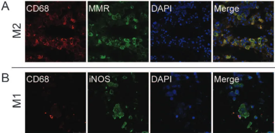
For the simultaneous visualization of CD68 and MMR or CD68 and inducible nitric oxide synthase (iNOS) on the same tissue section, double-label immunofluorescence staining was performed. The tumor tissues were acetone-fixed at 4
˚
C and then cut into 10-mm frozen sections. After being rinsed three times in PBS, the slides were treated with 3% H2O2 to inactivate endogenous peroxidases and then withnormal goat serum to block nonspecific binding sites. Next, slides were immunostained using a monoclonal mouse anti-human CD68 antibody (15100; Zhongshan Company, Beijing, China). Slides were also stained with rabbit anti-human MMR antibodies (15200; BioLegand, San Diego, CA, USA) or rabbit anti-human iNOS antibodies (15200; Santa Cruz Biotechnology, Santa Cruz, CA, USA). All slides were incubated overnight at 4
˚
C. Next, the slides were rinsed three times in PBS and incubated with TRITC-conjugated goat anti-mouse IgG for 30 min and then with FITC-conjugated goat anti-rabbit IgG for 30 min. Nuclei were counterstained with 49-6-diamidino-2-phenylindole (DAPI) for 5 min. All slides were examined under a laser confocal microscope (Leica TCS SP5, Leica, German) fitted with TRITC (red fluorescence) and FITC (green fluorescence) filters. The excitation wavelength for FITC was 488 nm; the excitation wavelength for TRITC was 568 nm. Red CD68-positive fluorescence was located in the cytoplasm, and green MMR-positive fluorescence was located at the cell membrane and in the cytoplasm. However, green iNOS-positive fluorescence was only observed in the cytoplasm. If CD68 and MMR or CD68 and iNOS were coexpressed, the fluorescence was yellow. All nuclei were blue.Immunohistochemistry
exposed to a biotinylated secondary antibody for 20 min and then treated with streptavidin peroxidase. For color development, the slides were stained with 3, 39 diamino-benzidine (DAB). Hematoxylin and eosin (H&E) were used as a counterstain. A reddish-brown precipitate in the cytoplasm of LECs indicated a positive reaction.
After the immunostained sections were scanned at low magnification (6100), the regions with the greatest numbers of distinctly stained lymphatic foci (hot spots) were deter-mined simultaneously by two different observers. Then, two observers blinded to the tumor status or the stains used independently counted the slides for LMVD staining under 400x magnification (0.03 mm2 field) in three regions. An intratumoral compartment was identified as the area encompassing the cancerous tissue in the H&E sections. A peritumoral compartment was defined as a 1-mm-wide band surrounding the intratumoral compartment that included the edge of the tumor and just outside the tumor. Single immunoreactive endothelial cells and endothelial cell clus-ters separate from other microvessels were counted as vessels according to previously used procedures.24
Statistical analysis
The numbers of M1- or M2-polarized TAMs and intratumoral LMVD and peritumoral LMVD are expressed as the mean¡SD. Statistical differences between the means were analyzed using the independent samplest-test. Rates
were compared using a x2 test. The relationship between
TAM counts and LMVD was assessed using the Spearman correlation test. On the basis of TAM or M2-polarized TAM count, patients were classified into two groups, and the overall survival rate was then compared between the high and low groups, respectively. Overall survival time was calculated from the date of surgery until death or, if the patient was still alive, until the last follow-up visit. Death from any cause was considered in the calculations of overall survival. Two overall survival rates were calculated by the Kaplan-Meier method and compared using the log-rank test. Each prognostic factor was evaluated with regard to survival in a multivariate analysis using a Cox proportional hazards regression model. A p-value of ,0.05 was con-sidered statistically significant. All statistical analyses were performed withSPSS13.0 (SPSS Inc., Chicago, IL, USA).
RESULTS
Th2 shift in lung adenocarcinoma
To detect Th1/Th2 cytokines in the microenvironment of lung adenocarcinoma, we obtained fresh lung tumor tissues and benign lung lesions from surgical resections and assessed the expression of the Th1 cytokines IFN-cand IL-12 and the Th2 cytokines IL-4 and IL-10 in tissue homogenates using a cytokine-specific ELISA. As shown in Figure 1, IFN-cand IL-12 expression was lower in fresh Table 1 -The relationships between TAM, M1- or M2-polarized TAM counts and clinicopathologic features (xx¡s).
Clinicopathologic features Cases TAMs p-value M1-polarized TAMs p-value M2-polarized TAMs p-value
Gender
Male 38 104.19¡15.93 0.712 21.15¡6.11 0.948 83.04¡12.70 0.903
Female 27 103.07¡15.74 20.61¡5.73 82.15¡11.26
Age (yrs.)
$55 34 105.45¡18.96 0.895 21.29¡5.10 0.705 84.84¡12.04 0.681
,55 31 101.88¡14.11 20.58¡6.26 82.00¡10.61
Differentiation
Well-to-moderate 36 103.17¡15.15 0.984 20.63¡6.96 0.157 82.23¡10.43 0.645
Poor 29 103.98¡11.67 20.80¡6.16 82.87¡10.17
Lymph node metastasis
Positive 35 120.44¡35.83 0.042* 24.09¡4.56 0.882 95.99¡15.60 0.003*
Negative 30 94.26¡14.52 18.85¡7.27 70.26¡12.16
pTNM staging
I+II 39 96.38¡21.55 0.037* 20.31¡4.71 0.431 76.81¡10.32 0.029*
III+IV 26 115.49¡30.94 23.45¡5.35 92.05¡18.14
*Statistically significant.
lung adenocarcinoma homogenates than in benign lung lesion homogenates. However, fresh lung adenocarcinoma samples contained significantly more Th2 cytokines than benign lung lesion samples. IL-10 was the predominant Th2 cytokine present in the lung adenocarcinoma samples.
M2-polarized macrophages in lung adenocarcinoma
To identify and quantify the infiltrated TAMs associated with the M1 or M2 phenotype, we utilized iNOS, anti-MMR, and anti-CD68 antibodies for immunofluorescence staining. CD68-positive macrophages were detected in varying concentrations in all 65 lung adenocarcinoma cases and in all 20 benign lung lesion samples. In the lung adenocarcinoma samples, CD68-positive TAMs were pre-dominately located in the peritumoral stroma and tumor tissues, e.g., in the lumen of the lung adenocarcinoma; in particular, TAMs were observed along the invasive tumor front. MMR-positive signals predominately colocalized in macrophages, but iNOS expression was detected in both lung tumor cells and TAMs. M1-polarized TAMs were defined as CD68+iNOS+, whereas M2-polarized TAMs were distinguished by the expression of CD68 and MMR.15
M1-and M2-polarized TAMs were detected in each of the 65 lung adenocarcinoma tissue samples. Overall, the percen-tage of CD68+MMR+ TAMs was 79.71¡16.27% (Figure
2A), and the percentage of CD68+iNOS+ TAMs was 20.35¡5.31% (Figure 2B). CD68 and iNOS coexpression
was observed in 100% of the benign lung lesion samples; the percentage of CD68+iNOS+TAMs was 89.5%.
The correlation of M2-polarized TAMs with clinicopathologic features
Table 1 also depicts the correlations between TAMs, M1-or M2-polarized TAMs and clinicopathologic features. TAM number and M2-polarized TAM number were significantly associated with p-TNM staging (p =0.037, 0.029) and lymph node metastasis (p =0.042, 0.003); however, these factors were not associated with gender, age or tumor differentia-tion. Interestingly, the number of M2-polarized TAMs was more strongly correlated with lymph node metastasis than the number of infiltrating CD68-positive TAMs. The number of M1-polarized TAMs failed to correlate with any of the clinicopathologic features included in this study.
The correlation of M2-polarized TAMs with peritumoral LMVD
D2-40 positive lymph vessels were observed in each of the 65 lung adenocarcinoma examined. Lymphatic vessels with the characteristic irregular morphology, empty lumina without red blood cells, and thin endothelia were distinct and stained strongly with D2-40 when present. Vessels containing red blood cells did not stain positive with D2-40. In lung adenocarcinoma samples, D2-40-positive vessels were detected more frequently in the peritumoral stroma (Figure 3A) than in the intratumoral compartment (Figure 3B). The D2-40-positive peritumoral LMVD count was 11.56¡10.73, which was higher than the D2-40-positive
intratumoral LMVD count (2.96¡1.15) (Figure 3C,p,0.01). Because peritumoral LMVD is a significant prognostic factor for non-small-cell lung cancer (NSCLC),27we explored
the correlation between M2-polarized TAMs and peritumoral LMVD. We divided all of the cases into two groups according to their CD68 grade (cutoff value = 102); the low-TAM group included 25 cases, and the high-TAM group was comprised of 40 cases. There was a significant difference in peritumoral LMVD between the two groups (p =0.047). Next, we divided all of the cases into two groups according to their levels of CD68/MMR expression (cutoff values = 82): the low M2-polarized TAM group (16 cases) and the high M2-M2-polarized TAM group (49 cases). The peritumoral LMVD was significantly higher in the high M2-polarized TAM group than in the low M2-polarized TAM group (p =0.009). Lastly, we divided all of the cases into two groups according to their levels of CD68/iNOS expression: the low M1-polarized TAM group (31 cases) and the high M1-polarized TAM group (34 cases). There was no significant difference in peritumoral LMVD between the two groups.
The prognostic significance of M2-polarized TAMs
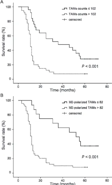
To assess the prognostic significance of M2-polarized TAM infiltration, patients were first classified into two groups based on their TAM count; patients in each group were then further divided into two subgroups based on their M2-polarized TAM count. . We used median values of 102 and 82 as the cutoff values for the TAM and M2-polarized TAM counts, respectively. The median survival times were 36 and 11 months for patients with TAM counts of#102 and.102, respectively. The five-year survival rates in the low TAM group and the high TAM group were 28.0%
and 7.5%, respectively. Figure 4A depicts the difference in the overall survival rates between the high-TAM-count group and the low-TAM-count group (p,0.001). The median survival times were 52 and 12 months for patients with M2-polarized TAM counts of #82 and .82, respec-tively. The five-year survival rates in the low M2-polarized TAM group and the high M2-polarized TAM group were 37.5% and 8.2%, respectively; the overall survival rate was significantly lower in the high M2-polarized TAM group than in the low M2-polarized TAM group (p,0.001) (Figure 4B).
Multivariate analysis
TAM count, M1-polarized TAM count, M2-polarized TAM count, intratumoral LMVD, peritumoral LMVD, and other factors, including gender, age, tumor differentiation, lymph node metastasis, and P-TNM staging, were analyzed using Cox proportional hazards regression models in all of the lung adenocarcinoma patients. TAM count, M2-polarized TAM count, peritumoral LMVD, lymph node metastasis, and P-TNM staging were independent prognostic factors for overall survival (Table 2).
DISCUSSION
Our data demonstrate that a shift toward the production of Th2 cytokines, a factor that induces alternative macrophage activation, occurs within the lung adenocarcinoma tumor microenvironment. Under normal conditions, Th1/Th2 cytokines in the body are in a dynamic balance. When a Th2 shift occurs, the immune system can become suppressed, making the host more susceptible to microbial infections, tumorigenesis and progression, allergic reactions, and graft
rejection reactions.28Human NSCLC cells express a type-2 cytokine pattern bothin situandin vitro, which may play an active immunoregulatory role in the lung cancer microenvir-onment.29In patients with several malignant tumors,
includ-ing lung tumors, a Th2 cytokine shift has been positively correlated with the degree of malignancy.28,30In our study,
IL-4 and IL-10 were detected in high amounts in lung adenocarcinoma tissue homogenates.
Macrophages are plastic cells; for example, they can switch between an activated M1 state and an activated M2 state depending upon specific signals within their micro-environment. Because IL-4 is an activator of M2-polarized macrophages and IL-10 is known to function to promote the development of M2 macrophages from monocytes,31 the
cytokine signals required for M2 activation were abun-dantly present within the microenvironment of the exam-ined lung adenocarcinoma. Recently, Ohtaki et al. found that IL-10 was significantly correlated with the number of CD204-positive macrophages (M2 macrophages) within the stroma of lung adenocarcinoma.32
M2-polarized TAMs were detected predominantly in lung adenocarcinoma and were significantly associated with p-TNM staging and lymph node metastasis. In a series of reviews, Martinez et al. proposed that TAMs might polarize toward an M2 phenotype.14 Hagemann et al.
demonstrated that when co-cultured, ovarian cancer cells switched co-cultured macrophages to an M2 phenotype similar to that of ovarian TAMs.15Initially, macrophages in
(62.73¡16.27%) and SR-A (66.28¡4.31%). Further,
co-culturing of human primary ovarian cancer cells with human macrophages increased macrophage MMR and SR-A surface expression. In murine colon adenocarcinoma-38 and GL261 murine glioma, over 90% of tumor-infiltrating myelomonocytoid cells were of the CD11b+F4/80+
mono-cyte/macrophage lineage. These cells also had a myeloid-derived suppressor cell phenotype, as they suppressed the proliferation of activated splenic CD8+T cells and exhibited
a CD11b+CD11c+Gr-1lowIL-4Ra+phenotype. In addition,
tumor-infiltrating myelomonocytoid cells expressed higher levels of MMR and lower levels of CXCL10, which are alternative and classical macrophage activation markers, respectively.33
In the present study, M1 and M2 macrophages coexisted in the tumor microenvironment, which showed the hetero-geneity of TAMs in lung adenocarcinoma. However, a greater number of M2-polarized TAMs were detected than M1-polarized TAMs. Further, M2-polarized TAMs were
more strongly correlated with lymph node metastasis than were CD68-positive TAMs. These data suggest the impor-tant role of M2-polarized TAMs in tumor progression and metastasis.
Our study also showed that M2-polarized TAMs were associated with poor prognosis in lung adenocarcinoma, perhaps resulting from accelerated lymphangiogenesis and lymph node metastasis. Because previous studies concern-ing the relationship between TAM infiltration and clinical outcome in various cancers did not describe TAM hetero-geneity and presented contradicting conclusions, the significance of macrophage infiltration in patient survival has been controversial. Recently, several studies have demonstrated that M2-polarized TAMs in gliomas,17 lung adenocarcinoma,32 intrahepatic cholangiocarcinomas,34 and angioimmunoblastic T-cell lymphoma adenocar-cinoma35 are associated with tumor progression and
prognosis. In the present study, we have also found M2-polarized TAMs to be associated with poor prognosis in lung adenocarcinoma patients. The patients with high M2-polarized TAM counts exhibited poor survival. Our multi-variate Cox proportional hazards analysis indicated that M2-polarized TAM density was a useful prognostic marker for overall survival in lung adenocarcinoma cases. Two research papers previously reported that in patients with extended survival, macrophages infiltrating the tumor islets in NSCLC were predominantly of the M1 pheno-type.36,37These results appear to differ from our findings. However, in our research, the types of lung cancer involved were restricted to lung adenocarcinoma, not NSCLC, which may be primarily responsible for the conflicting findings.
Metastatic tumors that are spread through the blood or lymphatic vessels occur in most forms of human cancer, with peritumoral lymphangiogenesis and regional lymph node metastasis often being the most important prognos-tic factors for carcinoma patients.38 In addition to tumor cells, TAMs and other inflammatory cells in the tumor stroma can express VEGF-C and VEGF-D and promote peritumoral lymphangiogenesis and lymph node metas-tasis.24,25 Previously, we reported that alternatively activated TAMs can induce peritumoral lymphangiogen-esis in a murine Lewis lung adenocarcinoma model.39 In the present study, for the first time, a positive correlation between M2-polarized TAM count and peritumoral LMVD, but not intratumoral LMVD, was found in patients with lung adenocarcinoma. Interestingly, the Figure 4 -Kaplan-Meier curves for overall survival by TAM count
(A) and M2-polarized TAM count (B) in patients with lung adenocarcinoma. A shows the difference in the overall survival rate between the high and the low TAM count groups (p,0.001). B shows the difference in the overall survival rate between the high and the low M2-polarized TAM groups (p,0.001).
Table 2 -Hazard ratios (HR), 95% confidence interval (CI) andp-value for the 65 patients with lung
adenocarcinoma.
Factor p-value HR 95% CI
Gender 0.303 0.574 0.200-1.649
Age 0.393 1.317 0.700-2.480
Differentiation 0.670 1.175 0.558-2.476
Lymph node metastasis 0.011* 2.778 1.260-6.123
P-TNM staging 0.039* 3.021 1.055-8.649
TAM count 0.015* 3.602 1.279-10.142
M1-polarized TAMs count 0.860 0.881 0.214-3.628 M2-polarized TAMs count 0.031* 4.280 1.146-15.984
Intratumoral LMVD 0.327 1.189 0.841-1.679
Peritumoral LMVD 0.038* 1.073 1.004-1.147
LMVD in cases with high numbers of infiltrating M2-polarized TAMs was significantly higher than in cases with low numbers of M2-polarized TAMs. These data suggest that in lung adenocarcinoma patients, M2-polar-ized TAM infiltration is correlated with poor survival, likely as a result of accelerated lymphangiogenesis and lymph node metastasis.
Our data indicate that M2-polarized TAMs in lung adenocarcinoma are a predictor of poor prognosis as a result of their promotion of lymphangiogenesis and lymphatic metastasis. Because M2-polarized TAMs appear to play an important role in peritumoral lymphangiogenesis and decreased survival in lung adenocarcinoma patients, they represent a potential target for therapeutic interven-tion. It is possible that the use of exogenous agents to switch the phenotype of the TAMs from M2 to M1 during tumor progression could decrease lymphatic metastasis and pro-long lung adenocarcinoma patient survival.
ACKNOWLEDGEMENTS
We thank Manli Qi (Department of Pathology, Wuhan General Hospital of Guangzhou Command, People’s Liberation Army, Wuhan, China) for her excellent technical assistance. This study was supported by the Natural Science Foundation of Hubei Province, China (Number 2010CDB09204).
AUTHOR CONTRIBUTIONS
Zhang B drafted the first manuscript and performed the experiments. Zhang Y performed the experiments, analyzed the data, prepared the figures, and performed the statistical analysis. Yao G, Gao J, Yang B, Rao Z and Gao J were responsible for the experiments. Zhang B and Yao G contributed equally to this work.
REFERENCES
1. Porta C, Subhra Kumar B, Larghi P, Rubino L, Mancino A, Sica A. Tumor promotion by tumor-associated macrophages. Adv Exp Med Biol. 2007;604:67-86, doi: 10.1007/978-0-387-69116-9_5.
2. Mantovani A. La mala educacio´n of tumor-associated macrophages: diverse pathways and new players. Cancer Cell. 2010;17:111-2, doi: 10. 1016/j.ccr.2010.01.019.
3. Ryder M, Ghossein RA, Ricarte-Filho JC, Knauf JA, Fagin JA. Increased density of tumor-associated macrophages is associated with decreased survival in advanced thyroid cancer. Endocr Relat Cancer. 2008;15:1069-74, doi: 10.1677/ERC-08-0036.
4. Lee CH, Espinosa I, Vrijaldenhoven S, Subramanian S, Montgomery KD, Zhu S, et al. Prognostic significance of macrophage infiltration in leiomyosarcomas. Clin Cancer Res. 2008;14:1423-30, doi: 10.1158/1078-0432.CCR-07-1712.
5. Shieh YS, Hung YJ, Hsieh CB, Chen JS, Chou KC, Liu SY. Tumor-associated macrophage correlated with angiogenesis and progression of mucoepidermoid carcinoma of salivary glands. Ann Surg Oncol. 2009;16:751-60, doi: 10.1245/s10434-008-0259-6.
6. Lewis CE, Pollard JW. Distinct role of macrophages in different tumor microenvironments. Cancer Res. 2006;66:605-12, doi: 10.1158/0008-5472. CAN-05-4005.
7. Mosser DM. The many faces of macrophage activation. J Leukoc Biol. 2003;73:209-12, doi: 10.1189/jlb.0602325
8. Edwards JP, Zhang X, Frauwirth KA, Mosser DM. Biochemical and functional characterization of three activated macrophage populations. J Leukoc Biol. 2006;80:1298-307, doi: 10.1189/jlb.0406249.
9. Stein M, Keshav S, Harris N, Gordon S. Interleukin 4 potently enhances murine macrophage mannose receptor activity: a marker of alternative immunologic macrophage activation. J Exp Med. 1992;176:287-92, doi: 10.1084/jem.176.1.287.
10. Gordon S. Alternative activation of macrophages. Nat Rev Immunol. 2003;3:23-35, doi: 10.1038/nri978.
11. Gratchev A, Kzhyshkowska J, Ko¨the K, Muller-Molinet I, Kannookadan S, Utikal J, et al. Mphi1 and Mphi2 can be re-polarized by Th2 or Th1 cytokines, respectively, and respond to exogenous danger signals. Immunobiology. 2006;211:473-86, doi: 10.1016/j.imbio.2006.05.017. 12. Mantovani A, Allavena P, Sica A. Tumour-associated macrophages as a
prototypic type II polarised phagocyte population: role in tumour progression. Eur J Cancer. 2004;40:1660-7, doi: 10.1016/j.ejca.2004.03.016.
13. Sica A, Schioppa T, Mantovani A, Allavena P. Tumour-associated macrophages are a distinct M2 polarised population promoting tumour progression: potential targets of anti-cancer therapy. Eur J Cancer. 2006;42:717-27, doi: 10.1016/j.ejca.2006.01.003.
14. Martinez FO, Sica A, Mantovani A, Locati M. Macrophage activation and polarization. Front Biosci. 2008;13:453-61, doi: 10.2741/2692.
15. Hagemann T, Wilson J, Burke F, Kulbe H, Li NF, Plu¨ddemann A, et al. Ovarian cancer cells polarize macrophages toward a tumor-associated phenotype. J Immunol. 2006;176:5023-32.
16. Kawamura K, Komohara Y, Takaishi K, Katabuchi H, Takeya M. Detection of M2 macrophages and colony-stimulating factor 1 expression in serous and mucinous ovarian epithelial tumors. Pathol Int. 2009;59:300-5, doi: 10.1111/j.1440-1827.2009.02369.x.
17. Komohara Y, Ohnishi K, Kuratsu J, Takeya M. Possible involvement of the M2 anti-inflammatory macrophage phenotype in growth of human gliomas. J Pathol. 2008;216:15-24, doi: 10.1002/path.2370.
18. Niino D, Komohara Y, Murayama T, Aoki R, Kimura Y, Hashikawa K, et al. Ratio of M2 macrophage expression is closely associated with poor prognosis for Angioimmunoblastic T-cell lymphoma (AITL). Pathol Int. 2010;60:278-83, doi: 10.1111/j.1440-1827.2010.02514.x.
19. Alitalo K, Tammela T, Petrova TV. Lymphangiogenesis in develop-ment and human disease. Nature. 2005;438:946-53, doi: 10.1038/nature 04480.
20. Birner P, Schindl M, Obermair A, Breitenecker G, Kowalski H, Oberhuber G. Lymphatic microvessel density as a novel prognostic factor in early-stage invasive cervical cancer. Int J Cancer. 2001;95:29-33, doi: 10.1002/1097-0215(20010120)95:1,29::AID-IJC1005.3.0.CO;2-W. 21. Kim HS, Sung W, Lee S, Chang SG, Park YK. Lymphatic vessel densities
of lymph node-negative prostate adenocarcinoma in Korea. Pathol Res Pract. 2009;205:249-54, doi: 10.1016/j.prp.2008.10.005.
22. Renyi-Vamos F, Tovari J, Fillinger J, Timar J, Paku S, Kenessey I, et al. Lymphangiogenesis correlates with lymph node metastasis, prognosis, and angiogenic phenotype in human non-small cell lung cancer. Clin Cancer Res. 2005;11:7344-53, doi: 10.1158/1078-0432.CCR-05-1077. 23. Kadota K, Huang CL, Liu D, Ueno M, Kushida Y, Haba R, et al. The
clinical significance of lymphangiogenesis and angiogenesis in non-small cell lung cancer patients. Eur J Cancer. 2008;44:1057-67, doi: 10.1016/j. ejca.2008.03.012.
24. Schoppmann SF, Birner P, Sto¨ckl J, Kalt R, Ullrich R, Caucig C, et al. Tumor-associated macrophages express lymphatic endothelial growth factors and are related to peritumoral lymphangiogenesis. Am J Pathol. 2002;161:947-56, doi: 10.1016/S0002-9440(10)64255-1.
25. Schoppmann SF, Fenzl A, Nagy K, Unger S, Bayer G, Geleff S, et al. VEGF-C expressing tumor-associated macrophages in lymph node positive breast cancer: impact on lymphangiogenesis and survival. Surgery. 2006;139:839-46, doi: 10.1016/j.surg.2005.12.008.
26. Kahn HJ, Marks A. A new monoclonal antibody, D2-40, for detection of lymphatic invasion in primary tumors. Lab Invest. 2002;82:1255-7. 27. Anagnostou VK, Tiniakos DG, Fotinou M, Achimastos A, Syrigos KN.
Multiplexed analysis of angiogenesis and lymphangiogenesis factors predicts outcome for non-small cell lung cancer patients. Virchows Arch. 2011;458:331-40, doi: 10.1007/s00428-010-1015-4.
28. Kidd P. Th1/Th2 balance: the hypothesis, its limitations, and implica-tions for health and disease. Altern Med Rev. 2003;8:223-246. 29. Huang M, Wang J, Lee P, Sharma S, Mao JT, Meissner H, et al. Human
non-small cell lung cancer cells express a type 2 cytokine pattern. Cancer Res. 1995;55:3847-53.
30. Ito N, Nakamura H, Tanaka Y, Ohgi S. Lung carcinoma: analysis of T helper type 1 and 2 cells and T cytotoxic type 1 and 2 cells by intracellular cytokine detection with flow cytometry. Cancer. 1999;85:2359-67, doi: 10. 1002/(SICI)1097-0142(19990601)85:11,2359::AID-CNCR10.3.0.CO;2-A. 31. Mantovani A, Sica A, Sozzani S, Allavena P, Vecchi A, Locati M. The
chemokine system in diverse forms of macrophage activation and polarization. Trends Immunol. 2004;25:677-86, doi: 10.1016/j.it.2004.09. 015.
32. Ohtaki Y, Ishii G, Nagai K, Ashimine S, Kuwata T, Hishida T, et al. Stromal macrophage expressing CD204 is associated with tumor aggressiveness in lung adenocarcinoma. J Thorac Oncol. 2010;5:1507-15, doi: 10.1097/JTO.0b013e3181eba692.
33. Umemura N, Saio M, Suwa T, Kitoh Y, Bai J, Nonaka K, et al. Tumor-infiltrating myeloid-derived suppressor cells are pleiotropic-inflamed monocytes/macrophages that bear M1- and M2-type characteristics. J Leukoc Biol. 2008;83:1136-44, doi: 10.1189/jlb.0907611.
34. Hasita H, Komohara Y, Okabe H, Ashimine S, Kuwata T, Hishida T, et al. Significance of alternatively activated macrophages in patients with intrahepatic cholangiocarcinoma. Cancer Sci. 2010;101:1913-9, doi: 10. 1111/j.1349-7006.2010.01614.x.
35. Niino D, Komohara Y, Murayama T, Aoki R, Kimura Y, Hashikawa K, et al. Ratio of M2 macrophage expression is closely associated with poor prognosis for Angioimmunoblastic T-cell lymphoma (AITL). Pathol Int. 2010;60:278-83, doi: 10.1111/j.1440-1827.2010.02514.x.
phenotype associated with extended survival. Eur Respir J. 2009;33:118-26, doi: 10.1183/09031936.00065708.
37. Ma J, Liu L, Che G, Yu N, Dai F, You Z. The M1 form of tumor-associated macrophages in non-small cell lung cancer is positively associated with survival time. BMC Cancer. 2010;10:112, doi: 10.1186/1471-2407-10-112.
38. Pepper MS. Lymphangiogenesis and tumor metastasis: myth or reality? Clin Cancer Res. 2001;7:462-8.