Metastasis and Poor Prognosis in Chinese Patients with
Gastric Cancer
Zu-Yan Luo1, Yuan-Yu Wang2, Zhong-Sheng Zhao3*, Bo Li1, Jun-Fa Chen1
1Department of Radiology, Zhejiang Provincial People’s Hospital, Hangzhou, Zhejiang, People’s Republic of China,2Department of Gastrointestinal Surgery, Zhejiang Provincial People’s Hospital, Hangzhou, Zhejiang, People’s Republic of China,3Department of Pathology, Zhejiang Provincial People’s Hospital, Hangzhou, Zhejiang, People’s Republic of China
Abstract
Purpose: The present study investigated the clinical significance of transmembrane protease, serine 4(TMPRSS4) and extracellular signal-regulated kinases 1 (Erk1) in the development, progression and metastasis of gastric cancer.
Methods:Immunohistochemistry was employed to analyze TMPRSS4 and Erk1 expression in 436 gastric cancer cases and 92 non-cancerous human gastric tissues.
Results: Protein levels of TMPRSS4 and Erk1 were up-regulated in gastric cancer lesions compared with adjacent noncancerous tissues. High expression of TMPRSS4 correlated with age, size, Lauren’s classification, depth of invasion, lymph node and distant metastases, regional lymph node stage and TNM stage, and also with expression of Erk1. In stages I, II and III, the 5-year survival rate of patients with high TMPRSS4 expression was significantly lower than in patients with low expression. Further multivariate analysis suggests that up-regulation of TMPRSS4 and Erk1 were independent prognostic indicators for the disease, along with depth of invasion, lymph node and distant metastasis and TNM stage.
Conclusions:Expression of TMPRSS4 in gastric cancer is significantly associated with lymph node and distant metastasis, high Erk1 expression, and poor prognosis. TMPRSS4 and Erk1 proteins could be useful markers to predict tumor progression and prognosis of gastric cancer.
Citation:Luo Z-Y, Wang Y-Y, Zhao Z-S, Li B, Chen J-F (2013) The Expression of TMPRSS4 and Erk1 Correlates with Metastasis and Poor Prognosis in Chinese Patients with Gastric Cancer. PLoS ONE 8(7): e70311. doi:10.1371/journal.pone.0070311
Editor:Rajeev Samant, University of Alabama at Birmingham, United States of America
ReceivedFebruary 20, 2013;AcceptedJune 18, 2013;PublishedJuly 29, 2013
Copyright:ß2013 Luo et al. This is an open-access article distributed under the terms of the Creative Commons Attribution License, which permits unrestricted use, distribution, and reproduction in any medium, provided the original author and source are credited.
Funding:This study was support by a grant from Zhejiang Provincial Department of Science and Technology Research Foundation (2008C33040) and National Special Funds for Health Foundation (200802112). The funders had no role in study design, data collection and analysis, decision to publish, or preparation of the manuscript.
Competing Interests:The authors have declared that no competing interests exist. * E-mail: zhaozhongsheng1950@163.com
Introduction
Type II transmembrane serine proteases (TTSPs) have been recognized as a new subfamily of serine proteases with a proteolytic domain, a transmembrane domain, a short cytoplasmic domain and a stem region of variable length that contains modular structural domains[1–2]. Most TTSPs have been implicated in tumor development and progression, mainly based on their dysregulated expression. Transmembrane protease, serine 4(TMPRSS4) is a TTSP that is highly expressed in pancreatic, thyroid, lung and colorectal cancers[3–4]. TMPRSS4 induced invasiveness and epithelial-mesenchymal transition (EMT) by activating FAK signaling and Erk [5]. Extracellular signal-regulated kinase 1 (Erk1) is a member of the MAP kinase family and acts in a signaling cascade that regulates various cellular processes such as proliferation, differentiation, and cell cycle progression in response to a variety of extracellular signals [6]. The biological functions and clinical significance of TMPRSS4 and Erk1 in gastric cancer are not understood. The current study examined the expression of TMPRSS4 and Erk1 in 436 surgical
specimens of gastric cancer by immunohistochemistry, to explore the possible correlation of TMPRSS4 and Erk1 expression with clinicopathological variables, and to determine the prognostic value of TMPRSS4 and Erk1 expression.
Materials and Methods
gastric cancer margins.5 cm. Routine chemotherapy was given to the patients with advanced-stage disease after operation, but no radiation treatment was administered to any of the patients included in our study.
Tissue Microarray Analysis
Blocks that contained a total of 436 tumour tissue samples and 92 samples of normal gastric mucosa were prepared as described previously [7–8]. Core tissue biopsies (2 mm in diameter) were taken from individual paraffin-embedded gastric tumors (donor blocks) and arranged in recipient paraffin blocks (tissue array blocks) using a trephine. Since it has been proven that staining results obtained from different intratumoral areas in various tumors correlate well [9], a core was sampled in each case. An adequate case was defined as a tumor occupying.10% of the core area [10]. Each block contained more than three internal controls that consisted of non-neoplastic gastric mucosa. Four-micrometer-thick sections were cut from each tissue array block, deparaffinized and dehydrated.
The project was approved by the ethics committee of Zhejiang Provincial People’s Hospital and written consent were obtained from all the participants.
Immunohistochemistry
Immunohistochemical analysis [11–12] was performed to study altered protein expression in 92 non-cancerous human gastric tissue samples and 436 human gastric cancer tissues. In brief, slides were baked at 60uC for 2 h followed by deparaffinization with xylene, and rehydrated. The sections were submerged in EDTA antigenic retrieval buffer and microwaved for antigen retrieval, after which they were treated with 3% hydrogen peroxide in methanol to quench endogenous peroxidase activity, followed by
incubation with 1% bovine serum albumin to block non-specific binding. Sections were incubated with rabbit anti-TMPRSS4 (ProteinTech Group, Inc) and rabbit anti-Erk1(Cell Signaling Technology), overnight at 4uC. Normal goat serum was used as a negative control. After washing, tissue sections were treated with secondary antibody. Tissue sections were then counterstained with hematoxylin, dehydrated, and mounted.
Evaluation of Results
TMPRSS4 and Erk1 were stained as buffy colored in the cytoplasm and nucleus. The degree of immunostaining was reviewed and scored independently by two observers based on the intensity of staining [11,13,14]. Staining intensity was graded according to the following criteria: 0 (no staining), 1 (weak staining = light yellow), 2 (moderate staining = yellow brown), and 3 (strong staining = brown). Moderate and strong staining were used to define tumors with high TMPRSS4 or Erk1 expression, and no and weak staining were used to indicate low TMPRSS4 or Erk1 expression.
Statistical Analysis
All statistical analyses were performed using SPSS 12.0 software. Measurement data were analyzed using Student’sttest, while categorical data were studied using thex2or Fisher exact test. Survival curves were estimated using the Kaplan–Meier method, and the log-rank test was used to calculate differences between the curves. Multivariate analysis using the Cox propor-tional hazards regression model was performed to assess the prognostic values of protein expression. Correlation coefficients between protein expression and clinicopathological findings were estimated using the Pearson correlation method. Statistical significance was set atP,0.05.
Figure 1. Immunohistochemical staining for TMPRSS4 and Erk1 in gastric cancer lesions and noncancerous tissues. ATMPRSS4 negative in noncancerous tissues, magnification6400. B TMPRSS4 was highly expressed in moderately differentiated adenocarcinoma,
magnification6400.CTMPRSS4 was highly expressed in poorly differentiated adenocarcinoma, magnification6400, respectively.DErk1 negative in noncancerous tissues, magnification6400.EErk1 was highly expressed in moderately differentiated adenocarcinoma, magnification6400.FErk1
was highly expressed in poorly differentiated adenocarcinoma, magnification6400.
Table 1.Correlation between TMPRSS4 expression and clinicopathological features of gastric cancer.
Clinical parameters TMPRSS4 expression
low high t/x2/r P
Age(yrs) 56.58611.14 61.89612.60 4.667 0.01
Gender 0.354 0.552
Male 169(54.3%) 142(45.7%) Female 64(51.2%) 61(48.8%)
Location 4.205 0.122
Proximal 24(43.6%) 31(56.4%) Middle 83(50.9%) 80(49.1%) Distal 126(57.8%) 92(42.2%)
Size 37.00 0.00
,5 cm 168(65.6%) 88(34.4%)
$5 cm 65(36.1%) 115(63.9%)
Lauren classification 141.0 0.001 Intestinal 181(81.2%) 42(18.8%)
Diffuse 52(24.4%) 161(75.6%)
Histology 1.956 0.582
Papillary adenocarcinoma 9(56.2%) 7(43.8%) Tubular adenocarcinoma 178(54.6%) 148(45.4%) Mucinous adenocarcinoma 12(41.4%) 17(58.6%) Signet-ring cell carcinoma 34(52.3%) 31(47.7%)
Histologic differentiation 7.33 0.062 Well 11(84.6%) 2(15.4%)
Moderately 74(57.8%) 54(42.2%) Poorly 147(50.2%) 146(49.8%) Others 1(50.0%) 1(50.0%)
Invasion depth 81.99 0.001
T1 54(94.7%) 3(5.3%)
T2 75(68.8%) 34(31.2%)
T3 101(41.4%) 143(58.6%)
T4 3(11.5%) 23(88.5%)
TNM Stages 180.6 0.00
I 86(95.6%) 4(4.4%)
II 83(79.8%) 21(20.2%)
III 60(34.7%) 113(65.3%)
IV 4(5.8%) 65(94.2%)
Vessel invasion 95.39 0.001
No 148(80.9%) 35(19.1%) Yes 85(33.6%) 168(66.4%)
Lymphatic metastasis 102.8 0.001
No 140(84.3%) 26(15.7%) Yes 93(34.4%) 177(65.6%)
Regional lymph nodes 136.4 0.001
PN0 140(84.3%) 26(15.7%) PN1 69(50.7%) 67(49.3%) PN2 23(23.2%) 76(76.8%)
Table 1.Cont.
Clinical parameters TMPRSS4 expression
low high t/x2/r P
PN3 1(2.9%) 34(97.1%)
Distant metastasis 62.65 0.001
No 229(61.1%) 146(38.9%)
Yes 4(6.6%) 57(93.4%)
doi:10.1371/journal.pone.0070311.t001
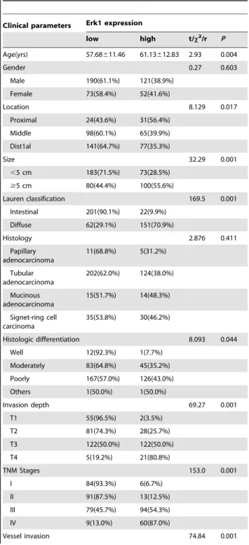
Table 2.Correlation between Erk1 expression and clinicopathological features of gastric cancer.
Clinical parameters Erk1 expression
low high t/x2/r P
Age(yrs) 57.68611.46 61.13612.83 2.93 0.004
Gender 0.27 0.603
Male 190(61.1%) 121(38.9%) Female 73(58.4%) 52(41.6%)
Location 8.129 0.017
Proximal 24(43.6%) 31(56.4%) Middle 98(60.1%) 65(39.9%) Dist1al 141(64.7%) 77(35.3%)
Size 32.29 0.001
,5 cm 183(71.5%) 73(28.5%)
$5 cm 80(44.4%) 100(55.6%)
Lauren classification 169.5 0.001 Intestinal 201(90.1%) 22(9.9%)
Diffuse 62(29.1%) 151(70.9%)
Histology 2.876 0.411
Papillary adenocarcinoma 11(68.8%) 5(31.2%) Tubular adenocarcinoma 202(62.0%) 124(38.0%) Mucinous adenocarcinoma 15(51.7%) 14(48.3%) Signet-ring cell carcinoma 35(53.8%) 30(46.2%)
Histologic differentiation 8.093 0.044 Well 12(92.3%) 1(7.7%)
Moderately 83(64.8%) 45(35.2%) Poorly 167(57.0%) 126(43.0%) Others 1(50.0%) 1(50.0%)
Invasion depth 69.27 0.001
T1 55(96.5%) 2(3.5%)
T2 81(74.3%) 28(25.7%)
T3 122(50.0%) 122(50.0%)
T4 5(19.2%) 21(80.8%)
TNM Stages 153.0 0.001
I 84(93.3%) 6(6.7%)
II 91(87.5%) 13(12.5%)
III 79(45.7%) 94(54.3%)
IV 9(13.0%) 60(87.0%)
Resuts
Expression of TMPRSS4 in Archived Gastric Tissue Samples and Non-tumor Mucosa
TMPRSS4 protein was detected in 14 of 92 (15.22%) human non-tumor mucosa samples, and all samples expressed the protein at a low level. TMPRSS4 protein was detected in 245 of 436 (56.19%) cases of human gastric cancer, high expression of TMPRSS4 protein was detected in 203 (46.56%) tumors. TMPRSS4 was localized mainly in the cytoplasm or nucleus of primary cancer (Figure 1A, B, and C).
Expression of Erk1 in Archived Gastric Tissue Samples and Non-tumor Mucosa
Erk1 protein was detected in 16 of 92 (17.39%) human non-tumor mucosa samples, and all samples expressed the protein at a low level. Erk1 protein was detected in 231 of 436 (52.98%) cases of human gastric cancer, high expression of Erk1 protein was detected in 173(39.68%) tumors. Erk1 was localized mainly in the cytoplasm or nucleus of primary cancer (Figure 1D, E and F).
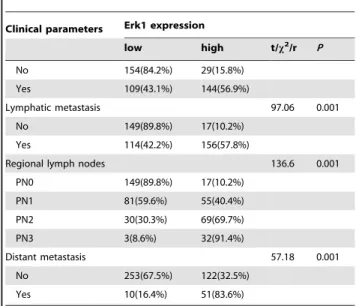
Table 2.Cont.
Clinical parameters Erk1 expression
low high t/x2/r P
No 154(84.2%) 29(15.8%) Yes 109(43.1%) 144(56.9%)
Lymphatic metastasis 97.06 0.001
No 149(89.8%) 17(10.2%) Yes 114(42.2%) 156(57.8%)
Regional lymph nodes 136.6 0.001
PN0 149(89.8%) 17(10.2%) PN1 81(59.6%) 55(40.4%) PN2 30(30.3%) 69(69.7%)
PN3 3(8.6%) 32(91.4%)
Distant metastasis 57.18 0.001
No 253(67.5%) 122(32.5%) Yes 10(16.4%) 51(83.6%)
doi:10.1371/journal.pone.0070311.t002
Figure 2. Kaplan-Meier curves with univariate analyses (log-rank) for patients with low TMPRSS4 expression versus high TMPRSS4 expression tumors.0: negative, 1: positive.
Correlation between TMPRSS4 Up-regulation and Clinical Features of Gastric Cancer
High expression of TMPRSS4 correlated with age, size, Lauren’s classification, depth of invasion, lymph node and distant metastases, regional lymph node stage and TNM stage (P,0.05) (Table 1). TMPRSS4 expression did not correlate with sex, tumor location, differentiation, or histological classification (P.0.05) (Table 1).
Correlation between Erk1 Up-regulation and Clinical Features of Gastric Cancer
High expression of Erk1 correlated with age, tumor location, size, depth of invasion, differentiation, Lauren’s classification, lymph node and distant metastases, regional lymph node stage and TNM stage (P,0.05) (Table 2). Erk1 expression did not correlate with sex, or histological classification (P.0.05) (Table 2). Correlation between TMPRSS4 Expression and Prognosis
In stage I, II and III tumors, the 5-year survival rate in patients with high expression of TMPRSS4 was significantly lower than that in patients with low expression (P,0.05) (Figure 2). In stage IV tumors, the expression of TMPRSS4 did not correlate with the 5-year survival rate (P.0.05) (Figure 2).
Correlation between Erk1 Expression and Prognosis In stage I, II and III tumors, the 5-year survival rate in patients with high expression of Erk1 was significantly lower than that in patients with low expression (P,0.05) (Figure 3). In stage IV tumors, the expression of Erk1 did not correlate with the 5-year survival rate (P.0.05) (Figure 3).
Multivariate Analysis of Clinicopathological Parameters and Prognosis
The factors with possible prognostic effects in gastric carcinoma were analyzed by Cox regression analysis. The study revealed that lymph node and distant metastases (P= 0.005), TNM stage (P= 0.005), expression of TMPRSS4 (P= 0.000) and Erk1 (P= 0.000) were independent prognostic factors of patients with gastric carcinoma. However, age, sex, tumor location and size, histological classification, tumor differentiation, invasion depth, Lauren’s classification, and regional lymph node stage had no prognostic value.
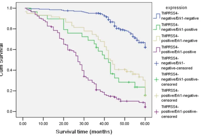
Association among Expression of TMPRSS4 and Erk1 Two hundred and four gastric cancer cases had low expression of both TMPRSS4 and Erk1, and one hundred and forty-four gastric cancer cases had high expression of both TMPRSS4 and
Figure 3. Kaplan-Meier curves with univariate analyses (log-rank) for patients with low Erk1 expression versus high Erk1 expression tumors in different TNM stage.0: negative, 1: positive.
Erk1. There was significant correlation between TMPRSS4 and Erk1 (x2= 155.1; P = 0.0001) (Table 3). We also detected the outcomes of TMPRSS4-positive/Erk1-negative, TMPRSS4-posi-tive/Erk1-positive, TMPRSS4-negative/Erk1-positive and TMPRSS4-negative/Erk1-negative gastric patients, and we found that there was a significant difference in overall comparisons (x2= 256.9; P = 0.000, Figure 4). The mean survival time were 42.862.23, months for TMPRSS4(+)/Erk1(2) group, 27.961.27 months for TMPRSS4(+)/Erk1(+), 54.760.72 months for TMPRSS4(2)/Erk1(2), 39.263.19 months for TMPRSS4(2)/ Erk1(+), respectively.
Discussion
Of the many molecules involved in the carcinogenesis and development of human tumors, proteases have been extensively studied as important participants in the carcinogenesis of many tumors. Recently, much attention has been focused on the role of type II transmembrane serine proteases (TTSPs) during tumor development [15–16]. TTSPs are members of the family of cell surface-associated proteases that mediate a variety of normal cellular functions as well as tumor invasion and metastasis.
TMPRSS4 is a TTSP that has been known to be upregulated in several cancers, particularly in pancreatic and thyroid cancers [4,17], and the expression of TMPRSS4 has been correlated with the metastatic potential of pancreatic cancer [3]. TMPRSS4 is necessary and sufficient for the human lung cancer cell line NCI-H322, the colon adenocarcinoma cell line Colo205, and the colorectal cancer cell line HCT15 to accomplish migration and invasion [18]. The study revealed that the expression of TMPRSS4 in gastric cancer lesions were closely associated with the age, size of tumor, location of tumor, depth of invasion, vessel invasion, lymph node and distant metastasis and TNM stage. The expression of TMPRSS4 was found to significantly correlate with the prognosis of gastric cancer. In stages I, II and III, the 5-year survival rates of patients with high expression of TMPRSS4 was significantly lower than those in patients with low expression. In stage IV, TMPRSS4 expression did not correlate with the 5-year
Figure 4. Kaplan-Meier curves with univariate analyses (log-rank) for patients withpositive/Erk1-negative, TMPRSS4-positive/Erk1-positive, TMPRSS4-negative/Erk1-positive and TMPRSS4-negative/Erk1-negative gastric patients.
doi:10.1371/journal.pone.0070311.g004
Table 3.Correlation between TMPRSS4 and Erk1 in gastric cancer.
Erk1 x2 Pvalue
Positive (%) Negative (%)
TMPRSS4
Positive (%) 144 (70.9%) 59 (29.1%)
Negative (%) 29 (12.4%) 204 (87.6%) 155.1 0.0001
survival rate. Further multivariate analysis suggested that the depth of invasion, lymph node and distant metastasis, TNM stage, and up-regulation of TMPRSS4 were independent prognostic indicators for gastric cancer. Overexpression of TMPRSS4 has a critical role in radiation-induced long-term dissemination and metastasis of residual hepatocellular carcinoma by facilitating epithelial-mesenchymal transition (EMT) [19]. Expression of TMPRSS4 in tumours with squamous cell carcinoma (SCC) histology was found to be significantly higher than those with adenocarcinoma (AC) histology. Kaplan-Meier curves demon-strated that high levels of TMPRSS4 were significantly associated with reduced overall survival in the patients with SCC histology, whereas no correlation was found for the AC histology [20].
Erk1 involved in the carcinogenesis through regulating various cellular processes such as proliferation, differentiation, and cell cycle progression in response to a variety of extracellular signals. Hepatoma-derived growth factor is involved in the gastric carcinogenesis process and promotes proliferation and metastasis via Erk1/2 activation [21]. ERK1 as an important mediator of lysophosphatidic acid signaling leading to upregulation of sphin-gosine kinase 1 (SphK1) and point to SphK1 and sphinsphin-gosine-1- sphingosine-1-phosphate production as potential therapeutic targets in gastric cancer [22]. The interaction between FVIIa and TF induces protease-activated receptor 2 activation, thereby triggers the
ERK1/2 and IkB-a/NF-kB signal transduction pathway to
regulate the gene expression of IL-8, TF, and caspase-7, and ultimately promotes SW620 cell proliferation and migration [23]. The expression of Erk1 in gastric cancer and its clinical significance are not understood. The current study was to examine the expression of Erk1 in 436 surgical specimens of gastric cancer by immunohistochemistry. The study showed that high expression of Erk1 protein was detected in 173(39.68%) tumors. High expression of Erk1 correlated with age, tumor location, size, depth of invasion, differentiation, Lauren’s classification, lymph node and distant metastases, regional lymph node stage, TNM stage and poor prognosis.
We further examined TMPRSS4 expression in gastric cancer specimens and its correlation with Erk1 expression. We found a positive correlation between TMPRSS4 and Erk1 expression, suggesting that TMPRSS4 may be involved in the carcinogenesis, progression and metastasis of gastric cancer through promotion of cell proliferation by Erk1 activation.
Author Contributions
Conceived and designed the experiments: ZL ZZ. Performed the experiments: YW BL. Analyzed the data: JC. Contributed reagents/ materials/analysis tools: YW BL. Wrote the paper: ZL YW.
References
1. Hooper JD, Clements JA, Quigley JP, Antalis TM (2001) Type II transmembrane serine proteases. Insights into an emerging class of cell surface proteolytic enzymes. J Biol Chem 276: 857–860.
2. Netzel-Arnett S, Hooper JD, Szabo R, Madison EL, Quigley JP, et al. (2003) Membrane anchored serine proteases: a rapidly expanding group of cell surface proteolytic enzymes with potential roles in cancer. Cancer Metastasis Rev 22: 237–258.
3. Wallrapp C, Ha¨hnel S, Mu¨ller-Pillasch F, Burghardt B, Iwamura T, et al. (2000) A novel transmembrane serine protease (TMPRSS3) overexpressed in pancreatic cancer. Cancer Res 60: 2602–2606.
4. Kebebew E, Peng M, Reiff E, Duh QY, Clark OH, et al. (2005) ECM1 and TMPRSS4 are diagnostic markers of malignant thyroid neoplasms and improve the accuracy of fine needle aspiration biopsy. Ann Surg 242: 353–361. 5. Kim S, Kang HY, Nam EH, Choi MS, Zhao XF, et al. (2010) TMPRSS4
induces invasion and epithelial-mesenchymal transition through upregulation of integrin alpha5 and its signaling pathways. Carcinogenesis 31: 597–606. 6. Lu Z, Xu S (2006) ERK1/2 MAP kinases in cell survival and apoptosis. IUBMB
Life 58: 621–631.
7. Lee HS, Lee HK, Kim HS, Yang HK, Kim WH (2003) Tumour suppressor gene expression correlates with gastric cancer prognosis. J Pathol 200: 39–46. 8. Lee HS, Lee HK, Kim HS, Yang HK, Kim YI, et al. (2001) MUC1, MUC2,
MUC5AC, and MUC6 expressions in gastric carcinomas: their roles as prognostic indicators. Cancer 92: 1427–1434.
9. Zhang D, Salto-Tellez M, Putti TC, Do E, Koay ES (2003) Reliability of tissue microarrays in detecting protein expression and gene amplification in breast cancer. Mod Pathol 16: 79–84.
10. Lee HS, Cho SB, Lee HE, Kim MA, Kim JH, et al. (2007) Protein expression profiling and molecular classification of gastric cancer by the tissue array method. Clin Cancer Res 13: 4154–4163.
11. Kolev Y, Uetake H, Iida S, Ishikawa T, Kawano T, et al. (2007) Prognostic significance of VEGF expression in correlation with COX-2, microvessel density and clinicopathological characteristics in human gastric carcinoma. Annals of Surgical Oncology 14: 2738–2747.
12. Mizokami K, Kakeji Y, Oda S, Irie K, Yonemura T, et al. (2006) Clinicopathologic significance of hypoxia-inducible factor 1 alpha overexpres-sion in gastric carcinomas. Journal of Surgical Oncology 94: 149–154.
13. Song LB, Liao WT, Mai HQ, Zhang HZ, Zhang L, et al. (2006) The clinical significance of twist expression in nasopharyngeal carcinoma. Cancer Lett 242: 258–265.
14. Chou YY, Jeng YM, Lee TT, Hu FC, Kao HL, et al. (2007) Cytoplasmic CD24 expression is a novel prognostic factor in diffuse-type gastric adenocarcinoma. Ann Surg Oncol 14: 2748–2758.
15. Szabo R, Bugge TH (2008) Type II transmembrane serine proteases in development and disease.Int J Biochem Cell Biol 40: 1297–1316.
16. Antalis TM, Bugge TH, Wu Q (2011) Membrane-anchored serine proteases in health and disease.Prog Mol Biol Transl Sci 99: 1–50.
17. Jarzab B, Wiench M, Fujarewicz K, Simek K, Jarzab M, et al. (2005) Gene expression profile of papillary thyroid cancer: sources of variability and diagnostic implications. Cancer Res 65: 1587–1597.
18. Jung H, Lee KP, Park SJ, Park JH, Jang YS, et al. (2008) TMPRSS4 promotes invasion, migration and metastasis of human tumor cells by facilitating an epithelial-mesenchymal transition. Oncogene 27: 2635–2647.
19. Li T, Zeng ZC, Wang L, Qiu SJ, Zhou JW, et al. (2011) Radiation enhances long-term metastasis potential of residual hepatocellular carcinoma in nude mice through TMPRSS4-induced epithelial-mesenchymal transition. Cancer Gene Ther 18: 617–626.
20. Larzabal L, Nguewa PA, Pio R, Blanco D, Sanchez B, et al. (2011) Overexpression of TMPRSS4 in non-small cell lung cancer is associated with poor prognosis in patients with squamous histology. Br J Cancer 105: 1608– 1614.
21. Mao J, Xu Z, Fang Y, Wang H, Xu J, et al. (2008) Hepatoma-derived growth factor involved in the carcinogenesis of gastric epithelial cells through promotion of cell proliferation by Erk1/2 activation. Cancer Sci 99: 2120–2127. 22. Ramachandran S, Shida D, Nagahashi M, Fang X, Milstien S, et al. (2010)
Lysophosphatidic acid stimulates gastric cancer cell proliferation via ERK1-dependent upregulation of sphingosine kinase 1 transcription. FEBS Lett 584: 4077–4082.