CLINICAL SCIENCE
In search of a tolerance-induction strategy for cow’s milk
allergies: significant reduction of beta-lactoglobulin
allergenicity via transglutaminase/cysteine polymerization
Celso Eduardo Olivier,I,IIRegiane Patussi dos Santos Lima,II Daiana Guedes Pinto,IIRaquel Aca´cia Pereira Gonc¸alves dos Santos,IIGrayce Katlen Moreno da Silva,IISoˆnia Letı´cia Silva Lorena,IIIMariana Battaglin Villas-Boas,IVFla´via Maria Netto,IVRicardo de Lima ZollnerIIUniversity of Campinas, Faculty of Medical Sciences, Department of Internal Medicine, Laboratory of Immunology and Experimental Allergy, Campinas/
SP, Brazil.IIInstituto Alergoimuno de Americana, Americana/SP, Brazil.IIIUniversity of Campinas, Department of Internal Medicine, Gastroenterology Unit, Campinas/SP, Brazil.IVUniversity of Campinas, Faculty of Food Engineering, Department of Food and Nutrition, Campinas/SP, Brazil.
OBJECTIVE: To explore the use of b-lactoglobulin polymerized using microbial transglutaminase and heating to identify whether protein polymerization could reduce in vivo allergenicity and maintain in vitro and ex vivo
immunoreactivity for use in tolerance-induction protocols.
METHODS:Based on previous protocols applied in mice and children, we performedin vivochallenges (using a skin prick test) with native and polymerized b-lactoglobulin in adult patients with an IgE-mediated allergy to b -lactoglobulin. In vitro humoral immunoreactivity was analyzed using immunoblotting. Cell-mediated immunor-eactivity was analyzed using ex vivo challenges with native and polymerized b-lactoglobulin and monitored by leukocyte adherence inhibition tests.
RESULTS:The skin tests demonstrated that there was a significant reduction in immediate cutaneous reactivity after polymerization. Polymerization did not decrease the immunoblotting detection of s-IgE specific tob-lactoglobulin. Cell-mediated immunoreactivity, as assessed byex vivochallenges and leukocyte adherence inhibition tests, did not
exhibit significant differences between leukocytes challenged with native versus polymerizedb-lactoglobulin.
CONCLUSIONS:The polymerization ofb-lactoglobulin decreasedin vivoallergenicity and did not decreasein vitro
humoral or ex vivocell-mediated immunoreactivity. Therefore, we conclude that inducing polymerization using
transglutaminase represents a promising technique to produce suitable molecules for the purpose of designing oral/ sublingual tolerance induction protocols for the treatment of allergies.
KEYWORDS: Beta-lactoglobulin; Milk Hypersensitivity; Leukocyte Adherence Inhibition Test; Polymerization; Immune Tolerancez.
Olivier CE, Lima RP, Pinto DG, Santos RA, Silva GK, Lorena SL, et al. In search of a tolerance-induction strategy for cow’s milk allergies: significant reduction of beta-lactoglobulin allergenicity via transglutaminase/cysteine polymerization. Clinics. 2012;67(10):1171-1179.
Received for publication onMarch 24, 2012;First review completed onApril 24, 2012;Accepted for publication onJune 19, 2012
E-mail: [email protected]
Tel.: 55 19 3289 3709
INTRODUCTION
The primary strategy currently used to manage cow’s milk allergies is to diagnose the offending protein (whey proteins, cheese caseins or both) and prescribe its dietary elimination (1-3). Nevertheless, avoidance strategies may also contribute to a loss of tolerance (4). Oral tolerance induction protocols using whole cow’s milk have been described (5,6). However, the use of a natural extract of an
allergen may elicit allergic reactions, thereby limiting the progress of desensitization. Modified allergens (allergoids) have been used in desensitization protocols to minimize the allergic reactions produced by the native allergen (7). Less allergenic immuno-equivalent proteins may be obtained, for example, using recombinant DNA technology (8), but this method is laborious and expensive. More accessible approaches, including extensive baking, have been used for milk tolerance induction based on the assumption that the destruction of conformational epitopes yields a safer protein for oral administration (9). However, this type of physical denaturation may also induce difficult-to-control chemical modifications in the allergen, making the char-acterization of the resultant allergen almost impossible while simultaneously destroying the linear epitopes neces-sary to prime tolerance induction (10). The rationale for Copyrightß2012CLINICS– This is an Open Access article distributed under
the terms of the Creative Commons Attribution Non-Commercial License (http:// creativecommons.org/licenses/by-nc/3.0/) which permits unrestricted non-commercial use, distribution, and reproduction in any medium, provided the original work is properly cited.
performing safe and efficient oral tolerance induction protocols ideally includes the use of a protein that is immunogenic (harboring all relevant T cell epitopes) but less allergenic (eliciting weaker clinical reactions) (11). The allergenicity of a modified antigen may be evaluated using in vivo challenge tests (cutaneous or oral) and/or ex vivo challenge tests, such as the basophil degranulation assay. The extent of the allergen-elicited cell-mediated immunor-eactivity responsible for priming the subsequent immune response and/or tolerance may be analyzed prior to treatment using T cell proliferation assays (12) or after treatment by detecting markers of immune tolerance from cultured PBMCs or searching for inducible Tregsexpressing
IL-10 and TGF-b (13). As a model for this study, we employed enzymatic polymerization, a process that is widely used to produce industrial foods. The use of biotechnology to modify the physical properties of dairy products is currently practiced and can alter the allergeni-city of native proteins, thereby resulting in the creation of new and troublesome allergens or less allergenic (hypoal-lergenic) products (14). Among bovine whey proteins, b -lactoglobulin (b-Lg) is a prevalent allergen, as demonstrated by oral challenges, skin scratch tests and skin prick tests (SPTs) (15). Because polymerization enzymes, such as microbial transglutaminase (TG), are applied to decrease syneresis, polymerizedb-Lg is found in industrialized dairy products, such as curd cheese and yogurt. TG catalyzes acyl-transfer reactions between the gamma-carboxyamide groups of glutamine residues and the epsilon-amino group of lysine, thereby causing the inter- or intramolecular crosslinking of proteins (16). Previous studies in mice have demonstrated that the polymerization ofb-Lg by TG in the presence of cysteine may reduce b-Lg antigenicity before and after digestion (17,18). SPTs have also indicated that polymerizedb-Lg may be less allergenic than nativeb-Lg in children with an IgE-mediated cow’s milk allergy (19). However,ex vivo cell-mediated immunoreactivity to poly-merizedb-Lg has not yet been studied in humans. Seeking an immunogenic but less allergenic molecule for use in desensitization protocols and in accordance with the directive of the FAO/WHO decision tree for analyzing the allergenicity of foods derived from biotechnological pro-cesses (20), we examined the allergenicity and immunor-eactivity of native and polymerizedb-Lg in adults with and without a confirmed diagnosis of an IgE-mediated allergy to b-Lg. The FAO/WHO directive recommends performing the diagnosis of a specific IgE-mediated allergy following SPTs using the native and bioprocessed proteins in people with proven allergies to the native protein. Because the detection of a specific IgE using SPT or ImmunoCAP assays is associated with certain pitfalls (21), we also performed confirmatory SDS-PAGE immunoblots to improve analyti-cal sensitivity and detect false-negative or false-positive results (22). This analysis was conducted in accordance with the GRADE approach (grades of recommendation, assess-ment developassess-ment and evaluation) for grading the quality of evidence of diagnostic tests (23). Therefore, as a secondary endpoint, we assessed the sensitivity of the ImmunoCAP and SPT assays for detecting specific IgE antibodies against b-Lg in patients who exhibited specific IgE sensitization, as confirmed by SDS-PAGE immunoblot-ting. Ex vivocell-mediated immunoreactivity for b-Lg has previously been evaluated with the leukocyte migration inhibition test (24). We used an equivalent but technically
simpler assay to assessb-Lg cell-mediated immunoreactiv-ity: the leukocyte adherence inhibition test (LAIT) (25-28).
MATERIALS AND METHODS
Ethics Statements
This study was submitted to and approved by the Institutional Research Ethics Board and was registered in the Brazilian National Ethics Research System (SISNEP 409/ 2008). In accordance with the Helsinki Declaration, signed consent forms were obtained from all subjects.
Study design and subjects
This descriptive case-control study was designed to examine the following parameters: A) thein vivo allergeni-city of nativeb-Lg and transglutaminase/cysteine-polymer-ized b-Lg (TgPolb-Lg) in symptomatic adult patients exhibiting specific IgE antibodies againstb-Lg (bs-IgE); B) thein vitro humoral immunoreactivity of nativeb-Lg and heated polymerizedb-Lg (HtPolb-Lg) in symptomatic adult patients exhibiting bs-IgE, as diagnosed by SDS-PAGE immunoblotting, SPT and ImmunoCAP assays; and C) the ex vivocell-mediated immunoreactivity ofb-Lg and TgPolb -Lg, independent of patientbs-IgE status. The participants were divided into three groups according to their clinical presentation and analytical results. The first group (group A) included 45 patients (17 males; mean age: 46.2 years, SD: 12.2 years) with convincing clinical histories of reproducible adverse reactions to bovine milk. All subjects exhibitedb s-IgE detectable by SDS-PAGE immunoblotting. The second group (group B) was used as a control for the immunoblot-ting analysis performed in the first group and included 20 individuals selected based on an evident tolerance to cow’s milk, an absence ofbs-IgE by ImmunoCAP assay and SPT non-reactivity tob-Lg or TgPolb-Lg (6 males; mean age: 21.9 years, SD: 17.6 years). The third group (group C) included 49 subjects with atopic respiratory and/or dermatological diseases (19 males; mean age: 28.7 years, SD: 20.6 years) regardless ofbs-IgE status. This group was used to compare the ex vivo cell-mediated immunoreactivity between b-Lg and TgPolb-Lg by comparing the mean ex vivo antigenic challenge results determined using the leukocyte adherence inhibition test (LAIT).
Allergens, antibodies and reagents
Chromatographically purified b-Lg (.95% pure) was provided by Davisco Foods, Inc. (Le Sueur, USA). A goat polyclonal secondary antibody to human IgE–epsilon chain (ab9159) and peroxidase (HRP)-conjugated rabbit polyclo-nal secondary antibody to goat IgG (ab6741) were acquired from Abcam, Inc. (Cambridge, MA, USA). A commercial preparation of transglutaminase from Streptoverticillium mobaraense(ActivaH) containing 99% maltodextrin and 1% transglutaminase (TG) with a declared activity of 100 U/g was donated by Ajinomoto Co. (Tokyo, Japan). Cysteine, sodium dodecyl sulfate (SDS) and 3,3’-diaminobenzidine tetrahydrochloride (DAB) were obtained from Sigma-Aldrich Chemie GmbH (Steinheim, Germany). Tween-20 and b-mercaptoethanol were obtained from Merck KgaA (Darmstadt, Germany).
b-lactoglobulin polymerization
forma-tion of tetramers (HtPolb-Lg) because the high-molecular weight polymers generated by transglutaminase activity did not penetrate the SDS-PAGE gel.
For skin tests, cysteine/transglutaminase b-Lg polymer-ization was performed as described previously (18).
Preparation of solutions for skin tests
To standardize skin tests with respect to the conditions of previous studies (2), b-Lg and TgPolb-Lg skin solutions were diluted to a concentration of 10,000 PNU/mL. Nitrogen levels were quantified by Kjeldahl’s method (AOAC 991.22), which indicated that each gram of lyophilized b-Lg contained 14.4 g of nitrogen and each gram of lyophilized TgPolb-Lg contained 11.1 g of nitrogen. For the skin tests, b-Lg, TgPolb-Lg and the control substances were solubilized at a pH of 7.0 and diluted to their final concentrations using a 20% w/v glycerol/saline solution (20% GS). The b-Lg solution was diluted to a concentration of 0.7 mg/mL using 20% GS, and the TgPolb -Lg solution was diluted to a final concentration of 0.9 mg/ mL using 20% GS. The TG and cysteine skin reactivities were tested simultaneously. The TG solution for the skin tests was diluted to a concentration of 0.2 mg/mL using 20% GS, and the cysteine solution for the skin tests was diluted to 0.3 mg/mL using 20% GS. Histamine (1 mg/mL in 20% GS) was used as a positive control, and the negative-control solution was 20% GS.
Skin test methodology
Skin tests were performed on the volar aspect of the patient’s forearms after withholding antihistamines for at least 10 days. Skin prick tests (SPTs) were performed using a disposable sterile acrylic needle consisting of a 1-mm lancet attached to a blunt base (PunctorH, Alko do Brasil Ltda, Rio de Janeiro, Brazil). The needle was introduced at a 90
˚
angle to the skin through an allergen solution droplet, removed after 1 second and discarded. The final reading was performed 15 minutes later, and the wheal’s greatest diameter and its perpendicular length at that point were measured to estimate the mean wheal diameter (MWD) (29). Because there was no reaction to the negative control, a MWD$3 mm was considered significant for sensitization.The samples were analyzed by SDS-PAGE under redu-cing conditions using a Mini Protean Tetra Cell apparatus (Bio-Rad, CA, USA) and 5%T/20%C gel gradients (30). Purifiedb-Lg samples (5 mg/mL) were prepared in a buffer containing 2% SDS and 5% b-mercaptoethanol and then heated for 10 min at 96
˚
C. An aliquot of 5mL, which was equivalent to 25mg of protein, was applied to each lane. A 10-170 kDa molecular mass pre-stained protein ladder (PageRulerH, Fermentas, Hanover, MD, USA) was used to identify approximate molecular weights.Following electrophoresis, the proteins were electro-blotted onto 0.45-mm pore-size nitrocellulose membranes (Bio-Rad) using a Mini Trans-Blot Cell apparatus (Bio-Rad). Membranes were incubated overnight at 4
˚
C with gentle agitation in 3 mL phosphate-buffered saline (PBS), pH 7.0, containing 3mL of the individual patient’s serum, followed by three washes in PBST (PBS containing 0.05% Tween-20). The membranes were incubated under gentle agitation for 2 hours in polyclonal goat IgG anti-human IgE (15 mg in 3 mL PBS), washed three times with PBST and incubated with horseradish peroxidase (HRP)-conjugated rabbit anti-goat IgG (30mg in 3 mL PBS) for 2 hours. After three washeswith PBST, HRP activity was stimulated by incubating the membranes in 10 mL PBS containing 0.05% DAB followed by the addition of 50mL developing solution (CoCl2.6H2O
plus NiSO4) and 20 mL of hydrogen peroxide (H2O2) (31).
When impregnation and color development were complete, the membranes were washed in distilled water and subse-quently dried. Images of the membranes were digitalized, and the relative densitometry of the bands was analyzed using ImageJ software (Windows version 1.44 of NIH ImageJ, http://rsb.info.nih.gov/ij/index.html) by measuring the average optical density (OD). Adjusted optical density (AOD) was calculated following background correction by subtracting the average OD from a user-defined area within the image from the average OD of a similar area defined around the measured bands (32).
Polymerization of b-Lg via heating created 72-kDab-Lg tetramers (HtPolb-Lg) exhibiting an electrophoretic mobility similar to BSA (66.7 kDa) (33). Although the b-Lg samples were purified, we performed parallel in vitro serum competition assays with BSA (1 mg/mL) to rule out the possibility of BSA interference in the assay.
The concentrations of serum IgE antibodies specific forb -Lg were measured by ImmunoCAP assays (Pharmacia, Uppsala, Sweden), and the results were expressed in kU/L. A concentration greater than 0.35 kU/L was considered significant for sensitization (34).
Leukocyte adherence inhibition test
Leukocytes from heparin-treated blood samples drawn by venipuncture from the forearm were separated from fresh blood by sedimentation at 37
˚
C for one hour. Aliquots of 100 mL of plasma were incubated with 10 mL of each antigen extract (the same solutions used in the skin tests: b-Lg at 0.7 mg/mL in 20% GS and TgPolb-Lg at 0.9 mg/mL in 20% GS) for 30 minutes under gentle agitation (200 rpm) at 37˚
C. Plasma without antigens was used as a control. After incubation, the plasma was placed into a standard Neubauer hemocytometer counting chamber with a plain, non-metallic glass surface and left to stand for 2 hours at 37˚
C in a humidified atmosphere to allow leukocytes to adhere to the glass. The leukocytes were then counted, the coverslip was gently removed, and the chamber was washed by immersion in a beaker with PBS at 37˚
C. A drop of PBS was subsequently added to the hemocytometer chamber, and a clean coverslip was placed over it. The remaining cells were counted in the same squares examined previously. The percentage of cell adherence (cell number after washing/cell number before washing) was calculated, and leukocyte adherence inhibition (LAI) was estimated based on the ratio between the percentage of adherent cells from the challenged groups versus the control groups: LAI = [1-(% adherent cells of challenged sample/% adherent cells of control sample)]x100.Statistical analyses
LAIT results betweenb-Lg and TgPolb-Lg. For all analyses, a p-value of less than 0.05 was considered significant.
RESULTS
Group A: ImmunoCAP s-IgE
The mean level of IgE antibodies specific forb-Lg was 1.5 kU L-1 (CI: 0.5 to 2.4; SD: 3.2 kU/L). Specific IgE antibodies were detected using ImmunoCAP assays in 19 patients (sensitivity = 42.2 %). The test results are presented in Table 1.
Group A: Skin Prick Test
The test results are presented in Table 1. The mean MWD for histamine (positive control) obtained by SPT was 6.8 mm
(CI: 6.2 to 7.3; SD: 1.8 mm).b-Lg was reactive in SPTs in 26 patients (sensitivity = 57.7%). The mean MWD for b-Lg obtained by SPT was 2.6 mm (CI: 1.9 to 3.4; SD: 2.5 mm). TgPolb-Lg was reactive in SPTs in 19 patients (sensitiv-ity = 42.2%). The mean MWD for TgPolb-Lg obtained by SPT was 1.6 mm (CI: 1.0 to 2.2; SD: 2.0 mm). The mean difference between the paired MWD means for b-Lg and TgPolb-Lg was 1.0 (p= 0.0075). There was no skin reactivity observed against TG, cysteine or the negative control solution in any patient (results not shown).
Comparison of categorical performance between assays
Analysis of the contingency table between the Im-munoCAP (19 positives) and SPT assays usingb-Lg (26
Table 1 -Test results from group A patients.
Patient
Age years
sIgE
bLg kU/L
SPT PC MWD
SPT
bLg MWD
SPT TgbLg MWD
AOD 72 kDa
AOD 36 kDa
AOD 18 kDa
1 57 0.6 10 5 0 37.6 16.4 0.4
2 45 0.6 7 4 4 10.0 0.0 0.0
3 51 0.0 8 5 0 20.4 0.0 0.0
4 48 0.5 8 4 4 10.7 9.6 2.1
5 32 0.0 7 0 0 16.5 13.5 1.8
6 34 0.0 6 3 3 21.0 8.0 6.1
7 47 12.6 9 0 0 46.6 7.0 0.0
8 26 0.0 11 0 3 18.8 9.9 0.0
9 18 2.1 6 0 0 48.8 10.2 0.0
10 53 0.0 5 0 4 24.1 12.1 0.0
11 39 0.0 8 0 0 10.4 13.4 8.1
12 46 1.2 5 0 0 13.2 5.9 0.0
13 55 5.2 5 0 0 23.2 0.0 0.0
14 42 0.0 9 0 3 24.1 7.8 3.4
15 65 0.0 5 0 0 21.1 6.8 0.0
16 53 0.0 6 5 0 23.1 12.5 0.0
17 48 15.4 5 3 0 35.9 0.0 0.0
18 49 4.0 6 0 0 7.6 0.0 0.0
19 58 0.0 6 0 0 24.8 0.0 0.0
20 59 0.4 6 4 5 15.6 0.0 0.0
21 43 0.5 6 4 4 19.2 0.0 0.0
22 28 0.0 9 6 0 19.4 0.0 0.0
23 39 4.0 6 3 3 14.6 0.0 0.0
24 55 0.0 7 0 0 25.9 0.0 0.0
25 51 0.0 7 3 0 16.4 0.0 0.0
26 42 9.2 9 9 6 21.0 0.0 0.0
27 52 1.8 8 5 5 24.3 12.2 12.5
28 36 0.0 8 3 3 10.5 9.2 0.0
29 22 0.0 7 4 0 7.1 0.0 0.0
30 41 1.2 7 6 3 12.0 0.0 0.0
31 56 0.0 7 6 4 12.2 12.2 12.5
32 58 0.0 4 0 4 17.0 0.0 0.0
33 56 0.0 8 4 0 19.9 0.0 0.0
34 29 0.0 6 4 4 10.9 0.0 0.0
35 44 0.4 7 0 0 9.8 0.0 0.0
36 48 0.0 6 5 3 20.4 0.0 0.0
37 70 0.0 7 4 0 8.2 0.0 0.0
38 64 4.6 5 0 0 10.0 0.0 0.0
39 40 1.2 0 0 0 11.7 5.8 4.4
40 61 0.0 7 4 0 14.6 0.0 0.0
41 50 3.8 10 6 4 5.5 5.4 0.0
42 63 0.0 8 6 0 5.0 0.0 7.3
43 23 0.0 6 5 5 14.1 0.0 0.0
44 46 0.0 8 0 0 6.0 3.5 13.9
45 38 0.0 7 0 0 7.2 0.0 0.0
positives) for the 45 patients with positive immunoblots via chi-squared (a,0.05 and p= 0.39) and Fisher exact tests (a,0.05 and p= 0.46) failed to reveal significant differences.
Combined categorical analytical sensitivity
Combined SPTs with b-Lg and TgPolb-Lg exhibited cutaneous reactivity in 30 patients (sensitivity = 66.6%). The combination of ImmunoCAP and SPT assays forb-Lg revealed reactivity in 34 patients (sensitivity = 75.5%). A combination of the three tests detected cutaneous reactivity in 38 patients (sensitivity = 84.4%).
Immunoblot assays
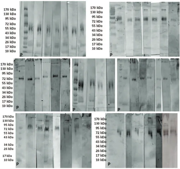
Figure 1 shows the purified beta-lactoglobulin electro-blotted 0.45-mm pore-size nitrocellulose membranes after incubation with patient’s serum, primary antibody (goat IgG anti-human IgE), secondary antibody (HRP-conjugated rabbit anti-goat IgG) and DAB revelation from subjects of group A. Our immunoblot analysis revealed 18-kDa bands
(b-Lg monomers) in 11 patients (24.4%) with a mean AOD of 1.6 (CI: 0.5 to 2.7). The immunoblots also revealed 36-kDa bands (b-Lg dimers) in 19 patients (42.2%) with a mean AOD of 4.0 (CI: 2.4 to 5.6). Additionally, there were bands at 72-kDa (b-Lg tetramers) in 45 patients (100%) with a mean AOD of 17.7 (CI: 14.73 to 20.6). The mean of the difference between the AODs of the 18-kDa bands and 36-kDa bands was 2.4 (p= 0.00027, CI: 0.8 to 3.9). The mean of the differences between the AODs of the 18-kDa bands and 72-kDa bands was 16.0 (p,0.0001, CI: 12.7 to 19.4). The mean of the difference between the AODs of the 36-kDa bands and 72-kDa bands was 13.6 (p,0.0001, CI: 10.7 to 16.5). The blot membranes incubated with the serum subjected to BSA competition did not exhibit any difference compared with those incubated with the corresponding serum not subjected to BSA competition (see Figure 2). Of the 20 patients from group B, representing milk-tolerant controls with non-reactive SPTs forb-Lg and TgPolb-Lg and absentbs-IgE based on ImmunoCAP, only one patient (5%) exhibited a 72-kDa band with an AOD = 10.5 (see Figure 3).
Leukocyte adherence inhibition tests
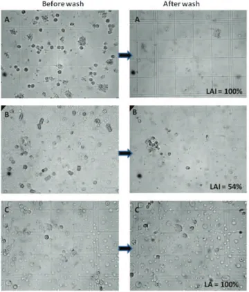
In group C, the mean leukocyte adherence inhibition (LAI) produced byex vivochallenge with b-Lg was 39.6% (SD: 36.8%; CI: 29.0% to 50.2%), whereas the LAI produced by exvivochallenge with TgPolb-Lg was 47.7 (SD: 38.4; CI: 36.6 to 58.7). The difference of the means based on a paired t-test was not significant (p= 0.07). The correlation between challenges was significant (Spearman r = 0.65 CI = 0.44 to 0.79p,0.0001). Results are presented in Table 1. Figure 4 depicts the microscope aspect before and after the PBS wash of two patients with LAI = 54% and 100%, respectively, and a control (without antigen challenge) with 100% leukocyte adherence.
DISCUSSION
Among the immunoreactive proteins found in whey in bovine milk,b-Lg is prominent because of its relatively high concentration (3 to 4 g/L on average) and low digestibility (35). The high incidence of sensitization to this protein (38) is understandable in light of the fact that b-Lg is not a component of human breast milk (36) and is usually absorbed in an unchanged form (37).b-Lg proteins naturally occur as dimers at a pH.3 (39). Heating b-Lg to 70
˚
C induces further polymerization (40). Purified heated b-Lg preparations generally contain monomers, dimers and tetramers when examined using reducing SDS-PAGE (33). Protein polymerization using TG in industrialized dairy products is a commonly employed technique to decrease whey syneresis and improve the gel strength/viscosity of set-type yogurt and curd cheese (41). Treatment with TG has been shown to decrease wheat allergenicity (42). Given the evidence that polymerization reduces the antigenicity ofb -Lg (18) and ovalbumin in mice (43), the application of polymerization techniques could potentially reduce the allergenicity of industrialized foods for humans and might be useful in producing ‘‘allergoids’’ for desensitization immunotherapy (7). A probable explanation for thisreduced allergenicity is that polymerization masks IgE antibody binding sites due to crosslinking of the protein. Polymerization may also create new epitopes, which may result in the development of hypersensitivities that are not related to the native protein.
The skin test results presented in this study indicated a significant reduction in the allergenicity of TgPolb-Lg
Figure 2 -Purified beta-lactoglobulin electroblotted on 0.45-mm pore-size nitrocellulose membranes after incubation with patient’s serum, primary antibody (goat IgG anti-human IgE), secondary antibody (HRP-conjugated rabbit anti-goat IgG) and DAB revelation. Immunoblot membranes exhibited bands of s-IgE against b-lactoglobulin (b-Lg). Lane 1: Molecular weight ladder (10 to 170 kDa); Lane 2: distinct band at 72 kDa in a patient from group A; and Lane 3: distinct band at 72 kDa from the same patient afterex vivoserum competition with BSA.
Figure 3 -Purified beta-lactoglobulin electroblotted on 0.45-mm pore-size nitrocellulose membranes after incubation with patient’s serum, primary antibody (goat IgG anti-human IgE), secondary antibody (HRP-conjugated rabbit anti-goat IgG) and DAB revelation. Immunoblot membranes exhibiting bands of s-IgE against b-lactoglobulin from four non-reactive patients in group B (Bn), from the single reactive patient from group B (Br), and from one reactive patient from group A (A) used as a control for the blotting technique. P = pre-stained 10-170 kDa molecular mass protein ladder (PageRulerH, Fermentas).
compared with native b-Lg. Polymerization changes the spatial distribution of allergenic epitopes, which may explain the decreased cutaneous reactivity of these epitopes. However, although the average skin reactivity to TgPolb-Lg was lower than the average reactivity tob-Lg, four patients exhibited no skin reaction tob-Lg but did exhibit significant skin reactivity to TgPolb-Lg. This finding indicates that transglutaminase polymerization may be deleterious for certain patients. Polymerization also increases the analytical detection ofbs-IgE antibodies by SDS-PAGE immunoblot-ting. Before subjecting b-Lg samples to SDS-PAGE, the samples were heated for 10 min at 96
˚
C. Heating stimulates the polymerization ofb-Lg and results in the detection of 72-kDa bands (tetramers) (33). Because b-Lg monomers are small proteins, they can be lost during the blotting process due to diffusion through the nitrocellulose membrane pores. Polymerization increases the molecular size of the protein, which results in greater membrane retention and better detection by specific antibodies. This effect was demon-strated by the significant increase in the number of 72-kDa bands compared with the number of 36-kDa and 18-kDa bands following polymerization. Polymerization also pro-duced a significant increase in the AOD of the 72-kDa bands. As a secondary objective, we used immunoblotting as a proxy for sensitization to study the accuracy of the SPTs and ImmunoCAP tests. Both tests were unable to detect a significant number of sensitized patients. The ImmunoCAP test demonstrated a false-negative rate of 57.8%, and the SPT demonstrated a false-negative rate of 42.3%. When the assays were combined, the false-negative rate decreased to 24.5%. These data demonstrate that these two analytical tests are complementary tools for diagnosing allergic sensitization, and a negative result from one assay does not necessarily indicate an absence of sensitization.Failure of leukocytes to adhere to glass after ex vivo challenge with specific antigens depends mostly on cellular mechanisms resulting from the interactions of T cells, B cells and macrophages (44-46). When activated in the presence of a specific antigen, E-rosette-forming cells release soluble supernatants that interfere with the glass adherence phenomenon observed in control, unchallenged plasma (27). The involvement of T cells was also suggested by the fact that anti-h(anti-theta) antibodies abolish the inhibition of adherence (47). Anti-hantibodies are specifically directed against the C3H antigen associated with T cells (48).
Leukotrienes, specifically LTC4 (49), are important factors in the process and are modulated by interferon secretion (50). As cysteinyl leukotriene generation by dendritic cells is a recognized step in the Dectin-2 recognition of allergens (51), the LAIT may be used to analyze this pathway indirectly.
The evaluation of cell-mediated immunoreactivity may allow the prediction of the overall ability of an allergoid to induce tolerance, but this subject remains an object of study. The actual generators of tolerance are the tolerogenic dendritic cells that prime naive T cells to become IL-10- and TGF-b1-releasing Treg cells (52,53). Dendritic cells collect
macromolecules from mucosal layers via transepithelial dendritic phagocytosis (54). Oral dendritic cells (oral Langerhans cells, oLC) constitutively express high levels of high affinity IgE receptors (FceRI) occupied by IgE antibodies that appear to be essential for the collection of antigens (55). The increase in molecular weight induced by enzymatic polymerization may theoretically promote phagocytosis and
interfere with the absorption ofb-Lg monomers by lipocalin receptors (56). Thus, the ability of IgE to bind an allergoid is essential and must be evaluated before the application of a tolerance-induction protocol in clinical trials. Trans-glutaminase may also be used to create covalent linkages between different proteins. Recombinant fusion of allergenic proteins and bacterial immunogenic proteins stimulates dendritic cell activity (57). This fusion ability of transgluta-minase may theoretically be used to increase the tolerogenic activity of allergoid polymers through covalent addition of coadjuvants. This topic warrants further study.
Additionally, the perception that cow’s milk proteins such as beta-lactoglobulin may be immunogenic should stimulate the creation of human milk banks to feed premature newborns with immature digestive and immune systems (58).
In conclusion, our data support the concept that enzy-matic polymerization is a tool that can decrease thein vivo allergenicity of b-Lg. As polymerization did not decrease thein vitrohumoral or ex vivocell-mediated immunoreac-tivity ofb-Lg, we conclude that inducing polymerization by transglutaminase represents a promising technique to produce suitable molecules for the purpose of designing oral/sublingual tolerance induction protocols for the treat-ment of allergies.
ACKNOWLEDGMENTS
We thank Ms. Michele Augusto Fernandes and Ms. Conceic¸a˜o Aparecida Vilella for their excellent technical assistance.
AUTHOR CONTRIBUTIONS
Olivier CE, Lorena SL, and Zollner RL conceived the study and manuscript, outlined the content and were integrally involved in the writing/rewriting of the final manuscript. Villa-Boas MB and Netto FM performed/supervised the transglutaminase-induced beta-lactoglobulin polymerization. Santos RA, Pinto DG, Lima RP, and Olivier CE performed/supervised the cutaneous tests. Lima RP, Pinto DG, and Olivier CE performed/supervised the immunoblotting. Lima RP, Pinto DG, Silva GK, and Olivier CE performed/supervised the leukocyte adherence inhibition tests.
REFERENCES
1. Fiocchi A, Schunemann HJ, Brozek J, Restani P, Beyer K, Troncone R, et al. Diagnosis and Rationale for Action Against Cow’s Milk Allergy (DRACMA): a summary report. J Allergy Clin Immunol. 2010;126(6):1119-28 e12, http://dx.doi.org/10.1016/j.jaci.2010.10.011
2. Goldman AS, Sellars WA, Halpern SR, Anderson DWJ, Furlow TE, Johnson CHJ. Milk Allergy. II. Skin Testing of Allergic and Normal Children with Purified Milk Proteins. Pediatrics. 1963;32:572-9. 3. Vlieg-Boerstra BJ, van der Heide S, Bijleveld CM, Kukler J, Duiverman
EJ, Wolt-Plompen SA, et al. Dietary assessment in children adhering to a food allergen avoidance diet for allergy prevention. Eur J Clin Nutr. 2006;60(12):1384-90, http://dx.doi.org/10.1038/sj.ejcn.1602468. 4. Barbi E, Berti I, Longo G. Food allergy: from the of loss of tolerance
induced by exclusion diets to specific oral tolerance induction. Recent Pat Inflamm Allergy Drug Discov. 2008;2(3):212-4, http://dx.doi.org/ 10.2174/187221308786241875.
5. Meglio P, Bartone E, Plantamura M, Arabito E, Giampietro PG. A protocol for oral desensitization in children with IgE-mediated cow’s milk allergy. Allergy. 2004;59(9):980-7, http://dx.doi.org/10.1111/j.1398-9995.2004.00542.x.
6. Longo G, Barbi E, Berti I, Meneghetti R, Pittalis A, Ronfani L, et al. Specific oral tolerance induction in children with very severe cow’s milk-induced reactions. J Allergy Clin Immunol. 2008;121(2):343-7, http:// dx.doi.org/10.1016/j.jaci.2007.10.029.
8. Valenta R, Niederberger V. Recombinant allergens for immunotherapy. J Allergy Clin Immunol. 2007;119(4):826-30, http://dx.doi.org/10.1016/ j.jaci.2007.01.025.
9. Kim JS, Nowak-Wegrzyn A, Sicherer SH, Noone S, Moshier EL, Sampson HA. Dietary baked milk accelerates the resolution of cow’s milk allergy in children. J Allergy Clin Immunol. 2011;128(1):125-31 e2, http:// dx.doi.org/10.1016/j.jaci.2011.04.036
10. Larche M, Akdis CA, Valenta R. Immunological mechanisms of allergen-specific immunotherapy. Nat Rev Immunol. 2006;6(10):761-71, http:// dx.doi.org/10.1038/nri1934.
11. Bird JA. Food Allergy: Update on Clinical Interventions Leading to Desensitization and Tolerance. Pediatric Allergy, Immunology, and Pulmonology. 2010;23(4):231-6, http://dx.doi.org/10.1089/ped.2010.0065. 12. Ferrari E, Breda D, Longhi R, Vangelista L, Nakaie CR, Elviri L, et al. In
Search of a Vaccine for Mouse Allergy: Significant Reduction of Mus m 1 Allergenicity by Structure-Guided Single-Point Mutations. Int Arch Allergy Immunol. 2012;157(3):226-37, http://dx.doi.org/10.1159/ 000327551.
13. Bohle B, Kinaciyan T, Gerstmayr M, Radakovics A, Jahn-Schmid B, Ebner C. Sublingual immunotherapy induces IL-10-producing T regulatory cells, allergen-specific T-cell tolerance, and immune deviation. J Allergy Clin Immunol. 2007;120(3):707-13, http://dx.doi.org/10.1016/ j.jaci.2007.06.013.
14. Lehrer SB, Bannon GA. Risks of allergic reactions to biotech proteins in foods: perception and reality. Allergy. 2005;60(5):559-64, http:// dx.doi.org/10.1111/j.1398-9995.2005.00704.x.
15. Costa AJF, Sarinho ESC, Motta MEFA, Gomes PN, Melo SMO, Silva GAP. Allergy to cow’s milk proteins: what contribution does hypersen-sitivity in skin tests have to this diagnosis? Pediatric Allergy and Immunology. 2011;22(1Pt 2):e133-e8, http://dx.doi.org/10.1111/j.1399-3038.2010.00988.x.
16. Cozzolino A, Di Pierro P, Mariniello L, Sorrentino A, Masi P, Porta R. Incorporation of whey proteins into cheese curd by using transglutami-nase. Biotechnol Appl Biochem. 2003;38(Pt 3):289-95, http://dx.doi.org/ 10.1042/BA20030102.
17. Villas-Boas MB, Fernandes MA, de Lima Zollner R, Netto FM. Effect of polymerization with transglutaminase on in vitro digestion and antigenicity of beta-lactoglobulin. International Dairy Journal. 2012;(In press).
18. Villas-Boas MB, Vieira KP, Trevizan G, Zollner RL, Netto FM. The effect of transglutaminase-induced polymerization in the presence of cysteine on beta-lactoglobulin antigenicity. International Dairy Journal. 2010;20(6):386-392, http://dx.doi.org/10.1016/j.idairyj.2010.01.004 19. Olivier CE, Villas-Boas MB, Netto FM, Zollner RL. Allergenicity of Bos d
5 in Children with Cow’s Milk Allergy is Reduced by Transglutaminase Polymerization. Ped Allergy Immunol Pulmonol. 2012;25(1)30-33, http://dx.doi.org/10.1089/ped.2011.0101
20. Expert Consultation on Allergenicity of Foods Derived from Biotechnology [database on the Internet]. Food and Agriculture Organization of the United Nations (FAO) and World Health Organization (WHO) 2001. Available from: http://www.fao.org/ag/ agn/food/pdf/allergygm.pdf.
21. Plebani M. Clinical value and measurement of specific IgE. Clin Biochem. 2003;36(6):453-69, http://dx.doi.org/10.1016/S0009-9120(03)00037-7.
22. Weusten JJ. A statistical approach to assess analytical sensitivity of diagnostic assays in the presence of false-positive results. J Virol Methods. 2008;151(2):308-10, http://dx.doi.org/10.1016/j.jviromet.2008.05.013. 23. Brozek JL, Akl EA, Jaeschke R, Lang DM, Bossuyt P, Glasziou P, et al.
Grading quality of evidence and strength of recommendations in clinical practice guidelines: Part 2 of 3. The GRADE approach to grading quality of evidence about diagnostic tests and strategies. Allergy. 2009;64(8):1109-16, http://dx.doi.org/10.1111/j.1398-9995.2009.02083.x. 24. Ashkenazi A, Levin S, Idar D, Or A, Rosenberg I, Handzel ZT. In Vitro
Cell-Mediated Immunologic Assay for Cow’s Milk Allergy. Pediatrics. 1980 September 1, 1980;66(3):399-402.
25. Bullen AW, Losowsky MS. Comparison of a leucocyte adherence test with the leucocyte migration inhibition test and skin reactivity to PPD. Clin Exp Immunol. 1978;31(3):408-13.
26. Halliday WJ, Miller S. Leukocyte adherence inhibition: a simple test for cell-mediated tumour immunity and serum blocking factors. Int J Cancer. 1972;9(3):477-83, http://dx.doi.org/10.1002/ijc.2910090304.
27. Halliday WJ. Historical Background and Aspects of the Mechanism of Leukocyte Adherence Inhibition. Cancer Res. 1979;39(2):558-63. 28. Kuratsuji T. Studies on leukocyte adherence inhibition test. Part II.
Clinical applications of LAI test to detect delayed type hypersensitivity in infants and children. Keio J Med. 1981;30(2):65-9, http://dx.doi.org/ 10.2302/kjm.30.65.
29. Bernstein IL, Li JT, Bernstein DI, Hamilton R, Spector SL, Tan R, et al. Allergy diagnostic testing: an updated practice parameter. Ann Allergy Asthma Immunol. 2008;100(3 Suppl 3):S1-148.
30. Weber K, Pringle JR, Osborn M. Measurement of molecular weights by electrophoresis on SDS-acrylamide gel. Methods Enzymol. 1972;26 PtC:3-27, http://dx.doi.org/10.1016/S0076-6879(72)26003-7.
31. Pukac LA, Carter JE, Morrison KS, Karnovsky MJ. Enhancement of diaminobenzidine colorimetric signal in immunoblotting. Biotechniques. 1997;23(3):385-8.
32. Gassmann M, Grenacher B, Rohde B, Vogel J. Quantifying Western blots: pitfalls of densitometry. Electrophoresis. 2009;30(11):1845-55, http:// dx.doi.org/10.1002/elps.200800720.
33. Lee DN, Moore EE, Merson RL. Electrophoresis of Cottage Cheese Whey Proteins and Their Polymers. Journal of Dairy Science. 1975;58(5):658-67, http://dx.doi.org/10.3168/jds.S0022-0302(75)84624-8.
34. Sicherer SH, Wood RA, Stablein D, Burks AW, Liu AH, Jones SM, et al. Immunologic features of infants with milk or egg allergy enrolled in an observational study (Consortium of Food Allergy Research) of food allergy. J Allergy Clin Immunol. 2010;125(5):1077-83 e8, http:// dx.doi.org/10.1016/j.jaci.2010.02.038
35. Maier I, Okun VM, Pittner F, Lindner W. Changes in peptic digestibility of bovine beta-lactoglobulin as a result of food processing studied by capillary electrophoresis and immunochemical methods. J Chromatogr B Analyt Technol Biomed Life Sci. 2006;841(1-2):160-7.
36. Bertino E, Prandi GM, Fabris C, Cavaletto M, Di Martino S, Cardaropoli S, et al. Human milk proteins may interfere in ELISA measurements of bovine beta-lactoglobulin in human milk. Acta Paediatr. 1996;85(5):543-9, http://dx.doi.org/10.1111/j.1651-2227.1996.tb14083.x.
37. Fluckinger M, Merschak P, Hermann M, Haertle T, Redl B. Lipocalin-interacting-membrane-receptor (LIMR) mediates cellular internalization of beta-lactoglobulin. Biochim Biophys Acta. 2008;1778(1):342-7. 38. Goldman AS, Anderson DW, Jr., Sellers WA, Saperstein S, Kniker WT,
Halpern SR. Milk Allergy: I. Oral Challenge with Milk and Isolated Milk Proteins in Allergic Children. Pediatrics. 1963;32(3):425-43.
39. Uhrinova´ S, Smith MH, Jameson GB, Uhrin D, Sawyer L, Barlow PN. Structural Changes Accompanying pH-Induced Dissociation of the Beta-Lactoglobulin Dimer. Biochemistry. 2000;39(13):3565-74, http:// dx.doi.org/10.1021/bi992629o.
40. Laboure´ H, Cases E, Cayot P. Heat induced [beta]-lactoglobulin polymer-ization: role of the change in medium permittivity. Food Chemistry. 2004;85(3):399-406, http://dx.doi.org/10.1016/j.foodchem.2003.07.017. 41. Lorenzen PC, Neve H, Mautner A, Schlimme E. Effect of enzymatic
cross-linking of milk proteins on functional properties of set-style yoghurt. Int J Dairy Technol. 2002;55(3):152-7, http://dx.doi.org/ 10.1046/j.1471-0307.2002.00065.x.
42. Watanabe J, Tanabe S, Watanabe M, Shinmoto H, Sonoyama K. The production of hypoallergenic wheat flour and the analysis of its allergy suppressive effects. Biofactors. 2004;22(1-4):295-7, http://dx.doi.org/ 10.1002/biof.5520220157.
43. HayGlass KT, Strejan GH. Antigen- and IgE class-specific suppression mediated by T suppressor cells of mice treated with glutaraldehyde-polymerized ovalbumin. Int Arch Allergy Appl Immunol. 1983;71(1):23-31, http://dx.doi.org/10.1159/000233357.
44. Dunn IS, Halliday WJ. Subpopulations of splenic T and B lymphocytes producing and regulating leukocyte adherence inhibition factor. Cell Immunol. 1980;56(2):465-77, http://dx.doi.org/10.1016/0008-8749(80)90121-5.
45. Dunn IS, Halliday WJ. Interactions between T and B lymphocytes and macrophages in the production of leukocyte adherence inhibition factor. Cell Immunol. 1980;52(1):48-61, http://dx.doi.org/10.1016/0008-8749(80)90399-8.
46. Powell AE, Sloss AM, Smith RN. Leukocyte-Adherence Inhibition: A Specific Assay of Cell-Mediated Immunity Dependent on Lymphokine-Mediated Collaboration between T Lymphocytes. The Journal of Immunology. 1978;120(6):1957-66.
47. Holt PG, Roberts LM, Fimmel PJ, Keast D. The L.A.I. microtest: a rapid and sensitive precedure for the demonstration of cell-mediated immu-nity in vitro. J Immunol Methods. 1975;8(3):277-88, http://dx.doi.org/ 10.1016/0022-1759(75)90122-2.
48. Greaves MF, Raff MC. Specificity of anti-theta sera in cytotoxicity and functional tests on T lymphocytes. Nat New Biol. 1971;233(42):239-41. 49. Fink A, Bibi H, Eliraz A, Tabachnik E, Bentwich Z. Leukotrienes (LTC4,
LTD4) confer glass non-adherence on leukocytes of asthmatic indivi-duals. Dependency on cyclooxygenase products and calcium ion. Immunol Lett. 1985;10(6):319-23, http://dx.doi.org/10.1016/0165-2478(85)90125-7.
50. Fink A, Shahin R, Eliraz A, Bibi H, Berkenstadt H, Levin S, et al. Interferon modulates the leukotriene C4-induced non-adherence proper-ties of leukocytes: acquisition of an asthmatic phenotype. Immunol Lett. 1985;10(3-4):159-63, http://dx.doi.org/10.1016/0165-2478(85)90071-9. 51. Barrett NA, Maekawa A, Rahman OM, Austen KF, Kanaoka Y. Dectin-2
Recognition of House Dust Mite Triggers Cysteinyl Leukotriene Generation by Dendritic Cells. J Immunol. 2009;182(2):1119-28. 52. Mahnke K, Schmitt E, Bonifaz L, Enk AH, Jonuleit H. Immature, but not
inactive: the tolerogenic function of immature dendritic cells. Immunol Cell Biol. 2002;80(5):477-83, http://dx.doi.org/10.1046/j.1440-1711.2002.01115.x.
54. Iwasaki A. Mucosal dendritic cells. Annu Rev Immunol. 2007;25:381-418, http://dx.doi.org/10.1146/annurev.immunol.25.022106.141634. 55. Allam JP, Novak N, Fuchs C, Asen S, Berge S, Appel T, et al. Characterization
of dendritic cells from human oral mucosa: a new Langerhans’ cell type with high constitutive FcepsilonRI expression. J Allergy Clin Immunol. 2003;112(1):141-8, http://dx.doi.org/10.1067/mai.2003.1607.
56. Allam J-P, Wu¨rtzen PA, Reinartz M, Winter J, Vrtala S, Chen K-W, et al. Phl p 5 resorption in human oral mucosa leads to dose-dependent and time-dependent allergen binding by oral mucosal Langerhans cells, attenuates their maturation, and enhances their migratory and TGF-[beta]1 and IL-10-producing properties. Journal of Allergy and Clinical
Immunology. 2010;126(3):638-45.e1, http://dx.doi.org/10.1016/ j.jaci.2010.04.039.
57. Gerstmayr M, Ilk N, Schabussova I, Jahn-Schmid B, Egelseer EM, Sleytr UB, et al. A novel approach to specific allergy treatment: the recombinant allergen-S-layer fusion protein rSbsC-Bet v 1 matures dendritic cells that prime Th0/Th1 and IL-10-producing regulatory T cells. J Immunol. 2007;179(11):7270-5.