Submitted24 October 2015
Accepted 21 January 2016
Published9 February 2016
Corresponding author
Tatsuya Nishii, [email protected]
Academic editor
Ferdinand Frauscher
Additional Information and Declarations can be found on page 12
DOI10.7717/peerj.1680 Copyright
2016 Nishii et al.
Distributed under
Creative Commons CC-BY 4.0
OPEN ACCESS
Four-dimensional noise reduction using
the time series of medical computed
tomography datasets with short interval
times: a static-phantom study
Tatsuya Nishii1, Atsushi K. Kono1, Wakiko Tani2, Erina Suehiro2, Noriyuki Negi2, Satoru Takahashi1and Kazuro Sugimura1
1Department of Radiology, Kobe University Graduate School of Medicine, Kobe, Japan
2Division of Radiology, Center for Radiology and Radiation Oncology, Kobe University Hospital, Kobe, Japan
ABSTRACT
Backgrounds. This study examines the hypothesis that four-dimensional noise reduction (4DNR) with short interval times reduces noise in cardiac computed tomography (CCT) using ‘‘padding’’ phases. Furthermore, the capability of reducing the reduction dose in CCT using this post-processing technique was assessed.
Methods.Using base and quarter radiation doses for CCT (456 and 114 mAs/rot with 120 kVp), a static phantom was scanned ten times with retrospective electrocardiogram gating, and 4DNR with short interval times (50 ms) was performed using a post-processing technique. Differences in the computed tomography (CT) attenuation, contrast-to-noise ratio (CNR) and spatial resolution with modulation transfer function in each dose image obtained with and without 4DNR were assessed by conducting a Tukey–Kramer’s test and non-inferiority test.
Results.For the base dose, by using 4DNR, the CNR was improved from 1.18±0.15 to 2.08± 0.20 (P=0.001), while the CT attenuation and spatial resolution of the image of 4DNR did not were significantly inferior to those of reference image (P<0.001). CNRs of the quarter-dose image in 4DNR also improved to 1.28±0.11, and were not inferior to those of the non-4DNR images of the base dose (P<0.001).
Conclusions.4DNR with short interval times significantly reduced noise. Furthermore, applying this method to CCT would have the potential of reducing the radiation dose by 75%, while maintaining a similar image noise level.
SubjectsCardiology, Radiology and Medical Imaging
Keywords Image quality, Computed tomography, Radiation dose, Temporal noise reduction, Cardiac CT
INTRODUCTION
Several filtering methods have been recently proposed for the 4DNR of medical images (Liu et al., 2009) and can be used for a four dimensional dataset with any medical imaging modality such as CT. For the CT datasets, because not only the volume dataset but also temporal axis data are required, only multi-data acquisition examination has been considered for the application of 4DNR. Thus, 4DNR is generally performed for time series of volume datasets having a relatively long interval time (e.g., 1000 ms), such as datasets of computed tomography (CT) perfusion examinations (Li et al., 2014a;Li et al., 2014b; Corcuera-Solano et al., 2014). However, the applicability of 4DNR to time series of CT volume datasets with short interval times has not been fully discussed (Otton et al., 2013; Tatsugami et al., 2015). In contrast to the case for datasets having long time intervals, the strong advantage of applying 4DNR in the case of short interval times is considered to be the ability to scan datasets constructed by single data acquisition. Thus, we developed a method of applying 4DNR to datasets having short interval times (≤50 ms), which we
refer to as the method of obtaining a noiseless image by adaptive phase-shifted topological coherence analysis orlegato.
Cardiac CT (CCT) usually acquires datasets that include ‘‘padding’’ phases centered on the mid-diastole. From these ‘‘padding’’ phases, time-series datasets with short interval times can be easily reconstructed (Tatsugami et al., 2015). However, the additional information provided by additional phases has largely been ignored in post-processing noise reduction for CCT.Legatocan be applied to such datasets to reduce noise and the radiation dose in CCT. The present study conducts quantitative image quality analysis using staticex vivophantoms andin vivoretrospective analysis to examine the hypothesis that post processing with legatoreduces the noise in CCT images and permits a lower radiation dose when using ‘‘padding’’ phases.
MATERIALS AND METHODS
Study design
The present study comprises from three ex vivoand fourin vivostudies. The following four analyses was performed; (1) theex vivopreliminary analysis oflegato, (2) theex vivo
quantitative analysis of images post processed withlegato, (3) theex vivoassessment of the ability to reduce the radiation dose usinglegato, and (4) thein vivoassessment of the ability to reduce the radiation dose usinglegato.
Image acquisition and reconstruction
Images were acquired using a 128-detector row dual-source CT device (SOMATOM Definition Flash, Siemens AG, Forchheim, Germany).
Ex vivo study
Table 1 Data acquisition parameters.
Ex vivo In vivo
Catphan Wire Water
Image acquisition
Collimation (mm) 0.6
Rotation speed (ms/rot) 280
Tube voltage (kVp) 120
Helical pitch 1.7
Tube-current time products (mAs/rot) 456, 114 456 456 AEC:ref. 390 mAs (CARE Dose4D)
Image reconstruction Half
Slice thickness (mm) 1 3
Slice interval (mm) 1 3
Field of view (mm) 250 50 200 240
Kernels FBP(B26f), IR(I26f) FBP(B26f) FBP(B26f) IR(I26f)
Cardiac phase (ms from R wave) −300,−250,−200 −300,−250,−200 −300,−250,−200 −800,−750,−700,−250
Notes.
AEC, auto exposure control; ref., reference mAs; FBP, filtered back projection; IR, iterative reconstruction.
detailed parameters are given inTable 1. In the case of scanning the Catphan phantom, two tube current-time products were used; i.e., 456 mAs/rot as a reference radiation dose and 114 mAs/rot as a quarter radiation dose. In the case of scanning the wire and water phantom, a current-time product of 456 mAs/rot was used. The Catphan and water phantoms were positioned at the isocenter of the gantry. The wire phantom was slightly offset from the isocenter of the gantry (Ichikawa et al., 2008).
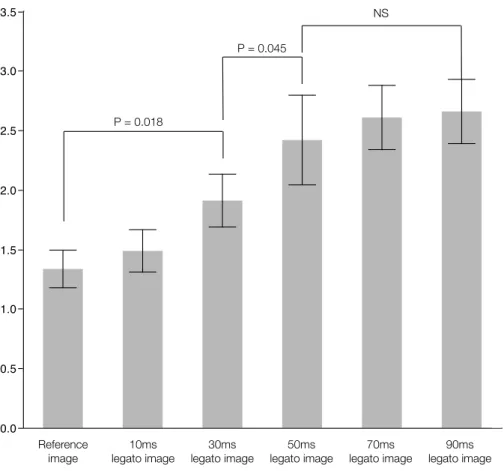
For theex vivopreliminary analysis, three phases separated by intervals of 10, 30, 50, 70 and 90 ms (each center phase was set as−250 ms relative to the R wave) were reconstructed
with convolution kernels for assessment of the coronary artery employing filtered back projection (FBP) (B26f).
For the other twoex vivostudies, three phases separated by intervals of 50 ms (−300,
−250, −200 ms relative to the R-wave) were reconstructed with convolution kernel
employing FBP and iterative reconstruction (IR) (SAFIRE, Sinogram Affirmed Iterative Reconstruction, Siemens AG, Forchheim, Germany) with strength 3 (B26f and I26f, respectively). The interval times were decided by the result of theex vivopreliminary study, that the CNR reached a plateau at an interval time of 50 ms.
In vivo study
75 bpm, a beta-blocker was used prior to the examination. Iopamidol (Iopamiron 370; Bayer Yakuhin, Osaka, Japan) was injected at a concentration of 370 mgI/mL via a 22-gauge catheter into the right antecubital vein at a flow rate of 22 mgI/s/kg over a period of 15 s, which was followed by a saline flush of 30 mL at the same rate. Bolus tracking was performed for a region of interest (ROI) in the ascending aorta. The scan automatically started 6 s after contrast enhancement of the ROI reached a threshold of+150 Hounsfield units (HU). The image dataset for the mid-diastolic phase (−250 ms relative to the R-wave) obtained with the reference dose and three image datasets for the systolic phase (200, 250, 300 ms relative to the R-wave) obtained with the quarter dose were reconstructed with the parameters given inTable 1.
Image post processing
Two board-certified Roentgen technologists who were blinded to the subjects’ identities performed further post image processing and image analyses. For the post processing including the implementation oflegato, a commercially available workstation (Ziostation 2.1.7.1 and PhyZiodynamics Technology, Ziosoft Inc., Tokyo, Japan) was used (Wai et al., 2013;Koike et al., 2014). From the three collected timeline datasets, three computed timeline datasets were calculated using ×1 for the generation of interpolated phases, a weight of 0.3 with two phases, and a non-cyclic algorithm as parameters, which we can set for the 4DNR with PhyZiodynamics Technology. PhyZiodynamics is software for non-rigid registration-based noise reduction, motion coherence and functional analysis. Noise is reduced through voxel-to-voxel mapping by tracking the temporal and spatial movement of individual voxels according to registration and interpolation algorithms (Brown, 2010). Image datasets for the center phase obtained from the collected and computed datasets are referred to as non-legatoandlegatoimages, respectively, and used in the following image analyses.
Image analysis
Ex vivo preliminary analysis of legato
The non-legatoimages scanned at 456 mAs/rot were set as reference images. The contrast-to-noise ratio (CNR) of each legatoimage with several interval times was obtained. The CNR assessments were performed using Module CTP515 of Catphan and Image J (Schneider, Rasband & Eliceiri, 2012). Circular ROIs were set for the 0.1%, 10-mm module and the neighboring background to obtain the mean and standard deviation (SD) of the CT attenuation within the ROI. The CNR was calculated as CNR=(ROIT−ROIB)/SDB, where ROITis the mean attenuation for the target module, ROIBis the mean attenuation for the background, and SDB is the SD of the background. In addition, to assess the difference between two-phase images for each interval form 10 to 90 ms, the mean squared error (MSE) (Zhou & Bovik, 2009) of each pixel was calculated using Image J.
Ex vivo quantitative image analysis of legato
The non-legatoimages scanned at 456 mAs/rot were set as reference images. Thelegato
terms of the CT attenuation, CNR, modulation transfer function (MTF), and noise power spectrum (NPS).
The CT attenuation and CNR were assessed for the images of Module CTP515 in Catphan. Both FBP and IR images were assessed. The measurement and calculation of the CT value and CNR were the same as previously described.
To assess the spatial resolution of an image, MTF and MTF10%were calculated employing the wire method and the software CTmeasure (http://www.jsct-tech.org/, 2012–2014) from the FBP image of the wire phantom.
To examine the variance and spatial frequency characteristics of image noise, the NPS was measured from the FBP image of the water phantom employing CTmeasure and the radial frequency.
Ex vivo assessment of the ability to reduce the radiation dose using legato
The CT value and CNR were assessed for the images of Module CTP515 in Catphan acquired at the reference radiation dose, quarter radiation dose, and quarter radiation dose using legato. The image datasets obtained with IR were used. The measurement and calculation of the CT number and CNR were the same as previously described. Furthermore, two board-certified radiologists (TN, with 7 years’ experience and AKK, with 12-years’ experience) was assessed the subjective image quality analysis by consensus reading in a blind manner. The image score was defined as the number of the modules that we could detect in each image with a slice thickness of 5 mm.
In vivo assessment of the ability to reduce the radiation dose using legato
Images of the diastolic phase (reference radiation dose) obtained withoutlegatoand the systolic phase (quarter radiation dose) obtained with and withoutlegatowere assessed. The circular ROI was set as the descending aorta at the level of the left atrium. The noise level as the SD of CT attenuation within the ROI was recorded.
Statistical analysis
The mean and SD values of metric variables were analyzed. Differences were assessed conducting a Tukey–Kramer’s honestly significant difference test. For the equivalence test, both bounds of 95% confidence intervals (CIs) of the difference from the reference image were compared with a margin set of a CT value of 2 HU. In the non-inferiority test, the lower bounds of 95% CIs of the difference from the reference image were compared with a margin set of a CNR of 0.1, MTF10%of 0.05 cycles/mm, and SD of 1 HU. The significance level was set atP=0.05.
For statistical analysis, the software JMP9.0 (SAS Institute Japan, Tokyo, Japan) and R (R Foundation for Statistical Computing, Vienna, Austria) was used.
RESULTS
Ex vivo preliminary analysis oflegato
Figure 1 The relationship with contrast noise ratio and interval times forlegato.Legato, the method of obtaining a noiseless image by adaptive phase-shifted topological coherence analysis.
time of 10 ms, 1.90±0.22 for 30 ms, 2.41±0.38 for 50 ms, 2.61±0.27 for 70 ms, and 2.65
±0.27 for 90 ms. In addition, the MSE for each interval time of 10, 30, 50, 70, and 90 was 14.0±0.7, 64.9±4.5, 114.2±5.1, 151.0±5.8, and 149.3±4.9 respectively. Although, the MSE increased significantly with the interval time until 70 ms (P<0.001), the MSEs for intervals of 70 and 90 ms were not significantly different.
Ex vivo quantitative image analysis oflegato
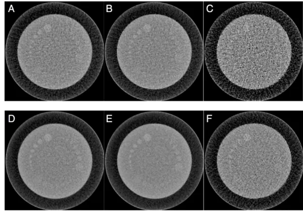
The CNR obtained with legatowas significantly better than that of the reference image (1.18±0.15 versus 2.08±0.19,P<0.001) (Figs. 2A,2Band3A). Even when employing the IR method, significant noise reduction was possible withlegato(1.25±0.15 versus 2.21
±0.22,P<0.001) (Figs. 2C,2Dand3B). In contrast, the CT attenuation did not change significantly when usinglegato(Table 2).
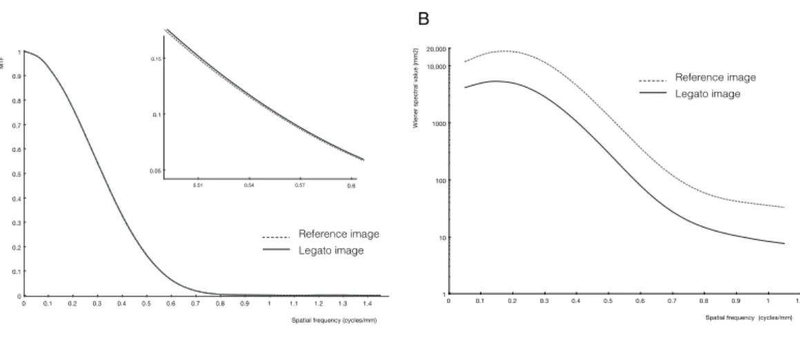
In the assessment of spatial resolution, MTF10%obtained withlegatowas not inferior to that of the reference image (Table 2). Each MTF curve is shown inFig. 4A.
Figure 2 Reference andlegatoimages.The original images obtained by filtered back projection (FBP) with reference dose (A), iterative reconstruction (IR) with reference dose (B), and IR with quarter radi-ation dose (C) are shown in upper column. By usinglegato, the image noise levels are significantly im-proved in images of FBP with reference dose (D), IR with reference dose (E), and IR with quarter radia-tion dose (F).Legato, the method of obtaining a noiseless image by adaptive phase-shifted topological co-herence analysis.
Table 2 Results on the equivalence test and non-inferiority test.
Reference Legato Margin Difference [95% CI] Pvalue
CT value (HU)
FBP 63.4±0.6 63.4±0.4 2 −0.02 [−0.51–0.46] <0.001
IR 63.6±0.6 63.9±0.4 2 −0.05 [−0.57–0.47] <0.001
MTF10%(cycles/mm) 0.55±0.01 0.56±0.02 0.05 0.005 [−0.010–0.019] <0.001
Reference Quarter dose+legato Margin Difference [95% CI] Pvalue
Ex vivo
CNR 1.25±0.15 1.37±0.09 0.1 0.113 [−0.003–0.230] <0.001
CT value (HU) 63.6±0.6 63.9±0.9 2 −0.3 [−0.96–0.40] <0.001
In vivo
SD (HU) 11.46±1.99 11.01±1.84 1 −0.455 [−1.89–0.980] 0.027
Notes.
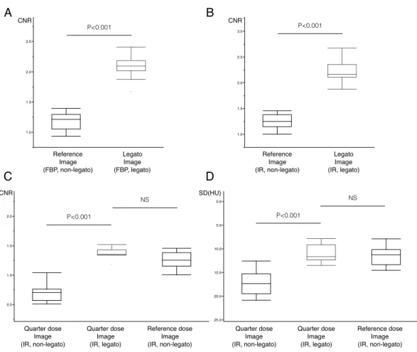
Figure 3 Box plots of the contrast-noise ratio (CNR) and image noise.Significant improvements in the CNRs of the images obtained by filtered back projection (FBP) (A), iterative reconstruction (IR) (B), quarter dose image-acquisition (C), were achieved usinglegato. Significant noise reduction of thein-vivo image with quarter dose (D), were also achieved usinglegato.Legato, the method of obtaining a noiseless image by adaptive phase-shifted topological coherence analysis.
Ex vivo assessment of the ability to reduce the radiation dose using
legato
Withlegato, CNRs for the quarter radiation dose significantly improved from 0.71±0.15 to 1.37±0.09 (P<0.001) (Figs. 2Eand2F) and were not inferior to those of the reference dose image (Table 2andFig. 3D). CT attenuation did not differ among reference dose image, quarter radiation dose image, and quarter radiation dose image withlegato(63.6±0.6 HU, 63.7±0.5 HU and 63.9±0.9 HU, respectively,P<0.001 for each). Furthermore, the subjective image score had a median value of 16 (range 13–18) for the reference, 6 (range 4–9) for the quarter radiation dose, and 15 (range 13–18) for the quarter radiation dose withlegato.
In vivo assessment of the ability to reduce the radiation dose using
legato
Figure 4 Results of modulation transfer function (MTF) analyses and noise power spectrum (NPS) analysis.The MTF curve (A) was almost the same for the image processed withlegato(black line) and the reference image not processed withlegato(gray dot line). The NPS curve (B) of thelegatoimage (black line) shows the uniform reduction in noise throughout the frequency band relative to the noise level in the reference image (gray dot line).Legato, the method of obtaining a noiseless image by adaptive phase-shifted topological coherence analysis.
Table 3 Subject characteristicsin-vivostudy.
Variables
Age(years) 66.3±13.8
Female/male (n) 2/13
Body mass index 22.4±3.97
Heart rate (bpm) 56.6±7.00
R–R interval time (ms) 1,066±147.7
CTDI (mGy) 50.2±8.49
Contrast material volume (mL) 60.4±13.0
Injection flow rate (mL/s) 3.60±0.61
Notes.
CTDI, computed tomography dose index.
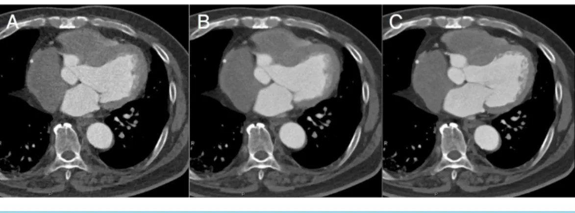
case. A representative image for the case of a 70-year-old male is shown in Fig. 5. The noise level in the image acquired at a quarter radiation dose and processed with legato
was significantly lower than that in the image not processed legato(17.11±2.73 versus 11.01±1.84,P<0.001) and no worse than that in the image acquired with the reference radiation dose (Table 2andFig. 3D).
DISCUSSIONS
Figure 5 Representative images ofin-vivostudy.Representative clinical images are shown; from the left, a quarter-radiation-dose image (systolic phase) withoutlegatopost processing (A), a quarter-radiation-dose image (systolic phase) withlegatopost processing (B), and a reference-dose image without legatopost processing (C).Legato, the method of obtaining a noiseless image by adaptive phase-shifted topological coherence analysis.
CCT images provide a good test of the performance of legatoin that several cardiac phases with short interval times are usually obtained as a padding phase (Tatsugami et al., 2015). In addition, the noise level would be higher than that in full reconstruction because there are fewer photons in the data acquisition of the half reconstruction that accounts for temporal resolution. Employing temporal filtering, this noise resulting from the half reconstruction could be compensated. Moreover, artifacts in half reconstruction are well known to mainly depend on the acquisition degree. Using a different phase means the use of a different acquisition degree. Thus, theoretically, these artifacts could also be minimized employed temporal noise reduction.
Interval times are important parameters when usinglegatoin CCT. Small movements might be better for the precise estimation of movement. Thus, datasets with relative short interval times are required. However, it is not easy to distinguish noise in datasets for which the intervals are too short because the images and the noise they contain are almost the same. Our preliminary study showed that noise reduction for interval times from 10 to 30 ms was relatively low. In contrast, the noise reduction reached a plateau at interval times exceeding 50 ms. Larger interval times can thus improve noise reduction but possibly only to a certain degree. If the interval time is set as 70 ms, the three phases are separated by 90◦
for our scanner (having a rotation time of 280 ms). In dual-source CT, two detectors are aligned at almost 90◦, and datasets having phases shifted by 90◦contain data of completely
Image filtering can reduce the resolution and produce artifacts by oversmoothing. There is thus a trade-off between such artifacts and the reduced rate of noise. However, ourlegato
can uphold the CT attenuation and the resolution with powerful noise reduction for static phantoms. Temporal filtering methods are known to avoid the oversmoothing of voxels because of their spatial independence (Brailean et al., 1995). Thus, legatoimproves the image quality by reducing noise while preserving structural detail, which is important for CCT. The importance of the spatial resolution has been well discussed in the detection of coronary stenosis (Leber et al., 2005). Recently, the CT attenuation of plaque has also been an important research topic. Plaque that is vulnerable to rupturing is known to have a low CT attenuation, and a small ulcer having a napkin-ring sign would be an important finding (Hoffmann et al., 2006;Motoyama et al., 2009). If the CT value changes, the definition of the plaque could be vague. Additionally, noise would easily affect these relatively low values. Noise-less images that maintain the CT attenuation and resolution would thus meet the clinical demands of CCT.
Our results also showed the possibility of combininglegatowith IR. Recently, powerful noise reduction using IR methods has been reported (Renker et al., 2011;Utsunomiya et al., 2012;Yin et al., 2013;Meyer et al., 2014). The IR methods are mainly used to reduce the radiation dose in clinical settings. Whereas the IR methods are used in the image reconstruction stage,legatois performed in the post-processing stage. Thus,legatocould be applied to the IR images and further reduce noise independently of the IR methods.
Another possibility oflegatois taking the same noise images with a lower radiation dose. Even at a quarter dose, the CNR obtained usinglegatowas not less than that of the reference image. In addition, our subjective image quality assessment supports this finding. The results are thus reasonable. Withoutlegato, the noise level would be twice that of the reference image for a quarter radiation dose. However, withlegato, the noise level can be reduced by half, as previously discussed. Our result theoretically suggests that a three-quarter reduction of the radiation dose is possible when we focus on the image noise level, but a larger investigation of clinical cases is needed to assess the diagnostic performance.
There were limitations to the present study. First, in theex vivostudy, a static phantom was used to investigate the potential of legato. However, the effect of motion and the analysis of temporal resolution need to be further investigated for the use of CCT in clinical cases. Second, the application oflegatoto other scanners, such as a single-source scanner, and with other reconstruction kernels and other IR methods needs to be investigated.
Abbreviations
CT Computed tomography
CCT Cardiac CT
CNR Contrast-to-noise ratio
4DNR Four- dimensional noise reduction
FBP Filtered back projection
IR Iterative reconstruction
MTF Modulation transfer function
NPS Noise power spectrum
Legato The method of obtaining a noiseless image by adaptive phase-shifted topological coherence analysis
ADDITIONAL INFORMATION AND DECLARATIONS
Funding
The authors received no funding for this work.
Competing Interests
The authors declare there are no competing interests.
Author Contributions
• Tatsuya Nishii conceived and designed the experiments, performed the experiments, analyzed the data, contributed reagents/materials/analysis tools, wrote the paper, prepared figures and/or tables, reviewed drafts of the paper.
• Atsushi K. Kono conceived and designed the experiments, reviewed drafts of the paper.
• Wakiko Tani, Erina Suehiro and Noriyuki Negi performed the experiments, contributed reagents/materials/analysis tools.
• Satoru Takahashi and Kazuro Sugimura reviewed drafts of the paper.
Human Ethics
The following information was supplied relating to ethical approvals (i.e., approving body and any reference numbers):
The authors have no conflicts of interest to declare. Institutional Review Board approval (No. 1372) was obtained. Written informed consent was waived by the Institutional Review Board.
Data Availability
The following information was supplied regarding data availability: Figshare:http://figshare.com/s/c6fda35678aa11e5b50906ec4b8d1f61.
REFERENCES
Brailean JC, Kleihorst RP, Efstratiadis S, Katsaggelos AK, Lagendijk RL. 1995.Noise reduction filters for dynamic image sequences: a review.Proceedings of the IEEE
Brown HA. 2010.PhyZiodynamics: a revolutionary approach for post-processed noise reduction, motion coherence and functional analytics. Redwood City: Ziosoft, Inc.
Available athttp:// www.qiimaging.com/ pdf/ phyzio.pdf.
Corcuera-Solano I, McLellan AM, Doshi AH, Pawha PS, Tanenbaum LN. 2014.
Whole-brain adaptive 70-kVp perfusion imaging with variable and extended sampling improves quality and consistency while reducing dose.American Journal of Neuroradiology35:2045–2051DOI 10.3174/ajnr.A4043.
Hoffmann U, Moselewski F, Nieman K, Jang IK, Ferencik M, Rahman AM, Cury RC, Abbara S, Joneidi-Jafari H, Achenbach S, Brady TJ. 2006.Noninvasive assessment of plaque morphology and composition in culprit and stable lesions in acute coronary syndrome and stable lesions in stable angina by multidetector computed tomography.Journal of the American College of Cardiology 47:1655–1662 DOI 10.1016/j.jacc.2006.01.041.
Ichikawa K, Hara T, Niwa S, Ohashi K. 2008.Method of measuring modulation transfer function using metal wire in computed tomography.Nihon H¯oshasen Gijutsu Gakkai zasshi64:672–680DOI 10.6009/jjrt.64.672.
Koike Y, Ishida K, Hase S, Kobayashi Y, Nishimura J-I, Yamasaki M, Hosaka N. 2014.Dynamic volumetric CT angiography for the detection and classification of endoleaks: application of cine imaging using a 320-row CT scanner with 16-cm detectors.Journal of Vascular and Interventional Radiology25:1172–1180 DOI 10.1016/j.jvir.2014.03.019.
Leber AW, Knez A, Von Ziegler F, Becker A, Nikolaou K, Paul S, Wintersperger B, Reiser M, Becker CR, Steinbeck G, Boekstegers P. 2005.Quantification of obstructive and nonobstructive coronary lesions by 64-slice computed tomog-raphy: a comparative study with quantitative coronary angiography and in-travascular ultrasound.Journal of the American College of Cardiology46:147–154 DOI 10.1016/j.jacc.2005.03.071.
Li Z, Li H, Zhang K, Li W, Chen X, Wu B, Song B. 2014b.Improvement of image quality and radiation dose of CT perfusion of the brain by means of low-tube voltage (70 KV).European Radiology24:1906–1913DOI 10.1007/s00330-014-3247-1.
Li H, Sun C, Xu Z, Miao F, Zhang D, Chen J, Li X, Wang X, Liu C, Zhao B. 2014a.
Low-dose whole organ CT perfusion of the pancreas: preliminary study.Abdominal Imaging 39:40–47DOI 10.1007/s00261-013-0045-1.
Liu X, Primak AN, Yu L, Li H, Krier JD, Lerman LO, McCollough CH. 2009. Quantita-tive evaluation of noise reduction algorithms for very low dose renal CT perfusion imaging.Proceeding of SPIE 7258, Medical Imaging 2009: Physics of Medical Imagining
72581T:1–8DOI 10.1117/12/813777.
Motoyama S, Sarai M, Harigaya H, Anno H, Inoue K, Hara T, Naruse H, Ishii J, Hishida H, Wong ND, Virmani R, Kondo T, Ozaki Y, Narula J. 2009.Computed tomographic angiography characteristics of atherosclerotic plaques subsequently resulting in acute coronary syndrome.Journal of the American College of Cardiology
54:49–57DOI 10.1016/j.jacc.2009.02.068.
Otton JM, Kühl JT, Kofoed KF, McCrohon J, Feneley M, Sammel N, Yu C-Y, Chiribiri A, Nagel E. 2013.Four-dimensional image processing of myocardial CT perfusion for improved image quality and noise reduction.Journal of Cardiovascular Computed Tomography7:110–116DOI 10.1016/j.jcct.2013.01.014.
Renker M, Ramachandra A, Schoepf UJ, Raupach R, Apfaltrer P, Rowe GW, Vogt S, Flohr TG, Kerl JM, Bauer RW, Fink C, Henzler T. 2011.Iterative image reconstruc-tion techniques: applicareconstruc-tions for cardiac CT.Journal of Cardiovascular Computed Tomography5:225–230DOI 10.1016/j.jcct.2011.05.002.
Schneider CA, Rasband WS, Eliceiri KW. 2012.NIH Image to ImageJ: 25 years of image analysis.Nature Methods9:671–675DOI 10.1038/nmeth.2089.
Tatsugami F, Higaki T, Nakamura Y, Yamagami T, Date S, Fujioka C, Kiguchi M, Kihara Y, Awai K. 2015.A new technique for noise reduction at coronary CT angiography with multi-phase data-averaging and non-rigid image registration.
European Radiology25:41–48DOI 10.1007/s00330-014-3381-9.
Utsunomiya D, Weigold WG, Weissman G, Taylor AJ. 2012.Effect of hybrid iterative reconstruction technique on quantitative and qualitative image analysis at
256-slice prospective gating cardiac CT.European Radiology22:1287–1294 DOI 10.1007/s00330-011-2361-6.
Wai B, Thai W-E, Brown H, Truong QA. 2013.Novel phase-based noise reduction strategy for quantification of left ventricular function and mass assessment by cardiac CT: comparison with cardiac magnetic resonance.European Journal of Radiology
82:e337–e341DOI 10.1016/j.ejrad.2013.02.023.
Yin WH, Lu B, Li N, Han L, Hou ZH, Wu RZ, Wu YJ, Niu HX, Jiang SL, Krazinski AW, Ebersberger U, Meinel FG, Schoepf UJ. 2013.Iterative reconstruction to preserve image quality and diagnostic accuracy at reduced radiation dose in coronary CT angiography: an intraindividual comparison.JACC: Cardiovascular Imaging
6:1239–1249DOI 10.1016/j.jcmg.2013.08.008.
Zhou W, Bovik AC. 2009.Mean squared error: love it or leave it? A new look at signal fidelity measures.IEEE Signal Processing Magazine26:98–117