CLINICAL SCIENCE
I Department of Thoracic Surgery, Hospital do Cancer AC Camargo - São Paulo/SP, Brazil.
II LIM-62, Faculdade de Medicina da Universidade de São Paulo - São Paulo, Brazil.
Email: [email protected] Tel: 55 11 3257-2808
Received for publication on January 26, 2009 Accepted for publication on March 13, 2009
SURGICAL RESECTION OF LUNG METASTASES:
RESULTS FROM 529 PATIENTS
Riad N. Younes,I,II Jefferson L. Gross,I Andrea M. Taira,I Andrea Aparecida C.
Martins,I Giuliana Sigolo NevesI
doi: 10.1590/S1807-59322009000600008
Younes RN, Gross JL, Taira AM, Martins AAC, Neves GS. Surgical resection of lung metastases: results from 529 patients. Clinics. 2009;64(6):535-41.
OBJECTIVE: The aim of this study is to determine clinical, pathological, and treatment-relevant variables associated with long-term (90-month) overall survival in patients with lung metastases undergoing pulmonary metastasectomy.
METHODS: A retrospective review was performed of patients who were admitted with lung metastases, and who underwent
thoracotomy for resection, after treatment of a primary tumor. Data were collected regarding demographics, tumor features, treat-ment, and outcome.
RESULTS: Patients (n = 529) were submitted to a total of 776 thoracotomies. Median follow-up time across all patients was 21.6 months (range: 0-192 months). The postoperative complication rate was 9.3%, and the 30-day mortality rate was 0.2%. The ninety-month overall survival rate for all patients was 30.4%. Multivariate analysis identified the number of pulmonary nodules detected on preoperative CT-scan, the number of malignant nodules resected, and complete resection as the independent prognostic factors for overall survival.
CONCLUSION: These results confirm that lung metastasectomy is a safe and potentially curative procedure for patients with treated primary tumors. A select group of patients can achieve long-term survival after resection.
KEYWORDS: Lung; Metastases; Resection, Metastasectomy; Prognostic factors.
INTRODUCTION
The presence of metastases in patients with malignant tumors is a sign of advanced systemic disease1.
Consequently, few patients survive more than one year after diagnosis.2 However, over 20% of patients with solid tumors,
including colorectal adenocarcinoma, may have metastases exclusively in the pulmonary parenchyma, with no detectable involvement of other organs. In these patients, surgical resection of lung metastases may significantly impact long-term survival.3 We have recently reported on metastatic lung
cancer in Brazil4 and reviewed possible sources of new
anti-tumoral drugs from Brazilian plant extracts.5
Pulmonary metastasectomy is currently indicated for patients with the following criteria: primary tumor controlled; no evidence of metastases outside lung parenchyma; possibility of complete resection as verified by chest imaging; pulmonary function compatible with the proposed lung resection procedure; and lack of another available treatment that would be more effective than metastasectomy.6 Using these criteria for metastasectomy,
several retrospective series have reported five-year survival rates ranging from 30% to 40%.7
a limited number of patients treated over a long period of time, and the heterogeneity of the studied population did not allow for definitive conclusions. Today, complete surgical resection, long disease-free intervals, and a suitably low number of metastases are considered the most important variables associated with better survival rates.8
Since the early 1990s, our institution has employed a continuous protocol-oriented approach to treat patients with resectable lung metastases. Our goal was to define factors related to outcome following lung metastasectomy in our patient population. All patients in the studied population underwent similar preoperative imaging, together with clinical and surgical evaluations. This was done to generate a more homogeneous and comparable group of patients, each of whom had detailed information registered in their individual records.
The goal of this study is to evaluate outcomes following the surgical treatment of lung metastases in patients treated at a single institution, as well as to identify prognostic factors that may significantly impact long-term overall survival. Our results will allow us to more precisely define the subgroups of patients who may benefit from surgical therapy.
METHODS
This is a retrospective analysis of patients previously diagnosed with primary malignant solid tumors and who subsequently underwent surgical resection of lung nodules in the context of suspected or diagnosed metastatic lesions. All patients were admitted and treated in the Department of Thoracic Surgery of AC Camargo Hospital from 1990 to 2006. This study was approved by the ethics committee at our institution.
Data were collected from medical record reviews of individual patients. The following characteristics of the primary tumor were recorded: primary site, date of surgical resection, histological type, and adjuvant or neoadjuvant treatment associated with the resection.
All patients were evaluated for lung nodules during the pretreatment evaluation and staging of a neoplasm or during routine follow-up after the treatment of various primary tumors. Disease-free interval was defined as the time period between the treatment of the primary tumor and the diagnosis of pulmonary metastases.
Patients were considered eligible for pulmonary metastasectomy if they presented with the following characteristics: 1) primary tumor controlled or controllable, 2) nodules confined to the lung parenchyma, 3) nodules that were amenable to surgical resection, 4) pulmonary function and clinical condition that were compatible with the planned operation, 5) predictable remaining lung function after
resection that would allow for adequate postoperative quality of life, and 6) non-availability of a more suitable treatment option for the metastases.
All patients underwent a chest CT scan in order to evaluate the resectability of their pulmonary nodules. Radiology reports were reviewed in each patient file, and the following characteristics were recorded: number of pulmonary nodules, size of the largest nodule, and laterality. The preoperative evaluation of the nodules was routinely performed by the radiologist and the surgeon. In cases of significant discrepancies between the number of nodules identified by the surgeon and by the radiologist, the evidence was discussed with a specialist. The number of nodules included in this retrospective study was the final number confirmed by the surgeon after complete preoperative evaluation.
In the case of bilateral nodules on admission, a staged thoracotomy was performed starting on the side with the lowest likelihood of a complete resection. Several factors determined which side to start with, including the number of nodules identified by chest CT, the anatomical position of one or more nodules relative to the pulmonary hilum, and the potential need for a more complex and extensive lung resection.
Patients were placed under general anesthesia with selective single lung ventilation. Surgical access to the thorax was performed by lateral thoracotomy even for patients with bilateral metastases. In all cases, we attempted to completely resect all lung nodules, preserving lung parenchyma by resecting the tumor with a small margin (5 mm to 10 mm). We did not routinely perform mediastinal lymph node dissection or sampling. In the presence of a single pulmonary nodule, frozen section analysis was mandatory. If histological evidence suggested a possible primary lung tumor, the patient underwent pulmonary lobectomy with radical mediastinal lymph-node dissection when possible. Lung nodules were identified individually according to the site of resection and sent for histopathological analysis.
Date of surgery, type of resection (complete or incomplete), number of malignant and benign resected nodules, size of the largest nodule, and type of lung resection (wedge, segmentectomy, lobectomy or pneumonectomy) were registered according to the surgical report in individual patient records.
Some patients received chemotherapy, based on the medical oncologist’s opinion, before or after a surgical resection of lung nodules. The type of treatment and radiological response (complete, partial, stable or progression of disease, as defined by RECIST9) were collected.
fever>38°C, bleeding, air leakage for more than three days, Intensive Care Unit stay for more than two days, cardiac complications (arrhythmia, ischemia, infarction), pulmonary atelectasis, embolism or respiratory failure, renal failure, neurological complications (coma, stroke, motor and sensitive dysfunctions), and infection (bacterioscopy or culture confirming the presence of a bacterial infection).
Beyond hospital discharge, patients were followed-up by clinical examination, with radiological evaluations (chest X-rays and CT scans) every three months during the first two years post-resection and every six months subsequently until the fifth year. Annual radiological follow-up was performed thereafter. Other ancillary tests were performed consistent
with symptoms or clinical suspicion of recurrence in organs other than the lungs. We recorded recurrence when new lesions were identified in the lungs or in other organs. When necessary, we performed a histological confirmation of a new tumor. When recurrence was again limited to the lung parenchyma, and if the disease was considered resectable by the attending thoracic surgeon, the patient underwent additional metastasectomies as needed.
After complete surgical resection, all patients were evaluated in the department of clinical oncology, and systemic treatment was administered by the clinical oncologist.
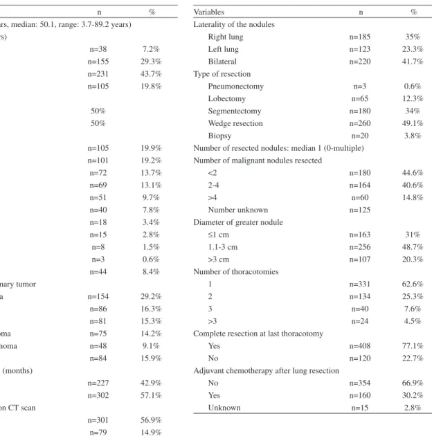
The characteristics of the patients included in this study are shown in Table 1.
Variables n %
Age (mean: 45.7 years, median: 50.1, range: 3.7-89.2 years) Age subgroups (years)
0-12 n=38 7.2%
13-40 n=155 29.3%
65 n=231 43.7%
70 n=105 19.8%
Gender
Male (n=267) 50%
Female (n=262) 50%
Primary tumor site
Bone n=105 19.9%
Head-Neck n=101 19.2%
Soft tissue n=72 13.7%
Breast n=69 13.1%
Colorectal n=51 9.7%
Skin n=40 7.8%
Uterus n=18 3.4%
Kidney n=15 2.8%
Stomach n=8 1.5%
Bladder n=3 0.6%
Other n=44 8.4%
Histology of the primary tumor
Adenocarcinoma n=154 29.2%
Osteosarcoma n=86 16.3%
Squamous cell n=81 15.3%
Soft tissue sarcoma n=75 14.2%
Malignant melanoma n=48 9.1%
Other n=84 15.9%
Disease-free interval (months)
≤ 12 n=227 42.9%
>12 n=302 57.1%
Number of nodules on CT scan
≤ 2 n=301 56.9%
3-4 n=79 14.9%
>4 n=149 28.2%
Variables n %
Laterality of the nodules
Right lung n=185 35%
Left lung n=123 23.3%
Bilateral n=220 41.7%
Type of resection
Pneumonectomy n=3 0.6%
Lobectomy n=65 12.3%
Segmentectomy n=180 34%
Wedge resection n=260 49.1%
Biopsy n=20 3.8%
Number of resected nodules: median 1 (0-multiple) Number of malignant nodules resected
<2 n=180 44.6%
2-4 n=164 40.6%
>4 n=60 14.8%
Number unknown n=125
Diameter of greater nodule
≤1 cm n=163 31%
1.1-3 cm n=256 48.7%
>3 cm n=107 20.3%
Number of thoracotomies
1 n=331 62.6%
2 n=134 25.3%
3 n=40 7.6%
>3 n=24 4.5%
Complete resection at last thoracotomy
Yes n=408 77.1%
No n=120 22.7%
Adjuvant chemotherapy after lung resection
No n=354 66.9%
Yes n=160 30.2%
Unknown n=15 2.8%
Statistical Analyses
Continuous data are presented as medians, and categorical data are presented as percentages. Overall and disease-free survival rates were estimated using Kaplan-Meier analysis10. Log-rank and Breslow analyses were used
to compare differences between variables. Overall survival was estimated from the date of surgery to the date of last follow-up or until death from any cause. The influence of each variable on overall and disease-free survival was calculated.
Multivariate analysis to determine the independent prognostic factors for overall survival was determined by the Cox proportional hazard model.11 Whenever more than
one procedure was performed, survival time was determined, with time zero denoting the first thoracotomy.
Statistical analyses were performed using SPSS 11.5 for Windows. Significant differences were defined as p<0.05.
RESULTS
The patients included in this retrospective study (n = 529) underwent a total of 776 thoracotomies. The average follow-up time for all patients was 40 months, with a median of 21.6 months (range: 0-192 months), and eight patients (1.5%) were lost to follow up. Most patients were discharged by the fifth postoperative day (length of stay in hospital ranged from two days to 27 days, with a median of four days). The postoperative complication rate was 9.3% for the 776 thoracotomies. More than one complication occurred in some patients (infection n=19, atelectasis n=29, cardiac arrhythmia n=18, stroke n=2, myocardial infarction n=3, prolonged air leak n=28), and the 30-day mortality rate was 0.2% (n=2, due to respiratory failure and stroke).
The ninety-month overall survival rate for all patients was 30.4% (Figure 1).
Figures 2-7 show the significant prognostic factors for overall survival, as identified by univariate analyses.
All other factors evaluated in the univariate analyses, including type of resection, response to preoperative chemotherapy, number of resected nodules, and the total number of thoracotomies in an individual patient, were not statistically significant (p>0.05).
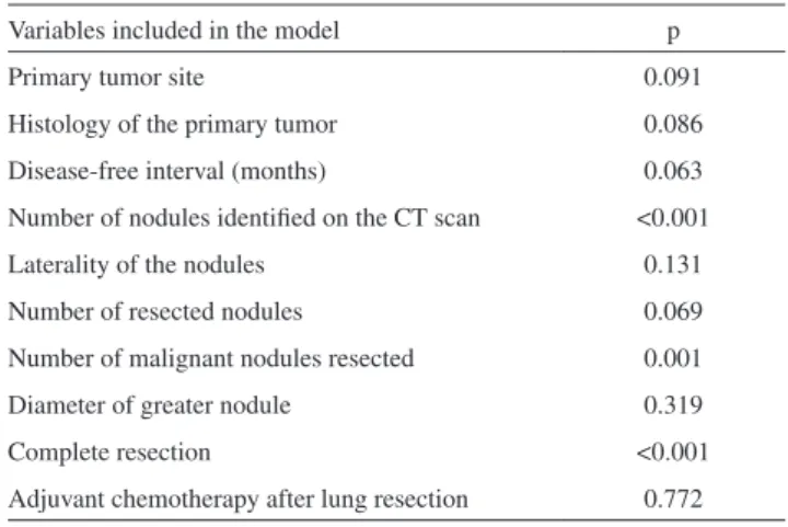
Multivariate analysis identified the number of pulmonary nodules (p<0.001), the number of malignant nodules resected (p=0.001), and complete resection (p<0.001) as the
Figure 1 - Overall survival rates for all patients included
Figure 2 - Overall survival according to primary tumor histology
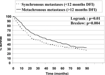
Figure 3 - Overall survival according to disease-free interval (DFI)
are confined to the lungs. Therefore, resectability should be evaluated preoperatively with great care. The recent introduction of positron emission tomography using 18F- fluorodeoxyglucose (FDG PET-CT) has proven to be of clinical value for the proper staging of cancer patients. The use of this technology during the preoperative evaluation of candidates for lung metastasectomy has been shown to eliminate 21% of patients otherwise deemed resectable.15
Unfortunately, our study population did not routinely undergo this imaging evaluation because the method was not available in our institution for most patients.
The 529 patients included in this study underwent 776 thoracotomies for the treatment of lung metastases. The overall survival of patients at 90 months in our study was 30.4%, with average follow-up of 40 months. Eight patients (1.5%) were lost to follow-up.
The histological type of a primary malignant tumor has been shown to influence the results of metastasectomy in previous studies.1 In this study, survival rates were
significantly better for patients with adenocarcinoma. Multivariate analysis, however, did not identify this variable as an independent prognostic factor. Better systemic therapy for patients with metastatic colorectal adenocarcinoma might explain the apparently favorable long-term outcome. Our study failed to provide sufficient evidence to support this observation.
On the basis of our univariate analysis, patients with synchronous metastases (DFI<12 months) exhibited significantly lower survival rates, compared to patients with metachronous disease, than reported in other studies.2,7
On the other hand, this factor was not significant in our multivariate analysis.
Several reports have demonstrated the prognostic importance of the number of nodules.7 The sensitivity
of preoperative imaging examinations of pulmonary metastases varies depending on the technique used. Dellai Figure 5 - Overall survival according to nodule laterality
Figure 6 - Overall survival according to the number of resected malignant nodules
Figure 7 - Overall survival according to the type of surgical resection (complete or incomplete)
independent prognostic factors for overall survival (Table 2).
DISCUSSION
Thirty to 40% of all extrathoracic cancers lead to secondary pulmonary lesions during the course of the disease, and approximately 20% of these cases feature metastases that are confined to the lungs.2,7,12-14 Improved
survival following resection of lung metastases has broadened the surgical indications for these lesions. It is likely that surgery itself impacts the outcome of patients who are candidates for a complete resection of nodules that
Table 2 - Multivariate analysis for overall survival
Variables included in the model p
Primary tumor site 0.091
Histology of the primary tumor 0.086
Disease-free interval (months) 0.063
Number of nodules identified on the CT scan <0.001
Laterality of the nodules 0.131
Number of resected nodules 0.069
Number of malignant nodules resected 0.001
Diameter of greater nodule 0.319
Complete resection <0.001
et al16 evaluated the precision of the diagnosis and the
exact quantification of lung nodules using computerized tomography. All of the nodules were subsequently resected, revealing a diagnostic accuracy of 51%. In a prospective study involving 182 patients in Brazil, the CT scan identified the exact number of nodules resected in 47% of all cases. However, the CT scan underestimated the actual number of lung nodules found during surgery in 28% of cases and overestimated the number in 24%.2
A recently published study by Cerfolio et al 17 showed
that in 37% of patients who were candidates for video-assisted thoracic surgery (VATS), intraoperative palpation during a formal thoracotomy detected more nodules than had been identified using preoperative imaging (CT scans). VATS has a definitive role in the management of solitary pulmonary lesions that are suspected to be metastatic, but the use of thoracoscopic techniques for pulmonary metastasectomy is controversial because small metastatic foci may be missed without thorough finger palpation of the lung. Prospective studies are currently evaluating this issue in several centers.
In our study, the number of nodules exerted a significant impact on long-term overall survival using both univariate and multivariate analyses. Similar observations have been previously reported in other publications7,18
In most studies published to date, there has been no significant difference in survival of patients with metastases confined to one lung compared to bilateral lesions.19-22
However, univariate analysis in this study suggested a significant difference in survival rates between patients presenting with unilateral or bilateral disease. However, this factor was found not to be significant under multivariate analysis.
Preoperative or postoperative systemic therapy had no significant impact on long-term survival rate in this patient population, consistent with data published elsewhere.3,7,8,23-26
Complete resection was the most significant prognostic factor associated with long-term overall survival, both in univariate and multivariate analyses, as has been consistently reported elsewhere.27-35 The 90-month overall survival rate
for patients who underwent complete resection in the context of metastatic disease was 35.1%, compared to 10.2% in the group who underwent incomplete resection.
Type of surgical resection and the total number of thoracotomies failed to influence the outcome of the patients, provided that complete resection was accomplished. Thus, we conclude that repeated resection may be beneficial in patients with relapses that are confined to the lung.36 As
reported elsewhere, limited resection with lung parenchyma preservation is adequate to achieve clear margins and local control of the disease.8 Recently, alternative options
like radiofrequency ablation have been evaluated for the management of lung metastases. Although promising, such methods are still considered experimental, since they are indicated only in limited numbers of patients, and they are usually considered clinically inappropriate for patients who can undergo a conventional resection.37
CONCLUSIONS
This study identified a group of patients who may benefit from pulmonary metastasectomy. Irrespective of other factors, all patients who present with fewer than four lung nodules should be considered candidates for surgical resection, after selection using the currently established inclusion criteria. Other patients should be individually evaluated in order to assess the impact of surgical resection on survival and long-term outcomes. Additional studies are needed to determine the role of adjuvant systemic therapy in patients stratified according to the histology of their primary tumors.
REFERENCES
1. Mountain CF, Khalil KG, Hermes KE, Frazier OH. The contribution of surgery to the management of carcinomatous pulmonary metastases. Cancer. 1978;41:833-40.
2. Younes RN, Haddad F, Ferreira F, Gross JL. Surgical removal of pulmonary metastasis: prospective study in 182 patients. Rev Assoc Med Bras. 1998;44:218-25.
3. Warwick R, Page R. Resection of pulmonary metastases from colorectal carcinoma. Eur J Surg Oncol. 2007;33Suppl 2:S59-63.
4. Metastatic non-small cell lung cancer in Brazil: treatment heterogeneity in routine clinical practice. Naime FF, Younes RN, Kersten BG, Anelli A, Beato CA, Andrade RM, Carrara MP, Gross JL. Clinics. 2007;62:397-404.
5. Discovery of new antitumoral and antibacterial drugs from Brazilian plant extracts using high throughput screening. Younes RN, Varella AD, Suffredii IB. Clinics. 2007;62:763-8.
7. Pastorino U. Lung metastasectomy: why, when, how. Crit Rev Oncol Hematol. 1997;26:137-45.
8. Friedel G, Pastorino U, Buyse M, Ginsberg RJ, Girard P, Goldstraw P, et al. Resection of lung metastases: long-term results and prognostic analysis based on 5206 cases-the International Registry of Lung Metastases. Zentralbl Chir. 1999;124:96-103.
9. Therasse P, Arbuck SG, Eisenhauer EA, Wanders J, Kaplan RS, Rubinstein L, et al. New guidelines to evaluate the response to treatment in solid tumors. European Organization for Research and Treatment of Cancer, National Cancer Institute of the United States, National Cancer Institute of Canada. J Natl Cancer Inst. 2000;92:205-16.
10. Kaplan EL, Meier P. Nonparametric estimation from incomplete observations. J Am Stat Assoc. 1958;53:457-81.
11. Cox DR. Regression models and life tables. J R Stat Soc. 1972;34:187-220.
12. Younes RN, Gross JL, Deheinzelin D. Surgical resection of unilateral lung metastases: is bilateral thoracotomy necessary? World J Surg. 2002;26:1112-6.
13. Younes RN, Gross JL, Silva JF, Fernandez JA, Kowalski LP. Surgical treatment of lung metastases of head and neck tumors. Am J Surg. 1997;174:499-502.
14. Farrell JT. Pulmonary metastases: A pathologic, clinical roentgenologic study based on 78 cases seen at autopsy. Radiology. 1935;24:444-9. 15. Pastorino U, Veronesi G, Landoni C, Leon M, Picchio M, Solli PG,
et al. Fluorodeoxyglucose positron emission tomography improves preoperative staging of resectable lung metastasis. J Thorac Cardiovasc Surg. 2003;126:1906-10.
16. Dellai RCA, Chojniak R, Marques E, Younes RN. Detecção de nódulos pulmonares por tomografia computadorizada em pacientes com metástases pulmonares submetidos a cirurgia. J. Pneumol. 1994;20:8. 17. Cerfolio R, Bryant AS. Is palpation of the nonresected pulmonary lobe(s)
required for patients with non-small cell lung cancer? A prospective study. J Thorac Cardiovasc Surg. 2008;131:261-8.
18. Casali C, Stefani A, Storelli E, Morandi U. Prognostic factors and survival after resection of lung metastases from epithelial tumours. Interact Cardiovasc Thorac Surg. 2006;5:317-21.
19. Brister SJ, DeVarennes B, Gordon PH, Sheiner NM, Pym J. Contemporary operative management of pulmonary metastases of colorectal origin. Dis. Colon Rectum 1998;31: 786-92.
20. Lanza LA, Miser JS, Pass HI, Roth JA. The role of resection and the treatment of pulmonary metastases from Ewing´s sarcoma. J Thorac Cardiovasc. Surg. 1987;94:181-7.
21. McAfee MK, Allen MS, Trastek VF, Ilstrup DM, Deschamps C, Pairolero PC. Colorectal lung metastases: Results of surgical excision. Ann Thorac Surg. 1992;53:780-6.
22. Pastorino U, Valente M, Gasparini M, Azzarelli A, Santoro A, Tavecchio L, et al. Lung resection as salvage treatment for metastatic osteosarcoma. Tumori. 1988;74:201-6.
23. Huth JF, Holmes EC, Vernon SE. Pulmonary resection for metastatic sarcoma. Am J Surg. 1980;140:9-16.
24. Neuman HB, Patel A, Hanlon C, Wolchok JD, Houghton AN, Coit DG. Stage IV melanoma and pulmonary metastases: factors predictive of survival. Ann Surg Oncol 2007; 14:2847-53.
25. Iizasa T, Suzuki M, Yoshida S, Motohashi S, Yasufuku K, Iyoda A, et al. Prediction of prognosis and surgical indications for pulmonary metastasectomy from colorectal cancer. Ann Thorac Surg. 2006;82:254-60.
26. Welter S, Jacobs J, Krbek T, Krebs B, Stamatis G. Long-term survival after repeated resection of pulmonary metastases from colorectal cancer. Ann Thorac Surg. 2007;84:203-10.
27. Ferretti S, Fornia S, Ampollini L, Rusca M, Salsi P, Vaglio A, et al. Lung metastasectomy in patients with renal cell cancer (RCC). A 17-year experience in Parma Hospital. Acta Biomed. 2007;78:41-5.
28. Gadd MA, Casper ES, Woodruff JM, McCormack PM, Brennan MF. Development and treatment of pulmonary metastases in adult patients with extremity soft tissue sarcoma. Ann Surg 1993;218:705-12. 29. Harpole DH, Johnson CM, Wolf WG, George SL, Seigler HF. Analysis
of 945 cases of pulmonary metastatic melanoma. J Thorac Cardiovasc. Surg. 1992;103:743-8.
30. Martini N, McCormack PM, Bains MS, Beattie EJ. Surgery for solitary and multiple pulmonary metastases. NY State J. Med. 1978;78:1711-4.
31. McCormack PM, Martini N. The changing role of surgery for pulmonary metastases. Ann Thorac Surg. 1979;28:139-45.
32. Aberg T, Malmberg KA, Nilsson B, Nou E. The effect of metastasectomy: fact or fiction. Ann Thorac Surg. 1980;30:378-84.
33 Mountain CF, McMurtrey MJ, Hermes KE. Surgery for pulmonary metastases: a 20-year experience. Ann. Thorac Surg. 1984;38:323-30. 34. Smith R, Pak Y, Kraybill W, Kane JM 3rd. Factors associated with
actual long-term survival following soft tissue sarcoma pulmonary metastasectomy. Eur J Surg Oncol. 2009;35:356-61.
35. Rama N, Monteiro A, Bernardo JE, Eugénio L, Antunes MJ. Lung metastases from colorectal cancer: surgical resection and prognostic factors. Eur J Cardiothorac Surg. 2009; 35:444-9.
36. Liebl LS, Elson F, Quaas A, Gawad KA, Izbicki JR. Value of repeat resection for survival in pulmonary metastases from soft tissue sarcoma. Anticancer Res 2007; 27:2897-902.