IntracranIal and spInal ependymoma
Series at Faculdade de Medicina, Universidade de São Paulo.
Fernanda Gonçalves de Andrade
1, Paulo Henrique Pires de Aguiar
1,
Hamilton Matushita
1, Mario Augusto Taricco
1, Sueli Mieko Oba-Shinjo
2,
Suely Kazue Nagahashi Marie
2, Manoel Jacobsen Teixeira
1abstract – Objective: Ependymomas are rare intracranial neuroepithelial tumors and the most common location is intramedullary. The aim was to analyze the characteristics of these tumors to determine the patients’ overall survival and the likelihood of recurrence. Method: Data of clinical presentation, tumor location, duration of symptoms, degree of resection and complementary treatment of 34 patients with intracranial ependymoma and 31 with intramedullary ependymoma who underwent surgery in the last ten years were collected and correlated with the recurrence time and overall survival. Results: There was statistically significant correlation between the degree of resection and intracranial tumor location, although it is not a hallmark of recurrence. Data analyses of intramedullary ependymoma did not show correlation with overall survival and likelihood of recurrence. Conclusion: The location of the intracranial tumor is connected with the degree of resection; however it is not a predictive factor to overall survival.
KEy worDS: brain tumor, intramedullary tumor, ependymoma.
ependimoma craniano e de medula espinhal: casuística da Faculdade de medicina da Universidade de são paulo resumo – Objetivo: os ependimomas são tumores neuroepiteliais raros na localização intracraniana, porém um dos mais freqüentes na medula espinhal. os autores analisaram as características destes tumores para determinar a sobrevida e probabilidade de recidiva nos pacientes. Método: Elementos da apresentação clínica, localização da lesão, duração de sintomatologia, grau de ressecção e tratamento complementar de 34 doentes com ependimoma intracraniano e 31 de medula espinhal operados nos últimos dez anos foram revisados e correlacionados com o período para a ocorrência da recidiva e a sobrevida. Resultados: Houve correlação estatística apenas entre o grau da ressecção e a localização dos ependimomas intracranianos, embora, este não se tenha mostrado um marcador de recidiva. A avaliação dos dados clínicos dos pacientes com ependimoma medular não permitiu definir correlação com a sobrevida e sobre a probabilidade de recorrência. Conclusão: A localização do tumor intracraniano está relacionada ao grau de ressecção, entretanto isso não foi um fator preditivo para a sobrevida. PAlAvrAS-cHAvE: tumor cerebral, tumor intramedular, ependimoma.
Faculdade de Medicina da Universidade de São Paulo, São Paulo SP, Brazil: 1Division of Neurosurgery, Department of Neurology and Neurosurgery,
Hospital das clínicas, Faculdade de Medicina da Universidade de São Paulo; 2laboratory of Neurological Investigation (lIM-15), Division of Neurology,
Department of Neurology, Faculdade de Medicina da Universidade de São Paulo. received 1 December 2008, received in inal form 20 May 2009. Accepted 16 June 2009.
Dra. Fernanda Gonçalves de Andrade – Rua Afonso Celso 1425 / 94 - 04119-062 São Paulo SP - Brasil. E-mail: [email protected]
Ependymomas are rare tumors of neuroectodermal origin. They may arise from the ependymal cells of ilum terminale, or from the cells that cover the central canal of the spinal cord or ventricular surface, or from the cells of the adjacent white matter to the ventricular surface, and fetal residual ependymal cells which migrate from periventricular regions1,2.
overall, they account for 6–9% of the intracranial
the patient’s age and the tumor location. Duration of the symptoms until diagnosis varies from one month to three years, generally being from three to six months5. Diagnosis is complemented by computerized tomography or mag-netic resonance scanning of the central nervous system with image samples varying because of calciication pres-ence, cystic or solid areas, microscopical areas of hemor-rhages, necrosis, etc.
The aim of this study is to deine the characteristics of the population of patients who had undergone surgery in
last the ten years and to correlate them with overall sur-vival and the probability of recurrence to determine hall-marks of prognosis.
metHod
Thirty-four patients with intracranial ependymoma and 31 with intramedullary spinal cord or cauda equine ependymoma that were consecutively submitted to surgery in the Hospital das clinicas at Faculdade de Medicina, Universidade de São Paulo from 1997 to 2007 have been assessed.
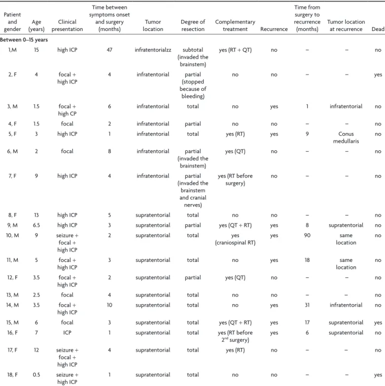
Table 1. Patients’ gender, age, clinical presentation, time between symptoms onset and surgery, tumor location, degree of resection, complementary treatment, time from surgery to recurrence, tumor location at recurrence and death status in patients with intracranial ependymoma at age between 0–15 years.
Patient and gender
Age (years)
clinical presentation
Time between symptoms onset
and surgery (months)
Tumor location
Degree of resection
complementary
treatment recurrence
Time from surgery to recurrence (months)
Tumor location
at recurrence Dead
Between 0–15 years
1,M 15 high IcP 47 infratentorialzz subtotal
(invaded the brainstem)
yes (rT+QT) no – – no
2, F 4 focal+
high IcP
4 infratentorial partial
(stopped because of
bleeding)
no no – – yes
3, M 1.5 focal+
high cP
6 infratentorial total no yes 1 infratentorial no
4, F 1.5 focal 2 infratentorial partial no no – – no
5, F 3 high IcP 1 infratentorial total yes (rT) yes 9 conus
medullaris no
6, M 2 focal 8 infratentorial partial
(invaded the brainstem)
yes (QT) no – – no
7, F 9 high IcP 4 infratentorial partial
(invaded the brainstem and cranial nerves)
yes (rT before
surgery) no – – no
8, F 13 high IcP 5 supratentorial total no no – – no
9, M 6.5 high IcP 3 supratentorial partial yes (QT+rT) yes 8 supratentorial no
10, M 9 seizure+
focal+ high IcP
2 supratentorial total yes
(craniospinal rT)
yes 90 same
location no
11, M 5 focal+
high IcP
3 supratentorial total no yes 18 same
location no
12, F 3.5 focal+
high IcP 2 supratentorial partial yes (QT) no – – no
13, M 2.5 focal 4 supratentorial total no no – – no
14, M 3.5 focal+
high IcP
10 supratentorial total no yes 31 infratentorial no
15, M 6 focal 3 supratentorial total yes (QT+rT) yes 17 supratentorial yes
16, F 7 IcP 1 supratentorial total yes (rT before
2nd surgery) yes 6 supratentorial no
17, F 12 seizure+
focal+ high IcP
4 supratentorial total yes (rT) no – – no
18, F 0.5 seizure+
high IcP
1 supratentorial total no no – – yes
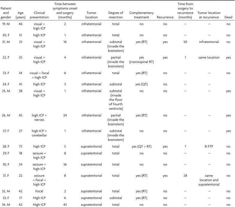
Table 1 (continuation). Patients’ gender, age, clinical presentation, time between symptoms onset and surgery, tumor location, degree of resection, complementary treatment, time from surgery to recurrence, tumor location at recurrence and death status in patients with intracranial ependymoma at age over 15 years.
Patient and
gender (years)Age presentationclinical
Time between symptoms onset
and surgery
(months) locationTumor Degree of resection complementary treatment recurrence
Time from surgery to recurrence
(months) Tumor location at recurrence Dead
19, M 46 visual +
high IcP 2 infratentorial total no no – – no
20, F 51 high IcP 1 infratentorial total no no – – no
21, M 23 visual +
high IcP
16 infratentorial subtotal
(invade the brainstem)
yes (rT) yes 50 infratentorial no
22, F 25 visual +
high IcP
4 infratentorial partial
(invade the brainstem)
yes (craniospinal rT)
yes 1 same location yes
23, F 34 visual + focal
+ high IcP 6 infratentorial total yes (rT) no – – no
24, F 41 high IcP 3 infratentorial subtotal yes (QT) no – –
25, M 28 visual +
high IcP 1 infratentorial subtotal (invade
the loor of fourth ventricle)
no no – – yes
26, M 45 high IcP +
nerves
24 infratentorial partial
(invade the brainstem)
yes (rT) no – – yes
27, F 27 high IcP +
cerebellar 1 infratentorial (invade the subtotal
brainstem)
no no – – yes
28, F 73 high IcP 5 supratentorial total yes (QT + rT) yes 1 r FTP no
29, F 18 seizure +
high IcP
8 supratentorial total no no – – no
30, F 24 seizure +
high IcP
36 supratentorial total no no – – no
31, F 22 seizure
+ focal + high IcP
8 supratentorial total yes (rT) yes 28 same
location and supratentorial
no
32, M 42 Focal 2 supratentorial total yes (rT) no – – no
33, F 17 High IcP 6 supratentorial subtotal yes (rT) no – – no
34, M 42 High IcP 43 supratentorial total no no – – no
M: male; F: female; IcP: increased intracranial pressure; focal: motor deicits (paresis); seizure: partial seizure or generalized seizure; rT: radiation therapy; QT: chemotherapy; rFTP: right fronto temporoparietal.
Data of the clinical presentation, tumor location, duration of the symptoms until the surgery, degree of resection, com-plementary treatment were collected and are presented in Ta-bles 1 and 2. Data of intracranial ependymomas in children and adults and spinal cord ependymomas are presented in separate tables and were correlated with the location of the tumor and the recurrence time and overall survival. SPSS v.15 was used for the statistical analyses of intracranial and intramedullary sub-groups to deine the possible prognostic factors.
This project is supported by FAPESP (number 04/12133-6) and cAPES (number 691/05) and their ethics commission.
resUlts
The mean age of the patients with intracranial
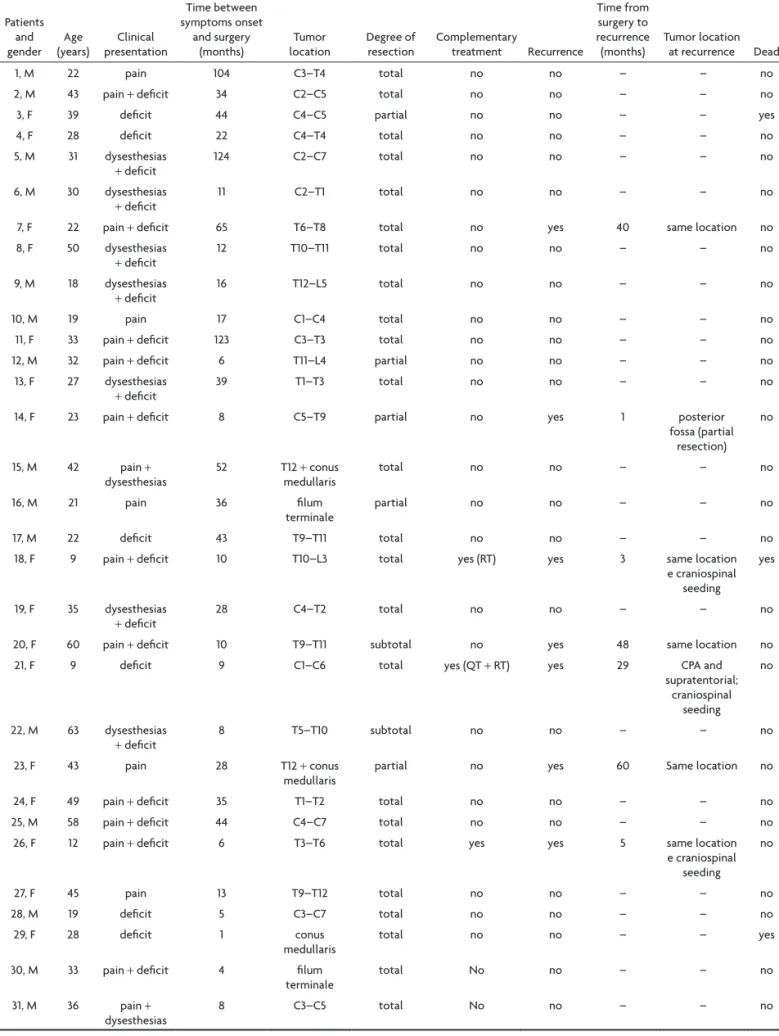
be-Table 2. Patients’ gender, age, clinical presentation, time between symptoms onset and surgery, tumor location, degree of resection, complementary treatment, time from surgery to recurrence, tumor location at recurrence and death status in patients with intramedullary and conus medullaris ependymoma.
Patients and
gender (years)Age presentationclinical
Time between symptoms onset
and surgery
(months) locationTumor Degree of resection complementary treatment recurrence
Time from surgery to recurrence
(months) Tumor location at recurrence Dead
1, M 22 pain 104 c3–T4 total no no – – no
2, M 43 pain + deicit 34 c2–c5 total no no – – no
3, F 39 deicit 44 c4–c5 partial no no – – yes
4, F 28 deicit 22 c4–T4 total no no – – no
5, M 31 dysesthesias
+ deicit 124 c2–c7 total no no – – no
6, M 30 dysesthesias
+ deicit
11 c2–T1 total no no – – no
7, F 22 pain + deicit 65 T6–T8 total no yes 40 same location no
8, F 50 dysesthesias
+ deicit 12 T10–T11 total no no – – no
9, M 18 dysesthesias
+ deicit
16 T12–l5 total no no – – no
10, M 19 pain 17 c1–c4 total no no – – no
11, F 33 pain + deicit 123 c3–T3 total no no – – no
12, M 32 pain + deicit 6 T11–l4 partial no no – – no
13, F 27 dysesthesias
+ deicit
39 T1–T3 total no no – – no
14, F 23 pain + deicit 8 c5–T9 partial no yes 1 posterior
fossa (partial resection)
no
15, M 42 pain +
dysesthesias
52 T12 + conus
medullaris
total no no – – no
16, M 21 pain 36 ilum
terminale
partial no no – – no
17, M 22 deicit 43 T9–T11 total no no – – no
18, F 9 pain + deicit 10 T10–l3 total yes (rT) yes 3 same location
e craniospinal seeding
yes
19, F 35 dysesthesias
+ deicit
28 c4–T2 total no no – – no
20, F 60 pain + deicit 10 T9–T11 subtotal no yes 48 same location no
21, F 9 deicit 9 c1–c6 total yes (QT + rT) yes 29 cPA and
supratentorial; craniospinal
seeding no
22, M 63 dysesthesias
+ deicit
8 T5–T10 subtotal no no – – no
23, F 43 pain 28 T12 + conus
medullaris
partial no yes 60 Same location no
24, F 49 pain + deicit 35 T1–T2 total no no – – no
25, M 58 pain + deicit 44 c4–c7 total no no – – no
26, F 12 pain + deicit 6 T3–T6 total yes yes 5 same location
e craniospinal seeding
no
27, F 45 pain 13 T9–T12 total no no – – no
28, M 19 deicit 5 c3–c7 total no no – – no
29, F 28 deicit 1 conus
medullaris total no no – – yes
30, M 33 pain + deicit 4 ilum
terminale
total No no – – no
31, M 36 pain +
dysesthesias 8 c3–c5 total No no – – no
tween symptoms onset and the surgery was not statisti-cally signiicant (p=0.805 and p=0.623, respectively).
The gross total resection was achieved in 58.8% of cases; supratentorial in 83.3% of cases and infratentori-al in only 31.5% of cases. Follow-up ranged from 1 to 120 months (mean of 38 months). The 2-year survival was 70%, and 5-year survival was 60%, calculated by Kaplan-Meier method (Fig 2A).
Twelve patients with intracranial ependymoma re-curred; 8 at the primary site, ranging from one to 90 months after surgery (mean of 21 months). The adjuvant therapy with chemotherapy or radiotherapy was indicated in 55.9% of the patients either when the tumor had subto-tal resection or if it recurred. No association was noticed between the degree of resection and the tumor location with recurrence or survival. Also, there was no statistical-ly signiicant correlation between survival and gender or patients’ age in cases of intracranial ependymoma.
The age of patients with intramedullary ependymoma ranged from 9 to 63 years (mean of 32 years) (Fig 1B). Fif-teen patients were female (48.4%). The time between the onset of symptoms and surgery ranged from one month to 10 years (mean of 31 months). The symptom of neuro-logical deicit (tetra- or paraparesis) was present in 80.7% cases, and associating painful aching sensations with neu-rological deicit was more common (38.7%). Neuneu-rological deicit was the only symptom in 19.4% cases; it was asso-ciated with other sensory symptoms, such as dysesthesias, in 22.6% of cases. The symptoms of sensory disturbances were only pain and association of pain and dysesthesias in 12.9% and 6.5%, respectively. The more commonly affect-ed site was thoracic (29%) and cervical cord (25.8%); only in 9.7% of the cases the location was exclusively in the co-nus medullaris and ilum terminale. The lesions were pres-ent in the cervico-thoracic and thoraco-lumbar transition in 19.4% and 16.1% of the population, respectively. No cor-relation was observed between the location and the clini-cal presentation with the duration of symptoms until sur-gery (p=0.362 and p=0.835, respectively).
Fig 1. Age frequency of the patients with intracranial [A] and in-tramedullary [B] ependymoma at the time of irst surgery.
The gross total resection was achieved in 77.4% cases of intramedullary ependymoma and there was no corre-lation between the location and the degree of resection (p=0.362). The cause of subtotal resection was the lack of tumor limits or the linkage between tumor and normal tissue. There were no sensory or motor evoked potentials (MEP) used throughout the procedure. The tumor grade was myxopapillary ependymoma (wHo grade I) in 5 cas-es in lumbar or thoraco-lumbar location; all other were ependymoma (wHo grade II). Follow-up ranged from one month to 10 years (mean of 53 months). The 5 and 10-year survival was about 90% and 70%, respectively (Fig 2B), ac-cording to Kaplan-Meier analysis. recurrence occurred in seven cases (31.8%), four of them being at the primary site. No correlation was identiied between the degree of re-section and recurrence (p=0.124). The time to recurrence ranged from 1 to 60 months (mean was 23 months). Adju-vant therapy was not routinely indicated, even in subto-tal resection. only three cases had radiation therapy in-dicated due to dissemination (two cases) and rapid recur-rence (one case, 3 months after irst surgery). The statisti-cal analysis failed to identify any factor related to recur-rence or survival.
dIscUssIon
Most reported series of ependymoma are retrospec-tive and include only a small number of patients. Addi-tionally, studies have been conducted over several de-cades, making it dificult to interpret results since classi-ication and treatment protocols have been modiied over time. As a consequence, the prognostic factors currently accepted are unsatisfactory5.
The signiicance of tumor grade is not always accepted, probably due to varying deinitions of anaplasia6, to dis-crepancies among pathologists7, and to the fact that histo-logical features of anaplasia seem to be unrelated to the biological behavior of ependymomas8. Another confound-ing factor is that most series do not distconfound-inguish cases with malignant ependymoma from those with ependymoblasto-ma, which have worse prognoses and distinct classiication.
A direct correlation between age and better prognos-tic was suggested. The small number of cases, the differ-ent deinitions of pediatric age among series (ranging from 12 to 20 years), and also, the heterogeneity of histological grade and tumor location have not allowed to draw dei-nite conclusions. Ependymomas are rare in adults. Adults present better prognosis and the 5-year survival is about 90%, while in the pediatric population it is around 60%. overall, the younger the child the worse the prognoses8. However, no correlation was observed between age and survival in our series.
The prognostic role of tumor location is also contro-versial. Intramedullary lesions present more favorable
prognosis but the intracranial tumors have uncertain prognoses. Some authors concluded that there was no relationship between prognosis and location, while oth-ers concluded that supratentorial lesions indicated worse prognosis due to their iniltrative characteristics in brain parenchyma, hindering total resection9,10. Furthermore, in-fratentorial tumors present a mitotic activity lower than supratentorial tumors11. Some authors observed a worse prognosis for ependymoma arising from posterior fossa, which occurs in younger patients and generally invades the brainstem, the loor of fourth ventricle, the cerebel-lopontine angle and the cranial nerves, making it difi-cult for a complete resection9,11. In this series, no corre-lation was noticed between the tumor location and the prognosis (correlation with recurrence, p=0.814; correla-tion with death, p=0.147), although it was observed that gross total resection was more statistically signiicant in the supratentorial tumors than in the infratentorial ones (83.3% × 31.5%; p=0.01).
The degree of resection was considered an indepen-dent prognostic factor; the complete resection presented better 5-year survival than subtotal removal or biopsy12,13. In some cases, the beneit of complete resection was lim-ited to low-grade tumors14. However, some authors failed to ind a correlation between improved survival and the extent of resection15-17. The lack of evidence for the im-pact of surgery on survival could be related to the unre-liability of subjective assessment of the degree of surgi-cal ablation, that is, when the degree of resection is not evaluated by postoperative imaging. It was proved that the surgeon evaluated the degree of resection in a differ-ent way than that observed in the postoperative imaging in 32% cases18. The degree of resection, assessed by post-operative imaging, has been related with an improved fol-low-up in 5-year survival18. In the present series no signii-cant correlation was identiied between the degree of re-section and improvement in follow-up, such as longer sur-vival or lower recurrence of tumors.
postoperative radiotherapy was indicated although gross total resection was achieved (these cases did not recur).
Intramedullary ependymomas present different prog-nosis from intracranial tumors. The strongest predictor of post-operative functional outcome is pre-operative func-tional ability. The aim of surgery is to preserve rather than restore neurological function. The morbidity and mortal-ity are directly related with postoperative deicits, while postoperative deicits correlate with the pre-operative status and the tumor location. The tumor growth is gen-erally slow; outcome and the risk of recurrence depend on the degree of resection in the irst surgery4. The post-operative radiotherapy is unnecessary when the resection is complete. when resection is incomplete the need of ra-diotherapy is dificult to interpret, because studies con-template small series with limited follow-up usually with no control groups19. In this series the radiotherapy was indicated only when dissemination was evident and in a case which recurrence was premature (three months after the irst surgery). Signiicant correlation was not observed between the lesion location and survival or the degree of resection (p=0.295 and p=0.362, respectively).
In conclusion, the study of ependymomas is of great interest. Many studies have been published throughout the years; however, handling these patients is still contro-versial. Surgery alone or combinations of resection with chemotherapy and radiotherapy have been proposed as forms of treatment. The absence of standard treatment and randomized studies hindered the establishment of the best treatment strategy20. Although the surgery has a well established role, adjuvant treatment procedures are still uncertain.
In the presented series, the degree of resection was connected with the intracranial tumor location with statis-tically signiicant correlation, although this inding was not a predictor for recurrence. The clinical data study of in-tramedullary ependymoma patients did not allow deining characteristics of survival and probability of recurrence. It is necessary to understand better the molecular bi-ology of ependymomas to improve the evaluation of the treatment and the outcome of patients.
reFerences
1. Oppenheim JS, Strauss RC, Mormino J, et al. Ependymomas of the third ventricle. Neurosurgery 1994;34:350-352.
2. Centero RS, Lee AA, Winter J, Barba D. Supratentorial ependymo-mas. Neuroimaging and clinicopathological correlation. J Neurosurg 1986;64:209-215.
3. Biegel JA. Cytogenetics and molecular genetics of childhool brain tu-mors. NeuroOncol 1999;1:139-151.
4. Schwartz TH, McCormick PC. Intramedullary ependymomas: clinical presentation, surgical treatment strategies and prognosis. J NeuroOn-col 2000;47:211-218.
5. Reni M, Gatta G, Mazza E, Vecht C. Ependymoma. Crit Rev Oncol/He-matol 2007;63:81-89.
6. Schiffer D, Chio A, Cravioto H, et al. Ependymomas: internal correla-tions among pathological signs: the anaplastic variant. Neurosurgery 1991;29:206-210.
7. Robertson PL, Zelter PM, Boyett JM, et al. Survival and prognostic fac-tors following radiation therapy and chemotherapy for ependymomas in children: a case report of the Children’s Cancer Group. J Neurosurg 1998;88:695-703.
8. Schiffer D, Chio A, Giordana MT, et al. Histologic prognostic factors. Radiother Oncol 1997;45:3-10.
9. Vanuytsel LJ, Bessell EM, Ashley SE, et al. Intracranial ependymoma: long-term results of a policy of surgery and radiotherapy. Int Radiat Oncol Bio Phys 1992;23:313-319.
10. MacLaughlin MP, Marcus Jr Rb, Buatti JM, et al. Ependymoma: results, prognostic factors and treatment recommendations. Int J Radiat Oncol Biol Phys 1998;40:845-850.
11. Goldwein JW, Leahy JM, Packer RJ, et al. Intracranial ependymomas in children. Int J Radiat Oncol Biol Phys 1990;19:1497-1502.
12. Rousseau P, Habrand JL, Sarrazin D, et al. Treatment of intracranial ependymomas of children: review of a 15-year experience. Int J Radiat Oncol Biol Phys 1994;28:381-386.
13. Sutton LN, Goldwein J, Perilongo G et al. Prognostic factors in child-hood ependymomas. Pediatr Neurosurg 1990;6:57–65.
14. Nazar GB, Hoffman HJ, Becker LE, et al. Infratentorial ependymomas in childhood: prognostic factors and treatment. J Neurosurg 1990;72:408-417. 15. Lyons MK, Kelly PJ. Posterior fossa ependymomas: report of 30 cases
and review of the literature. Neurosurgery 1991;28:659-664.
16. Pierre-Kahn A, Hirsch JF, Roux FX, et al. Intracranial ependymomas in childhood. Survival and functional results of 47 cases. Childs Brain 1983;10:145-156.
17. Paulino AC, Wen BC. The significance of radiotherapy treatment duration in intracranial ependymoma. Int J Radiot Oncol Biol Phys 2000;47:585-589.
18. Healey EA, Barnes PD, Kupsky WJ, et al. The prognostic signiicance of
postoperative residual tumor in ependymoma. Neurosurgery 1991;28: 666-671.
19. Wen BC, Hussey DH, Hitchon PW, et al. The role of radiation thera-py in the management of ependymomas of the spinal cord. Int J Radi-at Oncol Biol Phys 1991;20: 781-786.
![Fig 1. Age frequency of the patients with intracranial [A] and in- in-tramedullary [B] ependymoma at the time of irst surgery.](https://thumb-eu.123doks.com/thumbv2/123dok_br/15432290.594903/5.955.475.852.91.637/fig-age-frequency-patients-intracranial-tramedullary-ependymoma-surgery.webp)
