Orthopedic Oncology Group, Department of Orthopedics, Hospital das Clínicas, São Paulo University Medical School – São Paulo/SP, Brazil. Email: andre.baptista@uol.com.br
Received for publication on March 03, 2006. Accepted for publication on April 20, 2006.
CLINICAL SCIENCES
SYNOVIAL SARCOMA OF THE EXTREMITIES:
PROGNOSTIC FACTORS FOR 20 NONMETASTATIC
CASES AND A NEW HISTOLOGIC GRADING SYSTEM
WITH PROGNOSTIC SIGNIFICANCE
André Mathias Baptista, Olavo Pires de Camargo, Alberto Tesconi Croci, Cláudia Regina G.C.M. de Oliveira, Raymundo Soares de Azevedo Neto, Marcelo Abrantes Giannotti, Marcelo Tadeu Caiero, Telma Murias dos Santos, and Márcia Datz Abadi
Baptista AM, Camargo OP de, Croci AT, Oliveira CRGCM de, Azevedo Neto RS de, Giannotti MA et al. Synovial Sarcoma Of The Extremities: Prognostic Factors For 20 Nonmetastatic Cases And A New Histologic Grading System With Prognostic Significance. CLINICS. 2006;61(5):381-6.
PURPOSE: To evaluate 20 cases of nonmetastatic synovial sarcoma of the extremities regarding prognostic factors, and to propose a histologic grading system with prognostic significance.
METHODS: The cases of 20 patients (14 females and 6 males) with nonmetastatic synovial sarcomas of the extremities treated between 1985 and 1998, were retrospectively evaluated regarding prognostic factors. A histologic grading system with prognostic significance is proposed.
RESULTS: The mean follow-up period was 48.4 months (range, 16-116 months). There was local recurrence in 3 cases (15%), microscopic surgical margin being the only prognostic factor identified. Seven patients (35%) died of the disease in a mean postoperative period of 31.7 months (range, 16-53 months), all with pulmonary or brain metastasis. The survival rate was 65% in 48.4 months of follow-up.
CONCLUSION: The unfavorable prognostic factors identified regarding survival were high histologic grade, tumors proximal to the knee or elbow, and spontaneous tumor necrosis over 25%. Local recurrence did not have influence on survival in this study. The presence of mast cells appears to have a positive influence on survival, although statistical significance was not reached (P =
0.07). The oncologic and functional result was good in 6 cases (30%), regular in 7 (35%), and poor in 7 cases (35%).
KEYWORDS: Synovial, sarcoma. Extremities. Retrospective studies. Prognosis. Soft tissue neoplasms.
INTRODUCTION
Synovial sarcoma (SS) is the fourth most common soft tissue sarcoma,1 after malignant fibrous histiocytoma,
li-posarcoma, and rhabdomyosarcoma. It is more prevalent in males (1.2:1) between 15 and 40 years of age. About 75% of the cases of SS arise in the extremities, especially in the lower extremity, and there are 4 histologic subtypes
described: biphasic, monophasic fibrous, monophasic epi-thelial, and poorly differentiated. Five-year survival rates range from 30% to 74%,2,3 and several prognostic factors
have been described.
Most pathologists consider the SS a high-grade sarcoma, independent of its histologic characteristics. Nevertheless, some authors have tried to identify high- and low-risk groups, as well as prognostic factors regarding survival and local recurrence.
institu-tion. A histologic grading system with prognostic signifi-cance is proposed, dividing the tumors among low and high histologic grades.
MATERIALS AND METHODS
From 1950 to 1998, the Orthopedic Oncology Group of the Department of Orthopedics treated 351 patients with a diagnosis of soft-tissue sarcomas. Among them, 87 were SS, and 20 of them had complete data and were selected for this study. All patients underwent surgery, with 5 of them also undergoing radiation therapy and 1 undergoing chemotherapy postoperatively. There was no neoadjuvant therapy in this study.
The case index number, as well as age, gender, anatomic site, and date of the surgery are listed on Table 1.
The inclusion criteria for this study were as follows: 1) histologic diagnosis of SS, 2) anatomic site in the extremi-ties, 3) no evidence of metastasis on chest CT scan, and 4) a minimum follow-up of 24 months for the surviving patients.
The chest CT scan was used for the detection of lung metastasis, the most common site among soft-tissue sar-comas.
The average age was 27.4 years (range, 11-45 years), and there were 14 females and 6 males. All cases were at least partially deep tumors, with 7 occurring in the upper extremity and 13 in the lower extremity. Nine cases were proximal to the knee or the elbow, and 11 cases were distal.
Thirteen patients had tumors smaller than 10 cm, and 7 had tumors 10 cm or more at the longest axis. We chose the criterion, smaller and larger than 10 cm, because the mean size of the tumors in our study was 9.3 cm. Conventional radiographs and MRI of the tumor were made, as well as were chest radiographs and chest CT scans. Biopsy was percutaneous in all cases.
All patients underwent surgery, with 12 wide resections and 8 amputations.
The anatomopathologic study was made using hematoxylin-eosin stain, as well as an immunohistochem-istry study to confirm the diagnosis of SS. The monoclonal antibodies used were vimentin, keratin, EMA, actin, eno-lase, and S-100. Reticulin stain was used when the epithe-lial pattern was not evident.
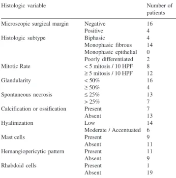
The following 10 histologic variables were evaluated: microscopic surgical margin, histologic subtype, mitotic rate, glandularity, spontaneous necrosis, presence of calci-fications, hyalinization, presence of mast cells, presence of a hemangiopericytic pattern, and presence of rhabdoid cells (Table 2).
The histologic grading system proposed considered high-grade tumors those with a mitotic rate of 5 or more mitosis figures in 10 high-power fields (HPF), more than 25% spontaneous necrosis, and less than 50% glandularity. Seven patients fulfilled these criteria. The remaining 13 cases were considered low-grade tumors.
Table 2 - Patients distributed according to histologic variables
Histologic variable Number of
patients
Microscopic surgical margin Negative 16
Positive 4
Histologic subtype Biphasic 4
Monophasic fibrous 14 Monophasic epithelial 0 Poorly differentiated 2
Mitotic Rate < 5 mitosis / 10 HPF 8
≥ 5 mitosis / 10 HPF 12
Glandularity < 50% 16
≥ 50% 4
Spontaneous necrosis ≤ 25% 13
> 25% 7
Calcification or ossification Present 7
Absent 13
Hyalinization Low 14
Moderate / Accentuated 6
Mast cells Present 9
Absent 11
Hemangiopericytic pattern Present 11
Absent 9
Rhabdoid cells Present 1
Absent 19
Table 1 - Patients distributed according to age, gender, anatomic site, and date of surgery
No AGE GENDER ANATOMIC DATE OF
SITE SURGERY
1 45 Female Right Wrist Dec / 1985
2 29 Male Right Ankle Oct / 1989
3 18 Female Right Ankle Dec / 1989
4 17 Female Right Thigh Jul / 1991
5 23 Female Right Forearm Jan / 1992
6 23 Female Left Leg Jun / 1993
7 41 Female Left Ankle Sep / 1993
8 42 Female Left Arm Oct / 1993
9 45 Male Left Thigh Aug / 1994
10 13 Female Right Wrist May / 1995
11 31 Male Left 3rd toe Jun / 1996
12 38 Female Right Knee Jul / 1996
13 20 Female Right Arm Aug / 1996
14 31 Female Right Thigh Nov / 1996
15 25 Male Left Arm Nov / 1996
16 11 Male Right Foot Jun / 1997
17 26 Female Left Foot Oct / 1997
18 24 Male Right Thigh Nov / 1997
19 11 Female Right 2nd Finger Dec / 1997
All the above mentioned variables were studied regard-ing local recurrence and disease-related survival.
The oncologic and functional result was considered (i) good when the patient was alive with no evidence of dis-ease (NED) after undergoing a limb-salvage procedure, (ii) medium when alive with NED but having undergone am-putation, and (iii) poor when the patient died of the dis-ease (DOD) or was alive with disdis-ease (AWD).
A statistical analysis was performed using the 2-tailed Fisher exact test, with 5% as the significance index (P =
0.05). The Kaplan-Meier method was used to estimate the survival and local recurrence rates during the follow-up period.4
RESULTS
The overall mean follow-up was 48.4 months (range, 16-116 months). Among the living patients at the last evalu-ation, the mean follow-up was 57.5 months (24-116). No patients were lost to follow-up.
Local recurrence
Four patients presented initially as having recurrences (Table 3). After surgery, 3 patients developed a local re-currence, with one of them being 1 of the 4 patients who presented with recurrence. Figure 1 illustrates the occur-rence of local recuroccur-rence during the follow-up period.
Survival
The overall survival was 65% in a mean follow-up pe-riod of 48.4 months. Seven patients died of the disease due to pulmonary metastases, one of them also having brain metastases (Table 4). Figure 2 illustrates the occurrence of disease-related death over the follow-up period.
The oncologic and functional result was good in 6 cases, medium in 7, and poor in 7 cases. There were no patients alive with disease; all individuals in the “poor” category died of the disease.
DISCUSSION
Efforts have been made in the last decades to establish prognostic factors for soft-tissue sarcomas (STS). Nonethe-less, approximately 30 different histologic entities are clas-sified as STS, many of them with different biologic behaviors. Malignant fibrous histiocytoma (MFH), for in-stance, has a different behavior when compared to SS. Due to the rarity of these lesions, many authors2, 5-7 2,7,12,22
di-vide them between high- and low-grade sarcomas, thus
Table 3 - Local recurrence according to all variables
Variable Local P
recurrence
Sex Female 3 / 14 0.521
Male 0 / 6
Age ≤ 20 1 / 6 1.000
> 20 2 / 14
Size < 10 cm 3 / 13 0.521
≥ 10 cm 0 / 7
Limb Upper 2 / 7 0.270
Lower 1 / 13
Location Proximal 1 / 9 1.000
Distal 2 / 11
Status at presentation Primary 2 / 16 0.509
Recurrence 1 / 4
Type of surgery Resection 3 / 12 0.242
Amputation 0 / 8
Adjuvant radiation therapy Used 2 / 5 0.140
Not used 1 / 15
Microscopic surgical margin Clear 0 / 16 0.004 (*)
Positive 3 / 4
Histologic subtype Monophasic fibrous 1 / 14 0.405
Biphasic 1 / 4
Poorly differentiated 1 / 2 Monophasic epithelial 0 / 0
Mitotic rate < 5 mitosis / 10 HPF 1 / 8 1.000 ≥ 5 mitosis / 10 HPF 2 / 12
Glandularity < 50% 2 / 16 0.509
≥ 50% 1 / 4
Spontaneous necrosis ≤ 25% 2 / 13 1.000
> 25% 1 / 7
Calcification or ossification Present 0 / 7 0.521 Not present 3 / 13
Hyalinization Mild 3 / 14 0.521
Moderate / Accent. 0 / 6
Mast cells Present 1 / 9 1.000
Not present 2 / 11
Hemangiopericytic pattern Present 2 / 11 1.000
Not present 1 / 9
Rhabdoid cells Present 0 / 1 1.000
Not present 3 / 19
Histologic grade High 1 / 7 1.000
Low 2 / 13
(*) = statistically significant
achieving an adequate number of cases for statistical analy-sis. We believe it is not safe to generalize the conclusions of these studies. Multicentric studies of each tumor would probably be the best choice. The present study is part of an overall project of the Department to systematically re-view our records concerning the treatment of musculoskel-etal tumors .8-10 Despite improvements in staging, surgical
technique, and adjuvant therapies, SS remains one of the most aggressive STS. Since 1936, when Knox115 first used
the term synovial sarcoma, the aggressiveness of this en-tity has been described. Five-year survival rates range from 30%3 to 74%,2 and the lungs are, as in most sarcomas, the
most affected organ by metastases. Lymph node metastases are uncommon in sarcomas, but SS may present them more frequently than other tumors. We had 2 patients with posi-tive regional lymph nodes in our study.
Several pathologists primarily consider SS a high-grade STS. Our impression, based on our experience, is that the biologic behavior in some cases is more benign than oth-ers. Thus the objective of separating the high-grade, more aggressive cases from the low-grade ones is to identify the more benign tumors that have better survival prognosis.
When considering local recurrence, our study showed that the only prognostic factor was the microscopic surgi-cal margin. When it is positive, most of the cases recur. Several studies have identified this variable as prognostic in terms of local recurrence.3,5,12,13 3,6-8 Many studies suggest
that adjuvant radiation therapy has good influence in re-currence rates 6,14,15 9-12. In fact, it is almost a consensus
among orthopedic oncology surgeons that adjuvant radia-tion therapy diminishes the local recurrence rates in STS, especially in high-grade tumors. Some authors indicate ra-diation therapy in high grade STSs larger than 5 cm, oth-ers in recurrent cases, and some preoperatively in tumors near neurovascular bundles. The indications vary, but most authors agree that radiation therapy lowers the chances of local recurrence. Nevertheless, serious complications may occur after radiation therapy, especially external beam ra-diation. These include dehiscence, limb length discrepancy in children, avascular necrosis, and pathologic fracture. 6,16 11,12 We did not have enough data to evaluate this issue. It
is interesting to observe, though, that local recurrence did not worsen the survival prognosis in our study. As other authors previously described, the influence of local recur-rence on survival remains controversial. 17 13 Some authors
believe that patients that present with local recurrence have a poorer survival prognosis. 17-19 13-15 Others believe, as we
do, that local recurrence does not influence survival.12, 20, 21 6,16,17
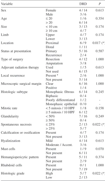
Regarding survival, the unfavorable prognostic factors identified in our study were spontaneous necrosis over 25%,
Table 4 - Disease-related survival according to all variables
Variable DRD P
Sex Female 4 / 14 0.613
Male 3 / 6
Age ≤ 20 1 / 6 0.354
> 20 6 / 14
Size < 10 cm 3 / 13 0.174
≥ 10 cm 4 / 7
Limb Upper 4 / 7 0.174
Lower 3 / 13
Location Proximal 6 / 9 0.017 (*)
Distal 1 / 11
Status at presentation Primary 5 / 16 0.587
Recurrence 2 / 4
Type of surgery Resection 4 / 12 1.000
Amputation 3 / 8
Adjuvant radiation therapy Used 1 / 5 0.613
Not used 6 / 15
Local recurrence Present # 2 / 6 1.000
Not present 5 / 14
Microscopic surgical margin Clear 6 / 16 1.000
Positive 1 / 4
Histologic subtype Monophasic fibrous 6 / 14 0.245
Biphasic 0 / 4
Poorly differentiated 1 / 2 -Monophasic epithelial 0 / 0 -Mitotic rate < 5 mitosis / 10 HPF 1 / 8 0.158
≥ 5 mitosis / 10 HPF 6 / 12
Glandularity < 50% 7 / 16 0.249
≥ 50% 0 / 4
Spontaneous necrosis ≤ 25% 2 / 13 0.022 (*)
> 25% 5 / 7
Calcification or ossification Present 4 / 7 0.174 Not present 3 / 13
Hyalinization Mild 4 / 14 0.613
Moderate / Accent. 3 / 6
Mast cells Present 1 / 9 0.070
Not present 6 / 11
Hemangiopericytic pattern Present 5 / 11 0.374
Not present 2 / 9
Rhabdoid cells Present 0 / 1 1.000
Not present 7 / 19
Histologic grade High 5 / 7 0.022 (*)
Low 2 / 13
# = including recurrences at presentation
(*) = statistically significant DRD = disease-related death
tumors proximal to knee or elbow, and high histologic grade tumors according to our criteria.
Some investigators have tried to establish high- and low-risk groups based on histologic variables. Skitting et al. 22 18 defined as favorable cases those presenting cellular
aty-pia, no necrosis and a mitotic rate under 10/10 HPF. These cases had a survival rate of 83%, whereas the remaining patients had only a 31% survival rate. Our criteria, although slightly different, present similar results. We defined high-grade tumors as those showing mitotic rate of 5 or more per 10 HPF, more than 25% of spontaneous necrosis, and less than 50% of glandularity. The remaining cases were considered low-grade cases. Patients with low-grade tumors had a 71% survival rate, compared to those with high-grade tumors, who had a 15% survival rate.
In most studies, proximal location is usually not an unfavorable prognostic factor. In our series, however, pa-tients with proximal tumors had a survival rate of 33%, ver-sus a survival rate of 91% for those with distal tumors. Mullen and Zagars6 12 and Hadju et al2319 also had similar
results.
Several prognostic factors have been described as sig-nificant for survival in SS, including primary size of the tumor, margin of resection, and histologic subtype. Size is one of the most described prognostic factors. Patients with tumors smaller than 5 cm had better survival prognoses in several studies. 2,6,7,13,17,20,24-28 2,8,12,13,16,20-25 Our study, despite
a tendency of patients with tumors larger than 10 cm to have worse survival prognoses, this association did not show statistic significance.
Another example of a prognostic factor is the presence of calcifications in simple radiographs, which is reported to be present in about 15% to 20% of the cases.1 In our
study, the patients who presented with tumor calcifications did not have better survival prognoses. Nevertheless, Varela-Duran and Enzinger 29 26 believed these calcifications to be
a favorable prognostic factor. Their series showed 82% sur-vival in cases presenting with heavy calcifications, better than all previously published papers.
Presence of mast cells also has been studied by some authors as a prognostic factor. It appears that mast cells have a positive influence on survival in SS cases. 27 24 In
our study, patients with tumors showing mast cells had bet-ter survival rates, ie, 11.0% versus 54.5%, but this differ-ence was not statistically significant (P = 0.07).
More controversial potential prognostic factors include sex, age, type of treatment, and tumor location.
In summary, we observed a survival rate of 65% and a local recurrence rate of 15% in our study, which seems comparable to most studies published in the last decade concerning only SS. The unfavorable prognostic factors that influenced survival were spontaneous necrosis above 25%, proximal tumors, and high histologic grade according to our criteria. Local recurrence was higher only when the microscopic surgical margin was positive. Although our study is limited in terms of patient numbers, we believe, based on our results, that our criteria for determining high-and low-histologic grade SS are valid high-and have prognostic significance.
RESUMO
Baptista AM, Camargo OP de, Croci AT, Oliveira CRGCM de, Azevedo Neto RS de, Giannotti MA et al. Sarcoma sinovial das extremidades: fatores de prognóstico em 20 casos não-metastáticos e um novo sistema de graduação histológica com significado prognóstico. CLINICS. 2006;61(5):381-6.
OBJETIVO: Avaliar casos de sarcoma sinovial não-metastático das extremidades no que se refere a fatores prognósticos, e propor um sistema histológico de pontua-ção com significado prognóstico.
MATERIAL E MÉTODO: Vinte casos (14 do sexo fe-minino e 6 do sexo masculino) de sarcomas sinoviais não-metastáticos das extremidades tratados entre 1985 e 1998 no departamento de Ortopedia foram avaliados retrospec-tivamente no que se refere a fatores prognósticos e está sen-do proposto um sistema de pontuação histológico com
sig-nificado prognóstico.
RESULTADOS: A média dos períodos de acompanhamen-to foi 48,4 meses (mínimo 16 meses, máximo 116). Hou-ve recorrência localizada em 3 casos (15%), sendo a mar-gem cirúrgica microscópica o único fator prognóstico iden-tificado. Sete pacientes (35%) morreram da doença, todos em período pós-operatório médio de 31,7 meses (mínimo 16 meses, máximo 53), todos com metástase pulmonar ou cerebral. A sobrevida foi de 65% em 48,4 meses de acom-panhamento.
oncológico e funcional foi bom em seis casos (30%), regu-lar em sete (35%) e insatisfatório em sete (35%).
UNITERMOS: Sarcoma sinovial. Extremidades. Estudos retrospectivos. Prognóstico. Neoplasmas em tecidos moles.
REFERENCES
1. Enzinger FM, Weiss SW.Soft tissue tumors. 3rd ed. St. Louis: Mosby; 1995. p.757-86.
2. Ladenstein R, Treuner J, Koscielniak E, Ladenstein R, Treuner J, Koscielniak E, et al. Synovial sarcoma in childhood and adolescence. Report of the German CWS-81 study. Cancer. 1993; 71:3647-55. 3. Van der Heide HJL, Veth RPH, Pruszczynski M, Wobbes T, van Hoesel
QG, Lemmens JA. Synovial sarcoma: oncological and functional results. Eur J Surg Oncol. 1998; 24:114-9.
4. Kaplan EL, Meier P. Nonparametric estimation for incomplete observations. J Am Stat Assoc. 1958; 53:457-81.
5. Rajpal S, Moore RH, Karakousis CP. Synovial sarcoma: a review of treatment and survival in 52 patients.NY State J Med. 1984; 84:17-9. 6. Mullen JR, Zagars GK. Synovial sarcoma outcome following conservation surgery and radiotherapy. Radiother Oncol. 1994;33:23-30.
7. Lewis JJ, Antonescu CR, Leung DHY, Blumberg D, Healey JH, Woodruff JM, et al. Synovial Sarcoma: a multivariate analysis of prognostic factors in 112 patients with primary localized tumors of the extremity. J Clin Oncol. 2000;18:2087-94.
8. Etchebehere M, Camargo OP, Croci AT Oliveira CRCM, Batista AM. Relationship between surgical procedure and outcome for patients with grade I chondrosarcomas. Clinics. 2005; 60(2):121-126.
9. Camargo OP de, Croci AT, Oliveira CRGMC de, Baptista AM, Caiero MT. Functional and radiographic evaluation of 214 aggressive benign bone lesions treated with curettage, cauterization, and cementation: 24 years of follow-up. Clinics. 2005; 60(6):439-444.
10. Vaz CES, Camargo OP de, Santana PJ de, Valezi AC. Accuracy of magnetic resonance in identifying traumatic intraarticular knee lesions. Clinics. 2005; 60(6):445-450.
11. Knox LC. Synovial sarcoma. Am J Cancer. 1936;28:461-80. 12. Buck P, Mickelson MR, Bonfiglio M. Synovial sarcoma: a review of 33
cases. Clin Orthop Relat Res. 1981; 156:211-5.
13. Wright PH, Sim FH, Soule EH, Taylor WF. Synovial sarcoma. J Bone Joint Surg Am. 1982; 64:112-22.
14. Bell RS, O’Sullivan B, Liu FF, Powell J, Langer F, Fornasier VL, et al. The surgical margin in soft-tissue sarcoma. J Bone Joint Surg Am. 1989; 71:370-5.
15. Dalton RR, Lanciano RM, Hoffman JP, Eisenberg BL. Wound complications after resection and immediate postoperative brachytherapy in the management of soft-tissue sarcomas. Ann Surg Oncol. 1996;3:51-6.
16. Gunderson LL, Petersen IA, Pritchard DJ, Haddock MG, Donohue J, Stafford S, et al. Role and methods of irradiation as a component of treatment for extremity and retroperitoneal soft tissue sarcomas. Probl Gen Surg. 1999;16:43-61.
17. Singer S, Baldini EH, Demetri GD, Fletcher JA, Corson JM. Synovial sarcoma: prognostic significance of tumor size, margin of resection, and mitotic activity for survival. J Clin Oncol. 1996;14:1201-8. 18. Rooser B, Willen H, Hugoson A, Rydholm A. Prognostic factors in
synovial sarcoma. Cancer. 1989;63:2182-5.
19. Shiu MH, McCormack PM, Hadju SI, Fortner JC. Surgical treatment of tendosynovial sarcoma. Cancer. 1979;43:889-97.
20. Brodsky JT, Burt ME, Hadju SI, Casper ES, Brennan MF. Tendosynovial sarcoma: clinicopathologic features, treatment, and prognosis. Cancer 1992;70:484-9.
21. Krall RA, Kostianovsky M, Patchefsky AS. Synovial sarcoma: a clinical, pathological, and ultrastructural study of 26 cases supporting the recognition of a monophasic variant. Am J Surg Pathol. 1981;5:137-51.
22. Skytting B, Meis-Kindblom JM, Larsson O, Virolainen M, Perfekt R, Akerman M, et al. Synovial sarcoma – identification of favorable and unfavorable histologic types: a Scandinavian sarcoma group study of 104 cases. Acta Orthop Scand. 1999;70:543-54.
23. Hadju SI, Shiu MH, Fortner JG. Tendosynovial sarcoma: a clinical pathological study of 136 cases. Cancer. 1977;39:1201-17.
24. Bergh P, Meis-Kindblom JM, Gherlinzoni F, Berlin O, Bacchini P, Bertoni F, et al. Synovial sarcoma: identification of low and high risk groups. Cancer. 1999;85:2596-607.
25. El-Naggar AK, Ayala AG, Abdul-Karim FW, McLemore D, Ballance WW, Garnsey L, et al. Synovial sarcoma: a DNA flow cytometric study. Cancer. 1990;65:2295-300.
26. Machen SK, Easley KA, Goldblum JR. Synovial sarcoma of the extremities. a clinicopathologic study of 34 cases, including semi-quantitative analysis of spindled, epithelial and poorly differentiated areas. Am J Surg Pathol. 1999;23:268-75.
27. Oda Y, Hashimoto H, Tsuneyoshi M, Takeshita S. Survival in synovial sarcoma: a multivariate study of prognostic factors with special emphasis on the comparison between early death and long-term survival. Am J Surg Pathol. 1993;17:35-44.
28. Thompson RC, Garg A, Goswitz J, Cheng EY, Clohisy DR, Dusenbery K. Synovial sarcoma. Large size predicts poor outcome. Clin Orthop Relat Res. 2000;373:18-24.