Submitted8 September 2016
Accepted 21 November 2016
Published21 December 2016
Corresponding author
Arthur de Lima Silva, [email protected]
Academic editor
Richard Bateman
Additional Information and Declarations can be found on page 25
DOI10.7717/peerj.2811
Copyright
2016 Silva et al.
Distributed under
Creative Commons CC-BY 4.0
OPEN ACCESS
Floral development and vascularization
help to explain merism evolution in
Paepalanthus
(Eriocaulaceae, Poales)
Arthur de Lima Silva1, Marcelo Trovó2and Alessandra Ike Coan1
1Departamento de Botânica, Universidade Estadual Paulista ‘‘Júlio de Mesquita Filho’’—UNESP, Rio Claro,
São Paulo, Brazil
2Departamento de Botânica, Universidade Federal do Rio de Janeiro, Rio de Janeiro, Brazil
ABSTRACT
Background. Flowers in Eriocaulaceae, a monocot family that is highly diversified in Brazil, are generally trimerous, but dimerous flowers occur in Paepalanthus and a few other genera. The floral merism in an evolutionary context, however, is unclear.Paepalanthusencompasses significant morphological variation leading to a still unresolved infrageneric classification. Ontogenetic comparative studies of infrageneric groups inPaepalanthusand in Eriocaulaceae are lacking, albeit necessary to establish evolution of characters such as floral merism and their role as putative synapomorphies. Methods. We studied the floral development and vascularization of eight species of Paepalanthusthat belong to distinct clades in which dimery occurs, using light and scanning electron microscopies.
Results. Floral ontogeny in dimerous Paepalanthus shows lateral sepals emerging simultaneously and late-developing petals. The outer whorl of stamens is absent in all flowers examined here. The inner whorl of stamens becomes functional in staminate flowers and is reduced to staminodes in the pistillate ones. In pistillate flowers, vascular bundles reach the staminodes. Ovary vascularization shows ventral bundles in a commissural position reaching the synascidiate portion of the carpels. Three gynoecial patterns are described for the studied species: (1) gynoecium with a short style, two nectariferous branches and two long stigmatic branches, in most species; (2) gynoecium with a long style, two nectariferous branches and two short stigmatic branches, inP. echinoides; and (3) gynoecium with long style, absent nectariferous branches and two short stigmatic branches, inP. scleranthus.
SubjectsPlant Science, Taxonomy
Keywords Dimery, Floral anatomy, Monocotyledons, Floral ontogeny, Paepalanthoideae
INTRODUCTION
Eriocaulaceae is a well-represented monocot family in the Brazilian flora, with approximately 620 species in Brazil (BFG, 2015;Sano et al., 2015). Flowers in Eriocaulaceae are generally trimerous as in most other monocots (Ruhland, 1903;Stützel, 1998). However, dimerous species occur in genera representing both of the two recognized subfamilies, particularly inEriocaulonL. (Eriocauloideae),ComantheraL.B.Sm.,PaepalanthusMart., andSyngonanthusRuhland (Paepalanthoideae) (Giulietti et al., 2012). Ontogenetic studies suggest that dimerous flowers in Eriocaulaceae may have evolved from trimerous flowers whose median sepal has been suppressed, as it develops late in comparison to the lateral sepals (Stützel, 1984;Stützel, 1985).
Paepalanthus is the third largest genus among Brazilian angiosperms (BFG, 2015), comprising more than half of native Eriocaulaceae species (∼340 species) and featuring
a great variety of floral patterns and habits (Mabberley, 1987;Giulietti & Hensold, 1990;
Giulietti et al., 2012;BFG, 2015). In all species ofPaepalanthus, the flowers have either free sepals or sepals fused at the base; fused petals in staminate flowers and free petals in pistillate flowers; gynoecium with alternating nectariferous and stigmatic branches, inserted at the same point on the style; and a number of floral parts that depends on whether the flowers are dimerous or trimerous (Ruhland, 1903;Rosa & Scatena, 2003;Rosa & Scatena, 2007;
Giulietti et al., 2005;Trovó et al., 2013).
(Giulietti et al., 2012; Trovó et al., 2013) show that dimerous species ofPaepalanthus occur in five distinct groups:P. subg.Thelxinoë Ruhland,P. sect.ConodiscusRuhland, P. sect. Diphyomene Ruhland, P. sect. Eriocaulopsis Ruhland, and P. ser. Dimeri Ruhland. According toTrovó et al. (2013), dimery has probably evolved multiple times inPaepalanthus. However, the available anatomical and ontogenetic studies in dimerous categories ofPaepalanthusdo not help to explain the evolution of floral merism, as the studies are restricted to the anatomy of vegetative organs and the embryology of a few species (Coan & Scatena, 2004;Scatena et al., 2005;Alves, Scatena & Trovó, 2013) and to the floral anatomy ofP. flaccidus(P. sect.Eriocaulopsis) (Rosa & Scatena, 2007). Further studies in floral anatomy and ontogeny are required to elucidate the evolution of dimery in the genus and in the family. Furthermore, these studies may help to establish synapomorphies that may contribute to the taxonomy ofPaepalanthusin a given evolutionary scenario.
MATERIALS AND METHODS
We selected eight dimerous species of Paepalanthusbelonging to Paepalanthussubg. Thelxinoë Ruhland,P. sect.ConodiscusRuhland,P. sect.Diphyomene Ruhland,P. sect. Eriocaulopsis Ruhland and P. ser. Dimeri Ruhland, representing all of the clades of Paepalanthuswith dimerous flowers proposed byTrovó et al. (2013). The material examined is listed inTable 1, based on the following field permits: SisBio collecting permit no. 47742-1 (February 2015–March 2016) to AL Silva; SisBio permanent permit no. 34782-1 (from May 2012 to present) and collecting permit no. 42939-1 (May 2014–June 2015) to M Trovó; and SisBio permanent permit no. 14929-3 (December 2011 to present) and collecting permits no. 37568-1 and 37568-2 (January 2013–February 2015) to AI Coan.
Inflorescences of different developmental stages were collected in regions of ‘‘campos rupestres’’ (rocky outcrops) in the states of Minas Gerais and Goiás, Brazil. The material was fixed using FAA 50 (Johansen, 1940) and then stored in 70% ethanol with a few drops of glycerin.
For the developmental study, the capitula were cut in half or in quarters, and their trichomes were removed using precision tweezers for the observation of floral primordia and young flowers in the central region of the inflorescences. Mature flowers were removed from the peripheral regions of the capitula and then observed in isolation. Samples were dehydrated in ethanol series, critical-point dried, mounted on metal stubs and then coated with gold for observation. SEM images were obtained using a Hitachi TM3000 microscope. The study was carried out in the Laboratório de Microscopia Eletrônica (Instituto de Biociências de Rio Claro, UNESP).
For the study of vascularization, flowers and capitula were dehydrated in a butyl alcohol series and embedded in historesin (Leica Historesin Embedding Kit). Samples were sectioned using glass or steel knives at 3–8µm on a Leica RM2245 microtome. Sections
Table 1 Species ofPaepalanthusexamined and respective collections.
Taxa Collection
P.subg.Thelxinoë
P. scleranthusRuhland Santana do Riacho, Minas Gerais, Scatena et al. 220 (HRCB)
Santana do Riacho, Minas Gerais, Scatena et al. 267 (HRCB)
Diamantina, Minas Gerais, Scatena et al. 273 (HRCB)
P. sect. Conodiscus
P. echinoidesTrovó Alto Paraíso de Goiás, Goiás, Trovó & Silva 647 (RB)
P. sect. Diphyomene
P. chiquitensisHerzog Alto Paraíso de Goiás, Goiás, Trovó 384 (SPF)
P. cordatusRuhland Alto Paraíso de Goiás, Goiás, Silva & Trovó 5 (HRCB)
P. urbanianusRuhland Alto Paraíso de Goiás, Goiás, Borges 708 (SPF) Alto Paraíso de Goiás, Goiás, Silva & Trovó 1 (HRCB)
P. sect.Eriocaulopsis
P. flaccidus(Bong.) Kunth. Santana do Riacho, Minas Gerais, Scatena et al. 235 (HRCB)
Santana do Riacho, Minas Gerais, Scatena et al. 240 (HRCB)
Alto Paraíso de Goiás, Goiás, Silva & Trovó 10 (HRCB)
P.ser.Dimeri
P. elongatus(Bong.) Körn. Alto Paraíso de Goiás, Goiás, Borges 706 (SPF) Alto Paraíso de Goiás, Goiás, Silva & Trovó 8 (HRCB)
P. vaginatus(Bong.) Körn. Serra da Canastra, Minas Gerais, Echternacht 2596 (HUFU)
Notes.
HRCB, Universidade Estadual Paulista, Rio Claro, São Paulo, Brazil; HUFU, Universidade Federal de Uberlândia, Uberlân-dia, Minas Gerais, Brazil; RB, Instituto de Pesquisas Jardim Botânico do Rio de Janeiro, Rio de Janeiro, Rio de Janeiro, Brazil; SPF, Universidade de São Paulo, São Paulo, Brazil.
Laboratório de Morfologia Vegetal, Departamento de Botânica (Instituto de Biociências de Rio Claro, UNESP).
RESULTS
In all the dimerousPaepalanthusspecies studied, the flowers are born in bisexual capitula (Fig. 1A). The flowers are unisexual and subtended by an abaxial floral bract (Fig. 1B). The capitula exhibit centripetal development, so that the oldest flowers occur at the periphery and the youngest in the centre (Figs. 1Band1C). Trimerous flowers are extremely rare in these taxa, so these species are considered exclusively dimerous.
Organography of staminate flower
Figure 1 Aspects of the capitulum of dimerous species ofPaepalanthus. (A) General aspect of the ca-pitulum ofP. elongatus. (B) Detail of a longitudinal section (LS) of the capitulum ofP. scleranthus(light microscopy—LM). (C) Young flowers, with bracts removed, in the capitulum ofP. vaginatus(Scanning Electron Microscopy—SEM). Labels:•, inflorescence centre; br, floral bract. Scale bars: A=1.5 cm; B=
200µm; C=60µm.
with bithecous tetrasporangiate anthers (Figs. 2Aand2C). The gynoecium is modified into two nectariferous structures and a central bulge (Figs. 2B–2F). The nectariferous structures have long papillae inP. chiquitensis(Fig. 2D),P. cordatus,P. elongatus,P. flaccidus(Fig. 2D),P. urbanianus(Fig. 2E) andP. vaginatus. InP. echinoidesandP. scleranthus(Fig. 2E), those structures have less prominent papillae. The nectariferous structures have a globular shape inP. chiquitensis(Fig. 2D),P. cordatus,P. elongatus,P. urbanianus(Fig. 2C) andP. vaginatus, whereas inP. echinoides,P. flaccidus(Fig. 2E) andP. scleranthus(Fig. 2E), they have a clavate shape.
Organography of pistillate flower
The pistillate flower of dimerous species ofPaepalanthusare pedicellate and have perianth with two whorls (Fig. 3A). The calyx consists of two free sepals and the corolla consists of two free petals. The androecium is reduced to two scale-like staminodes (Fig. 3B). The gynoecium in all species is superior, eusynascidiate, with a bilocular ovary (Figs. 3C–3E). It consists of three distinct zones: a proximal one, synascidiate, a median symplicate, and a distal one, asynascidiate (Fig. 3E).
Figure 2 Organography of staminate flowers of dimerous species ofPaepalanthus(SEM).(A) Stami-nate flower ofP. elongatus. (B–C) Successive stages of mature flowers ofP. urbanianuswith corolla opened and one stamen removed. (D–F) Details of carpellodes ofP.chiquitensis(D),P. flaccidus(E) andP. scleran-thus(F). Labels: an, anthophore; pt, petal; sp, sepal. Scale bars: A=300µm; B=90µm; C, D=150µm;
E=60µm; F=40µm.
only in mature flowers ofP. scleranthus(Fig. 4B). InP. chiquitensis,P. urbanianusandP. vaginatus, mature pistillate flowers were not found in the collected capitula. InP. elongatus (Fig. 4C), some sterile flowers were also found, in which stigmatic branches do not arise, but nectariferous branches (nb) are completely developed and appear to be functional.
In almost all studied species, the nectariferous branches have long papillae (Figs. 4Cand 4D), except inP. echinoides(Fig. 4A), whose branches lack papillae; and inP. scleranthus (Fig. 4B), which lacks nectariferous branches.Paepalanthus echinoides(Fig. 4A),P. elongatus (Fig. 3C) andP. flaccidushave nectariferous branches with a thin base, whereas inP. cordatus (Fig. 4D), they have a broad base.
Figure 4 Organography of pistillate flowers of dimerous species ofPaepalanthus(SEM). (A) Gynoe-cium ofP. echinoideswith both nectariferous and stigmatic branches. (B) Gynoecium ofP. scleranthus
showing stigmatic branches. (C) Gynoecium ofP. elongatuswith aborted stigmatic branches and devel-oped nectariferous branches. (D) Detail of the apical region of the gynoecium ofP. cordatus. (E) Detail of the stigma ofP. echinoides. (F) Detail of both nectariferous and stigmatic branches ofP. elongatus. Labels: nb, nectariferous branch; sb, stigmatic branch. Scale bars: A, B=100µm; C=150µm; D, F=200µm;
E=15µm.
the stigma is bifid for half of their length. InP. echinoides(Fig. 4A) andP. scleranthus (Fig. 4B), the two bifid stigmas are held close together in the style apex.
Ontogeny
Figure 5 Floral primordia of dimerous species ofPaepalanthus(SEM).(A) Floral primordium ofP. chiquitensis. (B) Floral primordium ofP. flaccidus. Label: br, floral bract. Scale bars: A, B=30µm.
stages: the emergence of floral parts of each whorl from the floral primordium; the sex differentiation of both staminate and pistillate flowers; and the maturation of all floral parts until anthesis. In the initial floral developmental stages, there is no distinction between staminate and pistillate flowers. The flower development is centripetal; hence, the results are presented in the order in which the floral parts emerge from the primordium.
Ontogeny of staminate flower
In staminate flowers of dimerous species ofPaepalanthus, the first whorl to emerge from the floral primordium is the calyx, which is formed by two sepal primordia (sp) alternate with the floral bract (Fig. 6A). Development initiates simultaneously for both lateral sepals. The corolla develops late, and the outer stamen whorl (which would be expected to be opposite the sepals) is absent during the whole flower ontogeny. Therefore, sepal initiation is followed by the appearance of the common petal-stamen primordia alternate with the calyx (Fig. 6B). The gynoecium primordium appears in the central region of the floral primordium (Fig. 6C). Two petal and stamen primordia are initiated shortly after, by division of the common petal-stamen primordia (Figs. 6Dand6E). From the beginning of floral development, the sepals protect the young flower (Figs. 6Eand6F). During this process, the anther thecae differentiate within the stamen primordia (Figs. 6Eand6F). Each stamen primordium differentiates into a filament and anther (Fig. 6G). The gynoecium primordium divides into three bulges in a line (two lateral and one central), whose lateral ones alternate with the stamens (Fig. 6H).
Figure 6 Early developmental stages of staminate flowers of dimerous species ofPaepalanthus(SEM).
(A) Floral primordium ofP. chiquitensis(floral bract removed) showing sepals. (B, C) Floral primordia of
P. urbanianusat successive stages showing stamen and gynoecium initiation. (D, E) Developing flowers of
P. cordatusshowing late-developing petals. (F) Developing flower ofP. chiquitensis. (G) Developing flower ofP. flacciduswith sepals removed. (H) Developing flower ofP. vaginatuswith stamens removed to show the bulges (1, 2 and 3) in the gynoecium. Labels: br, floral bract; gp, gynoecium primordium; pt, petal; sp, sepal; st, stamen. Scale bars: A, B=30µm; C=50µm; D=60µm; E, F=80µm; G, H=20µm.
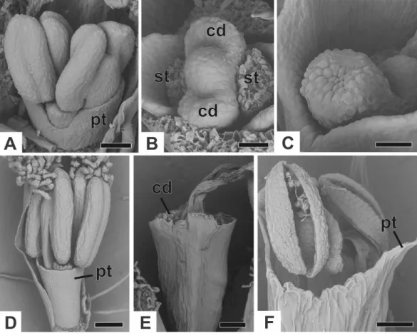
correspond to the nectariferous carpel walls, or carpellodes (cd), and each one soon acquires a cylindrical shape and papillate surface (Fig. 7C). Stamens and nectariferous carpellodes develop and the corolla tube elongates (Fig. 7D).
Figure 7 Successive developmental stages of staminate flowers of dimerous species ofPaepalanthus (SEM).(A) Developing flower ofP. chiquitensiswith sepals removed showing initiation of the corolla tube. (B) Detail of a developing flower ofP. chiquitensiswith sepals and stamens removed to show early development of carpellodes. (C) Detail of a developing carpellode ofP. chiquitensis. (D) Immature flower ofP. urbanianuswith sepal removed. (E) Flower ofP. cordatusat anthesis, with carpellodes at the same level of the corolla tube opening. (F) Flower ofP. scleranthusat anthesis. Labels: cd, carpellode; pt, petal; st, stamen. Scale bars: A, C=40µm; B=20µm; D, F=60µm; E=100µm.
height as, or exposed above, the corolla tube in P. chiquitensis,P. cordatus (Fig. 7E), P. elongatus,P. flaccidus,P. urbanianusandP. vaginatus. InP. echinoidesandP. scleranthus (Fig. 7F), carpellodes remain protected below the corolla opening.
Ontogeny of pistillate flower
Figure 8 Early developmental stages of pistillate flowers of dimerous species ofPaepalanthus. (A) Flo-ral primordium ofP. chiquitensisshowing initiation of sepals (SEM). (B) Floral primordium ofP. echi-noidesshowing sepals, stamens, gynoecium and late-developing petals (SEM). (C) Developing flower of
P. chiquitensisin frontal view (SEM). (D) Developing flower ofP. chiquitensiswith one sepal removed and detail of the ovarian septum (arrowhead) separating the locules (SEM). (E) Developing flower ofP. scler-anthuswith asymmetric sepals (SEM). (F) Developing flower ofP. cordatuswith one sepal removed show-ing petal, staminodia and gynoecium (SEM). (G) Longitudinal section of young gynoecium ofP. echi-noidesshowing ovules and ovarian septum (LM). Labels: arrowhead, ovarian septum; gp, gynoecium pri-mordium; pt, petal; sd, staminode; sp, sepal; st, stamen. Scale bars: A, C=40µm; B, E=20µm; D, F=
gynoecium (Figs. 8Dand8E). The central bulge of the gynoecium primordium corresponds to the septum (arrowhead) and divides the two locules, which are opposite the sepals (Fig. 8D). Early stages of carpel development were found only inP. chiquitensis(Fig. 8D), P. echinoidesandP. scleranthus(Fig. 8E). InP. scleranthus(Fig. 8E), some young flowers have sepals in a slightly asymmetric position, not exactly alternate to the floral bract.
After ovary differentiation, the sepals protect the young flower, and petals develop (Fig. 8F). During ovary development, the carpels form a ring around the septum, forming the symplicate zone (Fig. 8F). The stamens stop their development, becoming scale-like staminodes (sd) (Fig. 8F). The ovary septum raises the placenta, and the ovules become pendulous (Fig. 8G).
The apical portions of the carpels differentiate progressively into cylindrical structures with papillose epidermis (Figs. 9A–9C). These structures appear in carinal position and correspond to the nectariferous branches (nb) of the gynoecium. The stigmatic branches are formed later in the commissural position by the apical growth of the tissue on the boundary between the carpels (sb) (Figs. 9D–9F). During the formation of the stigmatic branches, the nectariferous branches elongate (Figs. 9D–9F). The style is formed by the intercalary growth of the region between the gynoecium branches and the ovary (Figs. 9Eand9F). The stigma is formed by the incomplete fusion of the adjacent carpel margins (Figs. 9D–9F).
InP. scleranthus, the primordia of the nectariferous branches may arise in some flowers (Fig. 9G), but in general they do not develop, becoming superficially absent during the gynoecium development (Figs. 9Hand9I). In this species, the style is formed by intercalary growth of the region between the ovary and the short stigmatic branches, resulting in a long columnar structure (Fig. 9I). InP. echinoides, the developmental stages associated with carpel closure and branch formation were not observed due to the absence of these stages in the collected capitula.
At female anthesis in all species, sepals reach the flower apex and petals develop, almost reaching the height of the sepals (Fig. 10A). Stigmatic and nectariferous branches elongate and are exposed above the sterile floral parts, along with the stigma surface (s) (Figs. 10Band10C).
Vascularization of staminate flower
InFig. 11A, we present the schematic representation of a staminate flower of a dimerous Paepalanthus, with details of its vascularization. Representations of cross-sections of the flower are also illustrated (Figs. 11B–11H), from the pedicel (Fig. 11B) to the floral apex (Fig. 11H).
Figure 9 Successive developmental stages of pistillate flowers of dimerous species ofPaepalanthus (SEM).(A) Developing flower ofP. urbanianuswith one sepal removed. (B) Developing gynoecium of
Figure 10 Anthesis of pistillate flowers of dimerous species ofPaepalanthus(SEM).(A) Pistillate flower ofP. flaccidus. (B) Detail of both stigmatic and nectariferous branches inP. cordatus. (C) Detail of the stigma ofP. echinoides. Labels: nb, nectariferous branch; st, stigma. Scale bars: A, B=300µm; C=60 µm.
finally divides into two bundles opposite to each other; these are the vascular bundles of the petals (Ptb) and stamens (Stb) (Figs. 11F–11H).
All floral parts are supplied by one single collateral bundle formed by 2–4 transport cells (Figs. 12A and12B). However, the nectariferous carpellodes are supplied only at their base (Fig. 12C) and may have more transport cells (Fig. 12B). The cross-sections of the carpellodes clearly show that they are not vascularized along their entire extent (Figs. 12Dand12E).
Vascularization of pistillate flower
In Fig. 13A, we present the schematic representation of a pistillate flower of dimerous Paepalanthus, with details of its vascularization. Representations of cross-sections are also illustrated (Figs. 13B–13I), from the region of the pedicel (Fig. 13B) to the style region (Fig. 13I). Details of the apical region of pistillate flowers are given inFig. 14.
At the flower base, the pedicel is vascularized by a single vascular strand (vs) (Fig. 13B). From the central strand, two vascular traces diverge, corresponding to the sepal traces (Spt) (Fig. 13C). Two traces that alternate with the sepals diverge above sepal insertion and correspond to the petal traces (Ptt) (Figs. 13Dand13E).
Above the petal insertions, two vascular traces emerge opposite the sepals, equivalent to the dorsal carpellary traces (Cpdt) (Fig. 13E). Then, the central strand divides into two vascular bundles that alternate with the sepals, corresponding to the heterocarpellar ventral bundles of the ovary (Cpvb) (Figs. 13Fand13G). At the synascidiate zone of the gynoecium (Fig. 9G), the two ventral bundles are in the commissural position, and the dorsal bundles are opposite the sepals. In the symplicate zone of the gynoecium (Figs. 9H,9Iand10A), only the two dorsal bundles of the carpels are present.
Figure 12 Details of staminate flowers of dimerous species ofPaepalanthus(LM).(A) Developing an-thers ofP. scleranthus, in cross-section (CS). (B) Flower ofP. flaccidusat the base of carpellodes, in CS. (C) Detail of nectariferous carpellodes ofP. chiquitensis, in longitudinal section (LS). (D) Flower ofP. flaccidus
in the median region of carpellodes, in CS. (E) Detail of the apical region of carpellodes ofP. flaccidus, in CS. Labels: cd, nectariferous carpellode; Cdb, carpellode vascular bundle; pt, petal; sp, sepal; st, stamen; Stb, stamen vascular bundle. Scale bars: A=400µm; B–E=500µm.
not vascularized in any of the studied species (Fig. 14C). In all species, the style has a stylar canal (sc) (Figs. 14Aand14B). InP. echinoides(Figs. 14Dand14E) andP. scleranthus, the stylar canal reaches the flower apex. InP. echinoides, no vascular bundles are found in the upper region of the style, above the nectariferous branches. InP. scleranthus, the vascular bundles reach only the median part of the style.
Figure 14 Details of pistillate flowers of dimerous species ofPaepalanthus(LM).(A) Detail of the style ofP. elongatus, in cross-section (CS). (B) Detail of the style ofP. chiquitensisat the base of the nec-tariferous branches, in CS. (C) Detail of free necnec-tariferous and stigmatic branches ofP. flaccidus, in CS. (D) Flower ofP. echinoidesat the insertion of nectariferous branches, in CS. (E) Flower ofP. echinoidesat the median region of the gynoecium branches, in CS. (F) Detail of vascular bundles reaching the base of staminodes inP. scleranthus, in longitudinal section (LS). Labels: Cpdb, carpel dorsal vascular bundle; nb, nectariferous branch; sb, stigmatic branch; sc, stylar canal; sd, staminode. Scale bars: A–E=50µm; F=
100µm; G=80µm.
DISCUSSION
Morphological (re)interpretation
In the dimerous species ofPaepalanthusstudied here, the floral parts emerge opposite to each other in the same whorl, with no evidence of a third set of parts. The two sepals of dimerous flowers arise simultaneously during floral development. This is similar to the condition in lateral sepals of trimerous flowers, in which the adaxial median sepal develops late (Stützel, 1984;Stützel, 1990). Thus, we hypothesize that sepals of dimerous flowers of Paepalanthusin fact correspond to the lateral sepals of trimerous flowers.
Figure 15 Putative steps leading to the dimerous pistillate flowers in species ofPaepalanthus. (A) Trimerous flower. (B) Trimerous flower with a suppressed sector comprising a lateral sepal and a lateral petal. (C) Trimerous flower with a suppressed sector comprising the median sepal and a lateral petal. (D) Trimerous flower with a suppressed sector comprising a lateral sepal and the median petal. (E) Dimerous flower. Labels: lpt, lateral petal; lsp, lateral sepal; mpt, median petal; msp, median sepal.
(due to mechanical pressure), the size of floral meristem and the available space for the floral whorls to develop (Ronse De Craene, 2015). The perianth is also known to be important in defining and fixing the floral merism (Ronse De Craene, 2015). Regarding the minimum dislocation of floral parts after the loss of one unit of each whorl (Fig. 15), the putative interpretation for the evolution of dimery inPaepalanthusis of the loss of a sector comprising the median sepal, one lateral petal, one lateral stamen and the median carpel (Figs. 15and16). In this context, our results on floral development and position of mature floral parts of dimerous flowers corroborateStützel’s (1990) hypothesis that the transition to dimery occurred from the suppression of the median sepal of trimerous flowers and help to explain merism evolution inPaepalanthus.
Particularly in some pistillate flowers ofP. scleranthus(P. subg.Thelxinoë), sepals are asymmetrical, which may be considered as evidence that a median sepal was suppressed. However, sepal position in these flowers is probably related to the reduced space in the region where the young flowers are found—the centre of the capitulum—and therefore to the accommodation of these flowers in this region.
In previous research,Stützel (1990)andStützel & Gansser (1995)suggested the absence of an outer whorl of stamens in early development stages of staminate and pistillate flowers of Lachnocaulon,PaepalanthusandSyngonanthus. Nonetheless,Rosa & Scatena (2003);
Figure 16 Hypothetical steps leading to the dimerous flowers in species ofPaepalanthus. (A) Trimer-ous staminate flower. (B) TrimerTrimer-ous staminate flower with a suppressed sector comprising the median sepal, a lateral petal, a lateral stamen and the median carpel. (C) Dimerous staminate flower. (D) Trimer-ous pistillate flower. (E) TrimerTrimer-ous pistillate flower with the median sepal, a lateral petal, a lateral stamin-ode and the median carpellstamin-ode suppressed. (F) Dimerous pistillate flower. Labels: lpt, lateral petal; lsp, lat-eral sepal; mpt, median petal; msp, median sepal.
Flower ontogeny of dimerous species ofPaepalanthuspresented in our study corroborates the results previously presented byStützel (1990)andStützel & Gansser (1995). In staminate flowers ofPaepalanthus, the staminodes reported byRosa & Scatena (2003);Rosa & Scatena (2007)in fact correspond to the apical portions of late-developing petals. We infer that similar structures were misinterpreted in other genera of Paepalanthoideae and require further investigation. Therefore, the results obtained here lead us to the conclusion that the common ancestor of the Paepalanthoideae probably had isostemonous flowers, and not diplostemonous flowers as concluded byRosa & Scatena (2007).
corroborated by the previously observation that the pistillodes are vascularized by dorsal carpellary bundles (Rosa & Scatena, 2007). Furthermore, some morphological features of the nectariferous structures, such as shape and type of epidermal cells, differ in mature flowers of some dimerous species ofPaepalanthus. These characteristics are important for species distinction and may also be an indicator of infrageneric delimitation in the genus. During gynoecium formation in pistillate flowers of Eriocaulaceae, the ovary septum raises the central placenta, and the ovules become pendulous (Coan, Stützel & Scatena, 2010). In dimerous species ofPaepalanthus, we observed that the septum does not fuse with the apical portion of the ovary in the mature flower, resulting in a proximal synascidiate zone and a short distal symplicate zone close to the stylar canal opening. This ovarian feature is common in angiosperms, and the occurrence of a distal symplicate zone, as the presence of commissural stigmatic branches, may be related to a regular distribution of pollen tubes in the locules, in case stigmas receive contrasting amounts of pollen grains (Endress, 2011). Pistillate flowers of Paepalanthoideae have a branched style, with nectariferous branches in the carinal (dorsal) position and stigmatic branches in the commissural position of the ovary (Stützel, 1990;Rosa & Scatena, 2003). In mature flowers of Eriocaulaceae, the gynoecium branches are free and inserted at the same point on the style in most genera (Stützel, 1990). The same organization was reported for the dimerous species of Paepalanthus studied here, except for P. echinoides (P. sect. Conodiscus) and P. scleranthus (P. subg. Thelxinoë). The stigmatic branches in these species are short and inserted terminally on a long style. The stigmatic branches divide into two bifid stigmas, which consequently are placed close together and may be misinterpreted as four simple stigmas. However, the presence of bifid stigmas is a common condition inPaepalanthus (Ruhland, 1903), whereas four simple stigmas is a character state that is absent from Eriocaulaceae as a whole.
In P. scleranthus, pistillate flowers generally have stylar and stigmatic branches, but no nectariferous branches. In Paepalanthoideae, pistillate flowers with only stigmatic branches were verified in a few species ofPaepalanthusandSyngonanthus(Ruhland, 1903;
Stützel, 1987;Stützel & Gansser, 1995). The style formation inP. scleranthusoccurs through intercalary growth, resulting in a columnar structure similar to that found in the gynoecium ofP. echinoides. In early development of the style ofP. scleranthus, nectariferous branch primordia are found in some pistillate flowers, but they generally do not develop, indicating this nectariferous structure was present in the species’ ancestor.
Vascularization and homologies
Anatomical details of dimerous flowers ofPaepalanthusshowed that there is no vestige of vascularization of a third floral part in any of the floral whorls. The vascular bundles were also dislocated following the dislocation of the floral parts during the transition from trimery to dimery.
and in staminate flowers ofPaepalanthus, the vacularization shared by these whorls is probably related to these species’ development, as these structures emerge from common primordia. In pistillate flowers ofPaepalanthus, staminodes share vascular traces with petals due to the homology of the staminodes and the functional stamens of staminate flowers.
The vascularization of the gynoecium of dimerous species ofPaepalanthusis similar to that reported in previously studied species of Paepalanthoideae (Rosa & Scatena, 2003;
Rosa & Scatena, 2007). However, the ventral bundles of the ovary in dimerous species are in the commissural position. Despite the position of the ventral bundles, they reach only the synascidiate portion of the ovary, and the stigmatic branches in these species lack vascularization, as is usual for trimerous species in the subfamily (Rosa & Scatena, 2003;
Rosa & Scatena, 2007). InP. echinoidesandP. scleranthus, the dorsal carpel bundles reach the median region of the style, whereas its upper region is not vascularized. We assume that the proximal region of the style of both species is homologous to the short style found in the other species ofPaepalanthusstudied here, which is vascularized. On the other hand, the upper region of the style is probably homologous to their stigmatic branches, which are non-vascularized. Thus, we can interpret the long style ofP. echinoidesandP. scleranthus as a short style on which are inserted two fused stigmatic branches.
Evolutionary and taxonomic implications
In recent phylogenetic studies, dimerous species of Paepalanthus are placed in five infrageneric categories, forming two distinct clades (Giulietti et al., 2012;Trovó et al., 2013).Paepalanthussubg.Thelxinoë appears as a sister group ofP. sect.Conodiscus, and both categories together form a sister group of the clade that includesActinocephalusas sister group ofP.sect.Diphyomene, P.ser.Dimeriand dimerous species ofP.sect.Eriocaulopsis
(previously circumscribed inP.sect.Diphyomene(Trovó & Sano, 2010;Trovó et al., 2013)
(Fig. 17). The occurrence of fused stigmatic branches inP. scleranthus(P.subg.Thelxinoë)
andP. echinoides(P. sect.Conodiscus) corroborates the phylogenetic proximity ofP.subg.
Thelxinoë andP. sect.Conodiscus(Fig. 17). Furthermore, the rise of nectariferous branch primordia and their subsequent suppression inP. scleranthusindicate that nectariferous branches may have been lost after the fusion of stigmatic branches in the clade that groups togetherP. subg.ThelxinoëandP. sect.Conodiscus(Trovó et al., 2013).
The morphology of the papilla in the carpellodes and their exposure in male anthesis is congruent with the topology found byTrovó et al. (2013)(Fig. 17). Although species of the cladeP. subg.Thelxinoë andP. sect.Conodiscushave protected carpellodes with few prominent papillae, species of the cladeP.sect.Diphyomene, P.ser.Dimeri, andP.sect.
Figure 17 Phylogenetic relationships (adapted fromTrovó et al., 2013) of the dimerous infrageneric cat-egories ofPaepalanthusand respective tridimensional floral diagrams of pistillate flowers compared at dif-ferent levels of gynoecium (A, ovary; B, gyonoeci; C, stigma).
syndrome, distinct from the previously studied species of Eriocaulaceae (Ramos, Borba & Funch, 2005;Oriani, Sano & Scatena, 2009).
For a more general panorama of floral characters in a phylogenetic context, we must expand our knowledge about the flower ontogeny inActinocephalusand its relationship to floral characters of species ofPaepalanthussect.Diphyomene, P.ser.Dimeriand dimerous species ofP.sect. Eriocaulopsis.Despite the suggestion that dimery has evolved more
than once inPaepalanthus,this study reveals a possible alternative interpretation—that dimerous flowers have appeared only once, followed by a reversal inActinocephalus(Fig. 17). It also seems clear that, although morphologically distinct, the exclusion of dimerous species fromP.sect.Diphyomene (Trovó & Sano, 2010) may be reconsidered after new
evidence.
FINAL REMARKS
Eriocaulaceae species. However, some development aspects and vascularization details of dimerous flowers ofPaepalanthusare new and important additions to the knowledge of the whole family. The results presented here contribute to the understanding of floral merism evolution inPaepalanthus, a genus of Eriocaulaceae whose morphology is unusually complex. In addition to corroborating the hypothesis proposed byStützel (1990)for dimery evolution in Eriocaulaceae, the sepal development, the position of floral parts, and the vascularization of dimerous species ofPaepalanthusalso helped to determine the position of the suppressed floral parts in each floral whorl. Furthermore, the correct interpretation of the incipient petal in staminate flowers ofPaepalanthus, and the consequent absence of staminodes in these flowers, reinforce the supposition that the ancestor of Paepalanthoideae had isostemonous flowers, rather than diplostemonous flowers as was inferred by previous anatomical studies in the family (Rosa & Scatena, 2003;Rosa & Scatena, 2007). Finally, the data obtained here show the importance of comparative ontogenetic studies for developing a better understanding of floral structures and their evolution in Eriocaulaceae.
ACKNOWLEDGEMENTS
We thank the Academic Editor, Dr. Richard Bateman, and both reviewers, Dr. Margarita Remizowa and Dr. Paula Rudall, for their generous comments on the manuscript.
ADDITIONAL INFORMATION AND DECLARATIONS
Funding
Financial support was provided to AL Silva by CAPES (MSc. Scholarship), to M Trovó by FAPERJ (E-26/110.031/2011, E-26/111.392/2012, E-26/010.001626/2014–BIOTA) and CNPq (proc. 470349/2013-1), and to AI Coan by CNPq (proc. 307515/2015-0). The funders had no role in study design, data collection and analysis, decision to publish, or preparation of the manuscript.
Grant Disclosures
The following grant information was disclosed by the authors: CAPES.
FAPERJ: E-26/110.031/2011, E-26/111.392/2012, E-26/010.001626/2014. CNPq: proc. 470349/2013-1, proc. 307515/2015-0.
Competing Interests
The authors declare there are no competing interests.
Author Contributions
• Arthur de Lima Silva, Marcelo Trovó and Alessandra Ike Coan conceived and
Field Study Permissions
The following information was supplied relating to field study approvals (i.e., approving body and any reference numbers):
The examined material is listed inTable 1based on the following field permits: SisBio collecting permit no. 47742-1 (February 2015–March 2016) to AL Silva; SisBio permanent permit no. 34782-1 (from May 2012 to present) and collecting permit no. 42939-1 (May 2014–June 2015) to M Trovó; and SisBio permanent permit no. 14929-3 (December 2011 to present) and collecting permits no. 37568-1 and 37568-2 (January 2013–February 2015) to AI Coan.
Data Availability
The following information was supplied regarding data availability: The raw data is included inTable 1.
REFERENCES
Alves PGM, Scatena VL, Trovó M. 2013.Anatomy of scapes, bracts, and leaves of Paepalanthussect.Diphyomene(Eriocaulaceae, Poales) and its taxonomic implica-tions.Brittonia65:262–272DOI 10.1007/s12228-012-9263-z.
Andrade MJG. 2007.Filogenia e taxonomia em Eriocaulaceae tropicais. Feira de Santana: Universidade Estadual de Feira de Santana, 1–172.
Andrade MJG, Giulietti AM, Rapini A, Queiroz LP, Souza Concei¸cão A, Almeida PRM, Van den Berg C. 2010.A comprehensive phylogenetic analysis of Eriocaulaceae: evidence from nuclear (ITS) and plastid (psbA-trnHandtrnL-F) DNA sequences. Taxon59:379–388DOI 10.2307/25677597.
BFG. 2015.Growing knowledge: an overview of seed plant diversity in Brazil.Rodriguesia 66:1085–1113DOI 10.1590/2175-7860201566411.
Coan AI, Scatena VL. 2004.Embryology and seed development ofBlastocaulon scirpeum andPaepalanthus scleranthus(Eriocaulaceae).Flora—Morphology, Distribution, Functional Ecology of Plants199:47–57DOI 10.1078/0367-2530-00124.
Coan AI, Stützel T, Scatena VL. 2010.Comparative embryology and taxonomic considerations in Eriocaulaceae (Poales).Feddes Repertorium121:268–284 DOI 10.1002/fedr.201000016.
Endress PK. 1995. Major traits of monocot flowers. In: Rudall PJ, Cribb PJ, Cutler DF, Humphries CJ, eds.Monocotyledons: systematics and evolution. Kew: Royal Botanic Gardens, 43–79.
Endress PK. 2011.Evolutionary diversification of the flowers in angiosperms.American Journal of Botany98:370–396DOI 10.3732/ajb.1000299.
Feder NED, O’Brien TP. 1968.Plant microtechnique.American Journal of Botany 55:123–142DOI 10.2307/2440500.
Giulietti AM, Harley RM, De Queiroz LP, Wanderley MGL, Van den Berg C. 2005. Biodiversity and conservation of plants in Brazil.Conservation Biology19:632–639 DOI 10.1111/j.1523-1739.2005.00704.x.
Giulietti AM, Hensold N. 1990.Padrões de distribui¸cão geográfica dos gêneros de Eriocaulaceae.Acta Botanica Brasilica4:133–158
DOI 10.1590/S0102-33061990000100010.
Jensen WA. 1962.Botanical histochemistry. New York: HH Freeman and Co. Johansen DA. 1940.Plant microtechnique. New York: McGraw–Hill.
Körnicke F. 1863. Eriocaulaceae. In: Martius CFP, Eichler AG, Urban NI, eds.Flora brasiliensis. Monachii: Typographia Regia, 273–307.
Mabberley DJ. 1987.The plant book. Cambridge: Cambridge University Press. Oriani A, Sano PT, Scatena VL. 2009.Pollination biology ofSyngonanthus elegans
(Eriocaulaceae—Poales).Australian Jornal of Botany57:94–105 DOI 10.1071/BT08119.
Oriani A, Scatena VL. 2012.Floral anatomy of xyrids (Poales): contributions to their reproductive biology, taxonomy, and phylogeny.International Journal of Plant Sciences173:767–779 DOI 10.1086/666664.
Ramos COC, Borba EL, Funch LS. 2005.Pollination in BrazilianSyngonanthus (Eri-ocaulaceae) species: evidence for entomophily instead of anemophily.Annals of Botany96:387–397 DOI 10.1093/aob/mci191.
Remizowa MV, Kuznetsov AN, Kuznetsova SP, Rudall PJ, Nuraliev MS, Sokoloff DD. 2012.Flower development and vasculature inXyris grandis(Xyridaceae, Poales); a case study for examining petal diversity in monocot flowers with a double perianth. Botanical Journal of the Linnean Society170:93–111
DOI 10.1111/j.1095-8339.2012.01267.x.
Ronse De Craene LP. 2015.Meristic changes in flowering plants: how flowers play with numbers.Flora—Morphology, Distribution, Functional Ecology of Plants221:22–37 DOI 10.1016/j.flora.2015.08.005.
Ronse De Craene LP, Smets EF. 1994.Merosity in flowers: definition, origin and taxonomic significance.Plant Systematics and Evolution191:83–104 DOI 10.1007/BF00985344.
Rosa MM, Scatena VL. 2003.Floral anatomy ofEriocaulon elichrysoidesand Syngonan-thus caulescens(Eriocaulaceae).Flora—Morphology, Distribution, Functional Ecology of Plants198:188–199DOI 10.1078/0367-2530-00090.
Rosa MM, Scatena VL. 2007.Floral anatomy of Paepalanthoideae (Eriocaulaceae, Poales) and their nectariferous structures.Annals of Botany99:131–139 DOI 10.1093/aob/mcl231.
Ruhland W. 1903. Eriocaulaceae. In: Engler A, ed.Das pflanzenreich, regni vegetabilis conspectus IV, 30. Leipzig: Engelmann.
Sano PT, Giulietti AM, Costa FN, Trovó M, Echternacht L, Tissot-Squalli ML, Watan-abe MTC, Hensold N, Andrino CO, Parra LR. 2015.Eriocaulaceae in Lista de Espécies da Flora do Brasil. Jardim Botânico do Rio de Janeiro.Available athttp: // floradobrasil.jbrj.gov.br/ jabot/ floradobrasil/ FB7506 (accessed on 28 July 2016). Scatena VL, Giulietti AM, Borba EL, Van den Berg C. 2005.Anatomy of Brazilian
Eriocaulaceae: correlation with taxonomy and habitat using multivariate analyses. Plant Systematics and Evolution253:1–22DOI 10.1007/s00606-004-0295-z.
Stützel T. 1984.Blüten- und infloreszenzmorphologische Untersuchungen zur System-atik der Eriocaulaceen. Dissertationes Botanicae, J. Cramer, Berlin.
Stützel T. 1985.Die Bedeutung monothecat-bisporangiater Antheren als systematisches Merkmal zur Gliederung der Eriocaulaceen.Botanische Jahrbücher fur Systematik 105:433–438.
Stützel T. 1987.On the morphological and systematic position of the genus Mold-enkeanthus(Eriocaulaceae).Plant Systematics and Evolution156:133–141 DOI 10.1007/BF00936068.
Stützel T. 1990.Appendices am Gynoeceum der Xyridaceen, Morphogenie, Funktion und Systematische Bedeutung.Beitrage zur Biologie der Pflanzen65:275–299. Stützel T. 1998. Eriocaulaceae. In: Kubitzki K, ed.The families and genera of vascular
plants IV—flowering plants: monocotyledons—alismatanae and commelinanae (except graminae). Berlin: Springer Heidelberg, 197–207.
Stützel T, Gansser N. 1995.Floral morphology of North American Eriocaulaceae and its taxonomic implications.Feddes Repertorium106:495–502
DOI 10.1002/fedr.4921060518.
Trovó M, Andrade MJG, Sano PT, Ribeiro PL, Van den Berg C. 2013.Molecular phylogenetics and biogeography of Neotropical Paepalanthoideae with emphasis on BrazilianPaepalanthus(Eriocaulaceae).Botanical Journal of the Linnean Society 171:225–243DOI 10.1111/j.1095-8339.2012.01310.x.