A Novel GTP-Binding Inhibitor, FX2149,
Attenuates LRRK2 Toxicity in Parkinson
’
s
Disease Models
Tianxia Li1☯, Xinhua He1,2☯, Joseph M. Thomas1, Dejun Yang1, Shijun Zhong3,
Fengtian Xue1*, Wanli W. Smith1*
1Department of Pharmaceutical Sciences, University of Maryland School of Pharmacy, Baltimore, MD, United States of America,2Beijing Institute of Pharmacology and Toxicology, Beijing, China,3School of Life Science and Biotechnology, Dalian University of Technology, Liaoning, China
☯These authors contributed equally to this work.
*[email protected](FX);[email protected](WWS)
Abstract
Leucine-rich repeat kinase-2 (LRRK2), a cytoplasmic protein containing both GTP binding and kinase activities, has emerged as a highly promising drug target for Parkinson’s dis-ease (PD). The majority of PD-linked mutations in LRRK2 dysregulate its GTP binding and kinase activities, which may contribute to neurodegeneration. While most known LRRK2 in-hibitors are developed to target the kinase domain, we have recently identified the first LRRK2 GTP binding inhibitor, 68, which not only inhibits LRRK2 GTP binding and kinase activities with high potencyin vitro, but also reduces neurodegeneration. However, thein vivoeffects of 68 are low due to its limited brain penetration. To address this problem, we re-ported herein the design and synthesis of a novel analog of 68, FX2149, aimed at increas-ing thein vivoefficacy. Pharmacological characterization of FX2149 exhibited inhibition of LRRK2 GTP binding activity by ~90% at a concentration of 10 nM usingin vitroassays. Fur-thermore, FX2149 protected against mutant LRRK2-induced neurodegeneration in SH-SY5Y cells at 50-200 nM concentrations. Importantly, FX2149 at 10 mg/kg (i.p.) showed sig-nificant brain inhibition efficacy equivalent to that of 68 at 20 mg/kg (i.p.), determined by mouse brain LRRK2 GTP binding and phosphorylation assays. Furthermore, FX2149 at 10 mg/kg (i.p.) attenuated lipopolysaccharide (LPS)-induced microglia activation and LRRK2 upregulation in a mouse neuroinflammation model comparable to 68 at 20 mg/kg (i.p.). Our results highlight a novel GTP binding inhibitor with better brain efficacy, which represents a new lead compound for further understanding PD pathogenesis and therapeutic studies.
Introduction
Parkinson’s disease (PD) is a progressive neurodegenerative disorder, affecting 2% of the popu-lation over the age of 60 [1,2]. PD patients display a loss of dopaminergic neurons in the sub-stantia nigra and the presence of Lewy bodies in their brains [1,2]. The current
a11111
OPEN ACCESS
Citation:Li T, He X, Thomas JM, Yang D, Zhong S, Xue F, et al. (2015) A Novel GTP-Binding Inhibitor, FX2149, Attenuates LRRK2 Toxicity in Parkinson’s Disease Models. PLoS ONE 10(3): e0122461. doi:10.1371/journal.pone.0122461
Academic Editor:Shaida A. Andrabi, Johns Hopkins University, UNITED STATES
Received:November 13, 2014
Accepted:February 12, 2015
Published:March 27, 2015
Copyright:© 2015 Li et al. This is an open access article distributed under the terms of theCreative Commons Attribution License, which permits unrestricted use, distribution, and reproduction in any medium, provided the original author and source are credited.
Data Availability Statement:All relevant data are within the paper.
Funding:This work is supported by internal fund from University of Maryland. The funders had no role in study design, data collection and analysis, decision to publish, or preparation of the manuscript.
pharmacotherapy for PD patients is limited to symptomatic treatment, which only temporarily reduces motor symptoms but does not prevent neurodegeneration. To date, there are no dis-ease modifying drugs to prevent dopaminergic neuron loss and abnormal protein deposition in the brains. There is a strong demand for neuroprotective therapies to prevent or attenuate do-paminergic neuron degeneration.
Recent genetic studies have identified that mutations in Leucine-rich repeat kinase-2 (LRRK2) cause a genetic form of PD and have implications in sporadic PD. LRRK2 is a large cytoplasmic protein (2527 aa) and contains both GTPase and kinase domains [1,2]. Most disease-linked mutations of LRRK2 have been reported to dysregulate its GTP binding and/or kinase activities [2–5]. The most common PD-linked mutation, G2019S, has abnormally ele-vated kinase activity [1–3,6]. A number of potential LRRK2 kinase inhibitors that target the kinase domain activity have been reported [7–11] and some of them can ameliorate neurode-generation [8,10]. However, none of these are available in the clinic yet due to poor specificity or lowin vivoefficacy.
The GTPase domain (ROC-COR) of LRRK2 contains the residues from amino acids 1335–1878, accounting for ~7% of the full length protein. PD-linked mutations within the GTPase domain (eg. R144C/G) alter either GTP binding or GTPase activity [1–3,6,12]. Abol-ished GTP binding by the K1347A mutation attenuates LRRK2 kinase activity [6]. This leads to suppression of mutant-LRRK2-induced neuronal degeneration [6], and suggests that the GTPase domain is a tractable target for therapeutic intervention. In addition, the crystal struc-ture of the LRRK2 GTPase domain is different from other small GTPases (eg, Ras, Rho) that could lead to development of potential inhibitors that only target LRRK2. Our recent studies have identified a GTP binding inhibitor, 68, that can reduce LRRK2 GTP binding activity but do not alter LRRK1 activity [13]. Moreover, 68 can reduce LRRK2 kinase activity and protect against mutant LRRK2 toxicity [13].
One of the challenges in developing therapeutics for neurodegenerative disorder is to im-prove both specific bioactive potency and blood-brain barrier penetration (BBB) simultaneous-ly [11]. Many agents have failed to be developed into clinical drugs due to their low efficacy in brains [11]. Compound 68 is a potent inhibitor of LRRK2 GTP binding activityin vitro, with inhibitory activity in the low nanomolar range. However, 68 displays weak BBB permeability that limits the application of these inhibitors in animal models [13]. Herein we reported the de-sign and synthesis of a novel analog of 68, compound FX2149, which not only kept the inhibi-tion of LRRK2 GTP binding and kinase activities but also showed improvedin vivoefficacy due to its enhanced BBB permeability. We further characterized the pharmacological effects of FX2149 usingin vitroandin vivoPD models. Our studies provided a novel LRRK2 GTP bind-ing inhibitor, FX2149, with a more efficient brain efficacy for future pathogenesis and
therapeutic studies.
Materials and Methods
Materials, reagents, and animals
from Jackson Laboratory and maintained in the animal facility at University of Maryland School of Pharmacy, and the animal procedure protocol was approved by the Animal Use and Care Committee of University of Maryland.
Synthesis of FX2149 [
16
,
25
]
3-(Pyridine-3-sulfonamido)benzoic acid, 4 was synthesized as following steps. To a solution of ethyl 3-aminobenzoate methanesulfonate (1, 2.80 g, 11 mmol) in THF (30 mL) was added pyridine-3-sulfonyl chloride,2(1.77 g, 10 mmol), followed by triethylamine (2.1 mL, 15 mmol). The reaction mixture was allowed to stir at room temperature for 24 h and then con-centrated. The crude product was purified with flash chromatography (EtOAc:hexanes, 1:4–1:1) to give ethyl 3-(pyridine-3-sulfonamido) benzoate,3, as a white solid (2.8 g, 9.2 mmol, 92%). The resulting compound3was dissolved in methanol (30 mL). To this solution was added NaON (1 N, 10 mL) drop wise. The reaction mixture was allowed to stir at 60°C for 16 hours and then cooled to room temperature. Methanol was removed by rotary evaporation, and the resulting bright yellow solution was acidified to pH 2 using HCl (4 N). Filtration under vacuum gave a white solid, which was further washed by HCl (1 N, 3 × 15 mL) to yield 3-(pyridine-3-sulfonamido)benzoic acid,4, as a white solid (2.45 g, 8.8 mmol, 88% for two steps):1H NMR (400 MHz, DMSO-d6)δ7.37–7.41 (m, 2H), 7.55–7.67 (m, 2H), 7.68 (s, 1H), 8.10–8.12 (d,J= 7.6 Hz, 1H), 8.77–8.78 (d,J= 4.0 Hz, 1H), 8.87–8.88 (d,J= 4.0 Hz, 1H), 10.20–11.20 (br s, 1H), 12.50–13.50 (br s, 1H);13C NMR (100 MHz, DMSO-d6)δ121.4, 124.9,
125.0, 125.9, 130.2, 132.3, 135.1, 136.0, 137.8, 147.4, 154.1, 167.0; LC-MS (M—H+) calcd for C12H10N2O4S 277, found 277.
N-Propyl-3-(pyridine-3-sulfonamido)benzamide (FX2149) was synthesized as following steps. To a mixture of carboxylic acid,4(556 mg, 2.0 mmol), EDC (575 mg, 3.0 mmol), and HOBt (460 mg, 3.0 mmol) was addedN,N-dimethylformamide (DMF, 8.0 mL), followed by propylamine (200μL, 2.4 mmol). The reaction mixture was heated at 40°C for 24 hours. DMF
was removed by rotary evaporation. To the resulting residue was added H2O (10 mL) to give a
white slurry. Filtration under vacuum gave a white solid, which was further washed using H2O
(4 × 10 mL). The product was further purified by recrystallization using CH2Cl2/hexanes give N-propyl-3-(pyridine-3-sulfonamido)benzamide (FX2149) as a white solid (515 mg, 1.61 mmol, 81%):1H NMR (400 MHz, CDCl3)δ0.94–0.98 (t,J= 7.2 Hz, 3H), 1.59–1.65 (m, 2H),
3.43–3.48 (q,J= 6.4 Hz, 2H), 6.37 (br s, 1H), 7.28–7.40 (m, 3H), 7.54–7.56 (d,J= 7.2 Hz, 1H), 7.75 (s, 1H), 8.09–8.11 (d,J= 8.0 Hz, 1H), 8.70 (s, 1H), 9.00 (s, 1H), 9.14 (s, 1H);13C NMR (100 MHz, CDCl3)δ11.4, 22.8, 42.0, 121.1, 122.5, 123.9, 124.0, 129.6, 135.5, 135.7, 137.4,
147.3, 152.4, 166.8; LC-MS (M + H+) calcd for C15H18N3O3S 320, found 320.
Cell culture, LRRK2 constructs, and transfection
Human embryonic kidney HEK293T and human neuroblastoma SH-SY5Y cells were from ATCC (Manassas, VA, USA) and grown in the media as described previously [6,13]. The Flag tagged wild type, G2019S, R1441C, Y1699C, and G2019S-K1347A constructs were described previously [6]. Transient transfections were performed using Lipofectamine and PLUS Re-agents (Invitrogen) according to the manufacturer's protocol.
Immunoprecipitation (IP) and Western blot analysis
primary antibodies followed by secondary antibody detection as described previously [6,13]. Enhanced chemiluminescence (ECL) reagents were used to detect proteins on the membranes.
LRRK2 GTP binding and phosphorylation (kinase) assays
GTP binding assays were performed using GTP-agarose beads (Sigma) as described previously [6,13]. Lysates of HEK 293T cells expressing LRRK2 proteins were incubated with 68 or FX2149 at various concentrations for 1h. The GTP-agarose beads were added for an additional 2 h. The samples were subjected to Western blot analysis using anti-Flag antibodies. LRRK2 ki-nase assays were performed using LRRK2 phosphorylation andin vitro32P incorporation methods as described previously [6,13].
For LRRK2 phosphorylation assays, HEK293T cells were transfected with various LRRK2 variants for 36 h, then were incubated in media without serum for 12 hours. The cells were left untreated or treated with 68, or FX2149 for 1 h, and then were harvested with lysis buffer (Cell Signaling). The resulting cell lysates were immunoprecipitated using anti-Flag antibodies to pull down Flag-tagged LRRK2. The immunoprecipitates were subjected to Western blot using anti-phospho-LRRK2 antibodies (S2032 or S935) as described previously [13].In vitro32P in-corporation was performed using purified LRRK2 that were left untreated or treated with 68 or FX2149 for 1 h. The samples were then incubated with the kinase reaction buffer containing 500μM ATP and 10μCi of [γ-32P]ATP (3,000 Ci/mmol) for 30 min. The LRRK2 autopho-sphorylation with32P incorporation was separated by SDS/PAGE gel and quantified with a phosphoimager (Bio-Rad Molecular Imager).
LRRK2 toxicity assays
SH-SY5Y cell viability assays were conducted as described [6,13]. Cells were co-transfected with GFP and various pcDNA3.1-LRRK2 plasmids at a 1:15 ratio for 24 h in 10% FBS OPTI-I media and then changed to DMEM with N2 supplement for 24 h. Compounds were added after 4-h transfection. Cell viability was measured by counting the healthy viable cells that con-tained at least one smooth extension (neurite) that was twice the length of the cell body from 20 randomly selected fields using fluorescence microscopy [6,13]. TUNEL assays were per-formed according to the manufacturer's instructions as described previously [6,13]. The experi-ments were repeated three times in duplicate. The quantification for LRRK2 toxicity was performed by an investigator who was blind to transfection groups.
LPS-based preinflammatory mouse model and immunohistochemical
analysis
avidin–biotin–peroxidase complex (Vector Laboratories), and detected by diaminobenzidine (DAB, Sigma). The images of brain sections were taken using a Zeiss Axioskop 2 microscope and a Zeiss Axiocam camera, and processed using Adobe Photoshop (VII) software. The con-tinuing middle brain section series from each mouse brain were sampled by 6 section intervals for fluorescent density quantification of the SN areas. The quantification of fluorescence densi-ty was performed by unbiased stereology with an investigator who was blind to
experiment groups.
Data Analysis
Quantitative data were shown as arithmetic means ± SEM from three separate experiments. Statistically significant differences among groups were analyzed by ANOVA using Sigmastart 3.1 statistical software (Aspire Software International, VA). Apvalue<0.05 was considered
significant.
Results
Design and synthesis of FX2149
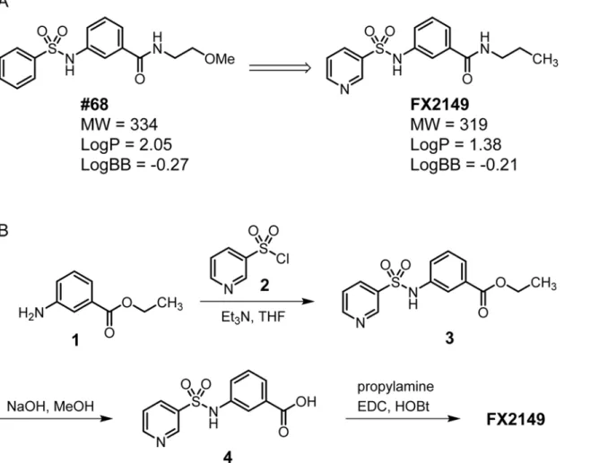
Given that 68 potently inhibits LRRK2 GTP binding and kinase activityin vitro[13], we kept its scaffold structure to retain the inhibition of GTP binding and kinase activity. A compound with good BBB permeability requires a Log P value between 1.0 and 3.0 and a Log BB value between -2.0 and 1.0 [17]. To optimize the BBB permeability of 68, we used a pyridine-3-sulfonamide group to replace the phenyl-sulfonamide head of 68 (Fig. 1A). Weakly basic groups, such as the pyridinyl group, are commonly present in therapeutic agents targeting the central nervous system [18]. Moreover, the 2-methoxy-ethyl tail of 68 was substituted by a pro-pyl group to reduce the number of H-bond acceptors to fit the binding site of the LRRK2 GTPase domain. Compound FX2149 was calculated to have increased hydrophilicity (LogP = 1.38 vs 2.05 for 68) and enhanced BBB permeability (LogBB = -0.21 vs -0.27 for 68, calculated by using ACD/Labs Suite 5.0).
The synthesis of compound FX2149 involved a three-step procedure as shown inFig. 1B. First, ethyl 3-aminobenzoate methanesulfonate,1, was treated with pyridine-3-sulfonyl chlo-ride,2, in the presence of triethylamine (Et3N) at room temperature to generate compound3 as a mixture of rotamers in excellent yields [19]. Next, saponification of ethylester in com-pound3using aqueous NaOH yielded carboxylic acid,4, in high yields. Finally, compound4 was coupled with propylamine usingN-ethyl-N0-(3-dimethylaminopropyl) carbodiimide
hy-drochloride (EDC) and 1-hydroxybenzotriazole hydrate (HOBt) to provide FX2149 in high yields [25]. Compound FX2149 was purified by flash chromatography with over 95% purity as described in the method section.
FX2149 reduced LRRK2 binding with GTP
FX2149 reduced LRRK2 kinase activity
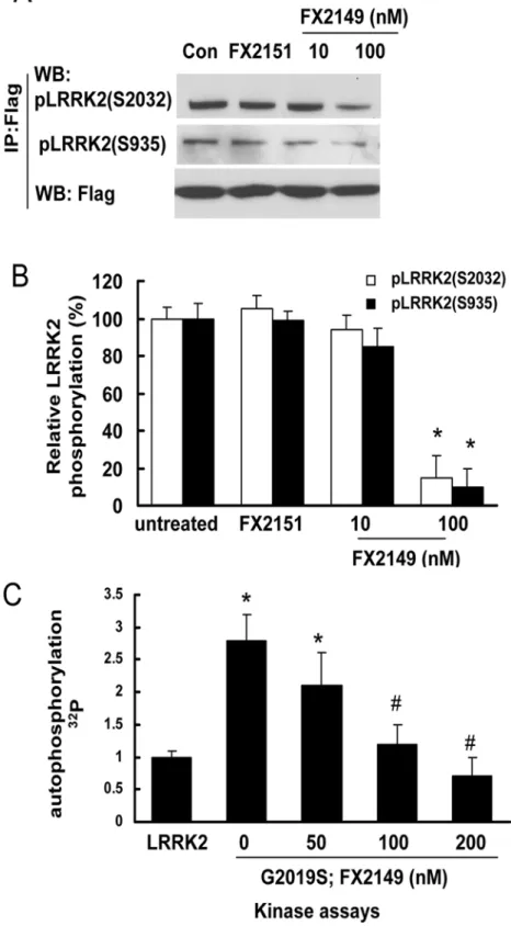
To further assess whether FX2149 alters LRRK2 phosphorylation (kinase activity), HEK293T cells expressing mutant G2019S-LRRK2 were treated with FX2149 at concentrations of 0, 10, and 100 nM for 1 h. Cell lysates were subject to LRRK2 autophosphorylation (kinase activity) assays. FX2149 at 100 nM concentration significantly reduced G2019S-LRRK2 phosphoryla-tion at residues S935 and S2032 by ~90% (Fig. 3A and 3B). We further validated these results byin vitrokinase assays showing the similar inhibition of G2019S-LRRK2 kinase activity by FX2149 (Fig. 3C). These findings indicated that FX2149 (100 nM) reduced LRRK2 kinase ac-tivity similar to that of 68 at 10 nM concentration [13]. An inactive analog of 68, FX2151, did not alter the LRRK2 phosphorylation at 10μM concentration, which is consistent with our pre-vious findings [13].
FX2149 attenuated mutant LRRK2-induced toxicity in SH-SY5Y cells
GTP binding activity and elevated kinase activities have been implicated in PD-linked mutant, G2019S-LRRK2, resulting in neurodegeneration [6,13]. SH-SY5Y cells contain dopamine and are often used as a PD cell model [6]. To assess whether FX2149 alters mutant LRRK2-induced neuronal degeneration, G2019S-LRRK2 construct transiently transfected into SH-SY5Y cells was used as a toxicity model as previously described [6]. Treatment of FX2149 significantly Fig 1. A. Chemical structures of 68 and FX2149; B. Synthesis of FX2149.
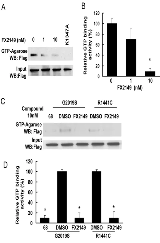
Fig 2. FX2149 inhibits LRRK2 GTP binding activity.WT or mutant LRRK2 was pulled down from lysates of transfected HEK293T cells using GTP-agarose in the absence or presence of FX2149 at 1 and 10 nM concentrations. The resulting precipitates were subjected to western blot analysis using anti-Flag antibodies. A and C. Representative blots from GTP binding assays. B and D. Quantification of A and C. K1347A-LRRK2, non GTP binding genetic control. All experiments were repeated three times with similar results.*p<0.05 by
ANOVA,vsvehicle control.
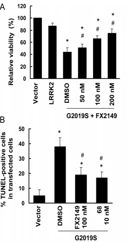
increased the viability of cells expressing G2019S-LRRK2 compared with vehicle treated cells (Fig. 4A). Moreover, FX2149 at 100 nM significantly reduced the TUNEL-positive cells ex-pressing mutant G2019S-LRRK2 and had effects equivalent to that of 68 at 10 nM (Fig. 4B).
FX2149 was more efficient in reducing LRRK2 GTP binding and kinase
activities in transgenic mice brains than 68
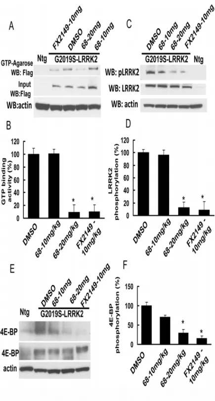
To assess the effects of FX2149 on LRRK2 in brains compared with 68, both 68 and FX2149 were injected intraperitoneally into G2019S-BAC-LRRK2 transgenic mice at 10 and 20 mg/kg doses. One hour after the injection, the mouse brain homogenates were subjected to LRRK2 GTP binding and kinase assays. Both 68 (20 mg/kg) and FX2149 (10 mg/kg) reduced LRRK2 GTP binding activity in mouse brains (Fig. 5A and 5B). While FX2149 at a 10mg/kg dose had the equivalent GTP binding inhibition as 68 at a 20 mg/kg dose, compound 68 at 10 mg/kg dose did not alter brain GTP binding activity 1 hour after injection. Moreover, both 68 (20 mg/ kg) and FX2149 (10 mg/kg) also significantly reduced brain LRRK2 kinase activity (Fig. 5C and 5D). To further confirm the effect of FX2149, we also assessed a LRRK2 downstream effec-tor, 4E-BP phosphorylation. 4E-BP is a transcription factor that can be phosphorylated by LRRK2 [20]. We found that both 68 and FX2149 reduced 4E-BP phosphorylation in mouse brains (Fig. 5E and 5F). FX2149 at a 10mg/kg dose reduced 4E-BP up to 15% of the untreated control group, while 68 at a 20 mg/kg dose reduced 4E-BP up to 15% of the untreated control group. These data indicated that FX2149 was taken up into brains at a greater extent and had more potent efficacy in inhibiting GTP binding and kinase activity in mouse brains when com-pared with 68.
FX2149 reduced LPS-induced microglia activation and LRRK2
upregulation in mice
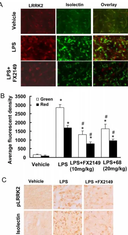
To further characterize the pharmacological effects of FX2149 in brains, a LPS-based mouse neuroinflammation model was used as described previously [13,16]. Consistent with previous findings [13,16], injection of LPS resulted in significant increases in LRRK2 expression, phos-phorylation, and microglial activation in the substantia nigra compared with vehicle controls (Fig. 6). Treatment of mice with FX2149 significantly reduced LPS-induced LRRK2-positive immunostaining compared with vehicle controls, but it did not alter LRRK2 cytoplasmic locali-zation. Moreover, the anti-phosphoryated-LRRK2 immunoactivity was also significantly re-duced in the FX2149 treated group. As in our previous study of 68 at 20 mg/kg [13], FX 2149 at a 10 mg/kg dose significantly reduced LPS-induced isolectin B4 (microglia marker) positive immunostaining in the substantia nigra. There was a ~43% isolectin B4 immunoactivity in the FX2149 treated group compared with the LPS alone treated group. In comparison, the immu-noactivity of isolectin B4 in the 68 treated group (20 mg/kg) was ~56% (Fig. 6B). Consistent with previous findings [13], anti-TH (dopaminergic neuron marker) immunostaining in the subtantia nigra did not change among various treatment groups and the control group, in-dicating that LPS treatment did not alter dopaminergic neuron degeneration in this acute inflammation condition.
repeated LRRK2 phosphorylation assays. B. Quantification of LRRK2 phosphorylation from A.*p<0.05 by
ANOVA compared with FX2151 treated group. C. LRRK2 and G2019S-LRRK2 were purified from cell lysates using anti-LRRK2 immunoprecipitation. The purified LRRK2 variants were incubated with FX2149 (0, 50, 100, or 200 nM) for 1 h and then subjected toin vitrokinase assays usingγ-32p -ATP incorporation method. LRRK2 autophosphorylation was quantified from three repeated experiments.*p<0.05 by ANOVA
compared to wild type LRRK2.#p<0.05 by ANOVA compared to G2019S-LRRK2 treated with vehicle.
Fig 4. FX2149 attenuated G2019S-LRRK2-induced neuronal degeneration in SH-SY5Y cells.SH-SY5Y cells were co-transfected with GFP and various pcDNA3.1-LRRK2 plasmids at a 1:15 ratio as described in the method section. After 4-h transfection, cells were treated with FX2149 for 48 hours. A. Cell viability was measured by counting the healthy viable GFP positive cells that contained at least one smooth extension (neurite) that was twice the length of the cell body.*p<0.05 by ANOVA compared to wild type LRRK2.
#p<0.05 by ANOVA compared to G2019S-LRRK2 treated with vehicle. B. TUNEL assays. The experiments
were repeated three times.*p<0.05 by ANOVA compared to vector control.#p<0.05 by ANOVA compared
to G2019S-LRRK2 treated with vehicle.
Fig 5. FX2149 improved the brain penetration and inhibition of LRRK2 GTP binding and kinase activities.FX2149 (10 mg/kg) and 68 (10 and 20 mg/kg) were injected intraperitoneally into G2019S-LRRK2 BAC transgenic mice at 6–12 weeks of age for 1 hour. There were 6 mice in each experimental group. The brain homogenates were used to detect LRRK2 binding and kinase activities. A and B, LRRK2 GTP-binding assays. C and D, LRRK2 phosphorylation assays using anti-phospho-LRRK2 antibodies. E and F, FX2149 reduced G2019S-LRRK2-induced 4E-BP phosphorylation determined by anti-phospho-4E-BP western blot analysis. Ntg: non-transgenic mouse.*p<0.05 by ANOVA compared with G2019S-LRRK2
transgenic mice treated with vehicle.
Fig 6. FX2149 reduced LPS-induced microglia activation and LRRK2-upregulation.G2019S-LRRK2 BAC transgenic mice (6–12 weeks) were injected with LPS (5μg) and FX2149 (10 mg/kg) as described in the methods section. Serial coronal sections through the substantia nigra were subjected to immunohistochemistry analysis. A. Representative immunofluorescent images with anti-isolectin (green) and anti-LRRK2 (red) staining. B. Quantification of immunofluorescence of A by unbiased stereology.*p<0.05 by ANOVA compared
with vehicle group.#p<0.05 by ANOVA compared with LPS treated group. C. Representative immunostaining
with anti-phospho-LRRK2-S935 and anti-isolectin B4 (marker for microglia) antibodies by DAB detection.
Discussion
In previous studies, we identified and characterized a LRRK2 GTP binding inhibitor, 68 [13]. To improvein vivoeffects of 68, we designed and synthesized a novel analog of 68, compound FX2149, to improve brain permeability. Thein vitrobiological characterization of FX2149 demon-strated that it inhibited LRRK2 GTP binding and kinase activity, and protected against mutant LRRK2 toxicity at 10–100 nM concentrations, which is a bit lower in efficacy than 68. However, FX2149 showed increasedin vivoefficacy, with a more than 2-fold improvement over 68. FX2149 significantly reduced LPS-induced microglia activation and LRRK2 upregulation at a significantly lower dose than 68. These findings demonstrated that FX2149 is a better lead GTP binding inhibi-tor with improved brain penetration for future drug development and pathogenesis studies.
The major pathology area of PD is in the substantia nigra [1,2]. Consequently, for therapeutic agents to prevent neurodegeneration, they must cross the BBB [11,17]. Most neuroprotective compounds fail further development due to either a lack of high potency in brains or poor BBB permeability [11,17]. Based on the chemical scaffold of 68, a new analog, FX2149, has been syn-thesized by substituting the phenylsulfonyl fragment of 68 with a pyridine-3-sulfonyl group, while replacing the methoxyethyl tail of 68 with a propyl tail [19,21]. FX2149 has similar effects in inhibition of LRRK2 GTP binding activity compared with 68, although it has less potency than 68 inin vitroassays. FX2149 at 100 nM inhibited LRRK2 kinase activity equivalent to 68 at 10 nM byin vitrophosphorylation assays. However, FX2149 had a potent efficacy in inhibiting LRRK2 GTP binding and kinase activities byin vivotesting with the LRRK2 transgenic mouse model. FX2149 at 10 mg/kg had an approximately equivalent GTP binding and kinase inhibi-tion effect as seen in 68 at 20 mg/kg. FX2149 at 10 mg/kg had a stronger effect in reducing mu-tant G2019S-induced 4E-BP phosphorylation compared with the treatment group of 68 at 20 mg/kg. 4E-BP is a stress-related transcription factor and increases in phosphorylation is believed to contribute to neuronal degeneration [20]. Taken together, these results demonstrated that FX2149 had better brain penetration efficacy for animal studies as required for PD intervention.
The loss of dopaminergic (DA) neurons in the substantia nigra is an early and key pathological hallmark of PD [21–23]. Disruption of LRRK2 GTP binding by genetic mutation reduces LRRK2 kinase activity, thereby suppressing neuronal degeneration [6,13]. Similar to the effects of 68 [13], our results showed that compound FX2149 reduced LRRK2 GTP binding and kinase activities, and significantly attenuated mutant LRRK2-induced neuron degeneration inin vitrocell culture models. Another feature of neurodegeneration is microglia activation and inflammation in brains [24]. Microglia activation releases various inflammatory cytokines which trigger or facilitate do-paminergic neuronal loss in PD [24]. Recent studies show that the preinflammatory agent, LPS, elevates LRRK2 expression and phosphorylation in activated microglia in mice [13,16,25]. Our results showed that FX2149 at 10 mg/kg significantly reduced LPS-induced microglia activation by 57% compared with the vehicle treated LPS mice group. In comparison, 68 at 20 mg/kg only reduced LPS-induced microglia by 44% (Fig. 5B). These results further validated the significantly improvedin vivoeffects of FX2149 compared to 68 in the LPS-based animal model.
In summary, these studies provided a novel GTP binding inhibitor, FX2149 (analog of 68), and further proved that GTP binding regulates LRRK2 kinase activity. Disruption of GTP binding activity may be an effective strategy to prevent neuron degeneration for PD and other LRRK2-related disorders.
Acknowledgments
Author Contributions
Conceived and designed the experiments: WWS FX TL XH. Performed the experiments: XH TL JMT DY SZ. Analyzed the data: TL XH FX WWS. Wrote the paper: WWS FX TL XH.
References
1. Li T, Yang D, Sushchky S, Liu Z, Smith WW. Models for LRRK2-Linked Parkinsonism. Parkinsons Dis. 2011; 2011: 942412. doi:10.4061/2011/942412PMID:21603132
2. Cookson MR. The role of leucine-rich repeat kinase 2 (LRRK2) in Parkinson's disease. Nat Rev Neu-rosci. 2010; 11: 791–797. doi:10.1038/nrn2935PMID:21088684
3. Tan EK, Schapira AH. LRRK2 as a therapeutic target in Parkinson's disease. Eur J Neurol. 2010; 18: 545–546. doi:10.1111/j.1468-1331.2010.03305.xPMID:21199179
4. Lee BD, Dawson VL, Dawson TM. Leucine-rich repeat kinase 2 (LRRK2) as a potential therapeutic tar-get in Parkinson's disease. Trends Pharmacol Sci. 2012; 33: 365–373. doi:10.1016/j.tips.2012.04.001 PMID:22578536
5. Liao J, Wu CX, Burlak C, Zhang S, Sahm H, Wang M, et al. Parkinson disease-associated mutation R1441H in LRRK2 prolongs the "active state" of its GTPase domain. Proc Natl Acad Sci U.S.A. 2014; 111: 4055–4060. doi:10.1073/pnas.1323285111PMID:24591621
6. Smith WW, Pei Z, Jiang H, Dawson VL, Dawson TM, Ross CA. Kinase activity of mutant LRRK2 medi-ates neuronal toxicity. Nat. Neurosci. 2006; 9: 1231–1233. PMID:16980962
7. Deng X, Dzamko N, Prescott A, Davies P, Liu Q, Yang Q, et al. Characterization of a selective inhibitor of the Parkinson's disease kinase LRRK2. Nat Chem Biol. 2011; 7: 203–205. doi:10.1038/nchembio. 538PMID:21378983
8. Lee BD, Shin JH, VanKampen J, Petrucelli L, West AB, Ko HS, et al. Inhibitors of leucine-rich repeat kinase-2 protect against models of Parkinson's disease. Nat Med. 2010; 16: 998–1000. doi:10.1038/ nm.2199PMID:20729864
9. Liu M, Dobson B, Glicksman MA, Yue Z, Stein RL. Kinetic mechanistic studies of wild-type leucine-rich repeat kinase 2: characterization of the kinase and GTPase activities. Biochemistry. 2010; 49: 2008–2017. doi:10.1021/bi901851yPMID:20146535
10. Liu M, Poulose S, Schuman E, Zaitsev AD, Dobson B, Auerbach K, et al. Development of a mechanism-based high-throughput screen assay for leucine-rich repeat kinase 2—discovery of LRRK2 inhibitors. Anal Biochem. 2010; 404: 186–192. doi:10.1016/j.ab.2010.05.033PMID:20566370
11. Deng X, Choi HG, Buhrlage SJ, Gray NS. Leucine-rich repeat kinase 2 inhibitors: a patent review (2006–2011). Expert Opin Ther Pat. 2012; 22: 1415–1426. doi:10.1517/13543776.2012.729041 PMID:23126385
12. Xiong Y, Coombes CE, Kilaru A, Li X, Gitler AD, Bowers WJ, et al. GTPase activity plays a key role in the pathobiology of LRRK2. PLoS Genet. 2010; 6: e1000902. doi:10.1371/journal.pgen.1000902 PMID:20386743
13. Li T, Yang D, Zhong S, Thomas JM, Xue F, Liu J, et al. Novel LRRK2 GTP-binding inhibitors reduced degeneration in Parkinson's disease cell and mouse models. Hum Mol Genet. 2014; 23: 6212–22. doi: 10.1093/hmg/ddu341PMID:24993787
14. Li Y, Liu W, Oo TF, Wang L, Tang Y, Jackson-Lewis V, et al. Mutant LRRK2(R1441G) BAC transgenic mice recapitulate cardinal features of Parkinson's disease. Nat Neurosci. 2009; 12: 826–828. doi:10. 1038/nn.2349PMID:19503083
15. Li X, Patel JC, Wang J, Avshalumov MV, Nicholson C, Buxbaum JD, et al. Enhanced striatal dopamine transmission and motor performance with LRRK2 overexpression in mice is eliminated by familial Par-kinson's disease mutation G2019S. J Neurosci. 2010; 30: 1788–1797. doi:10.1523/JNEUROSCI. 5604-09.2010PMID:20130188
16. Moehle MS, Webber PJ, Tse T, Sukar N, Standaert DG, DeSilva TM, et al. LRRK2 inhibition attenuates microglial inflammatory responses. J Neurosci. 2012; 32: 1602–1611. doi:10.1523/JNEUROSCI. 5601-11.2012PMID:22302802
17. Geldenhuys WJ, Van der Schyf CJ. Rationally designed multi-targeted agents against neurodegenera-tive diseases. Curr Med Chem. 2013; 20: 1662–1672. PMID:23410161
18. Fischer H, Gottschlich R, Seelig A. Blood-brain barrier permeation: molecular parameters governing passive diffusion. J Membr Biol. 1998; 165: 201–211. PMID:9767674
20. Imai Y, Gehrke S, Wang HQ, Takahashi R, Hasegawa K, Oota E, et al. Phosphorylation of 4E-BP by LRRK2 affects the maintenance of dopaminergic neurons in Drosophila. EMBO J. 2008; 27: 2432–2443. doi:10.1038/emboj.2008.163PMID:18701920
21. Dauer W, Przedborski S. Parkinson's disease. Mechanisms and models. Neuron. 2003; 39: 889–909. PMID:12971891
22. Forno LS. Neuropathology of Parkinson's disease. J Neuropathol Exp Neurol. 1996; 55: 259–272. PMID:8786384
23. Dawson TM, Dawson VL. Molecular pathways of neurodegeneration in Parkinson's disease. Science. 2003; 302: 819–822. PMID:14593166
24. Hamza TH, Zabetian CP, Tenesa A, Laederach A, Montimurro J, Yearout D, et al. Common genetic var-iation in the HLA region is associated with late-onset sporadic Parkinson's disease. Nat Genet. 2010; 42: 781–785. doi:10.1038/ng.642PMID:20711177