Resumo Casos atípicos de P. vivax relatados no Município de Manaus nos levou a detectar um isolado genético de P. vivax. Regiões variáveis do SSUrRNA foram examinadas no momento inicial da infecção e nas duas recrudescências de um mesmo paciente exibindo recrudescência/recidiva à cloroquina e primaquina. Um único isolado, encontrado em todos os estágios da infecção, sugere a presença de uma expansão clonal.
Palavras-chaves:P. vivax. Expansão clonal. Resistência.
303
Description of a possible clonal ex pansion of
Plasmodium vivax
in Manaus - Amazonas - Brazil
1. Fundação de Medicina Tropical do Amazonas da Universidade do Amazonas, Núcleo de Medicina Tropical e Nutrição da Universidade de Brasília, Brasília, DF. 2. Núcleo de Medicina Tropical e Nutrição da Universidade de Brasília, Brasília, DF. 3. Department of Preventive Medicine and Biometrics - Uniformed Services University of the Health Sciences - 4301 Jones Bridge Road, Bethesda, Maryland 20814-4799, USA. 4. Growth and Development Section - Laboratory of Parasitic Diseases - National Institutes of Health - Bethesda, Maryland 20892-0452, USA
Endereço para correspondência:Drª Maria das Graças Costa Alecrim. Av. Pedro Teixeira 25, D. Pedro I, 696040-000 Manaus, AM, Brazil. Tel: 55 92 238-2801; Fax: 55 92 238-7220.
E-mail [email protected] Recebido para publicação em 29/4/99.
Re vista d a Socie d ad e Brasile ira d e Me d icina Trop ical 32(3):303-305, m ai-jun, 1999.
NOTA PRÉVIA
Abstract Atypical P. vivax cases reported in Manaus municipality led us to detect a genetic isolate of P. vivax. Variable regions of SSUrRNA were examined from the initial time of infection and in the two recrudescences/relapses from a patient exhibiting chloroquine and primaquine resistance. A unique isolate, found at all stages of infection, suggests the presence of a clonal expansion.
Key-words:P. vivax. Clonal expansion. Resistance.
M aria das G raças Costa Alecrim1, Wilson Duarte Alecrim1, Vanize M acêdo2,
Caroline T. Korves3, Donald R. Roberts3, Jun Li4, M argery Sullivan4
and Thomas F. M cCutchan4
Descrição de uma possível expansão clonal do
Pla sm o dium viva x
em Manaus - Amazonas - Brasil
The nature and prevalence of Plasmodium vivax infection in Manaus Brazil seem to have changed over the past few years. Overall, the proportion of cases in Brazil due to P. vivax infection have increased from less than 50% in 1988 to 70% in 19965 6 7 8 Likewise, the ratio of reportable cases of P. vivaxversus P. falciparum in local clinics and hospitals around Manaus has increased, e.g., P. vivax accounted for 15,427 diagnoses in Manaus in 19982. Chloroquine resistant P. vivax in one patient in Manaus municipality (Amazonas State) was reported by
Ale crim MGC e t al
304
subset(s) of the P. vivax population is disease associated and that it maintains a degree of genetic isolation.
In most malaria species, there is intra-species D N A sequence polymorphism in variable regions of the small subunit rRNA3 4 9. We have used sequence variation in the rRNA to distinguish clones of P. falciparum in the past. Initially one may wonder why variation in this gene would be an indicator of genetic similarity between organisms. There are three different rRNA g e n e s on three different chromosomes from which rRNA is simultaneously transcribed during asexual blood-stage development9. Why would all three genes be identical to each other if chromosomes are constantly independently reassorting? The data to date, using P. falc iparu mclones as a model, has consistently revealed homogeneity within the clone but not between clones. Presumably this is because mechanisms designed to maintain homogeneity among the different alleles and the extent of self-matings maintain similarity in spite of whatever chromosomal reassortment that is occurring. If ribosomal sequence identity is an indicator of genetic relatedness between P. vivax isolates, as it appears to be with P. falciparum, one could use ribosomal sequence identities to follow the spread of a particular genotype.
Working on the above assumption we compared the sequence of the initial infection, a recrudescence/relapse (35 days later), and a subsequent relapse/recrudescence (180 days after the first recrudescence) of a single patient with vivax malaria from Manaus with sequences obtained from multiple randomly selected P. vivax patients. The initial infection was treated with chloroquine. The first recrudescence/relapse was treated with both chloroquine and primaquine.
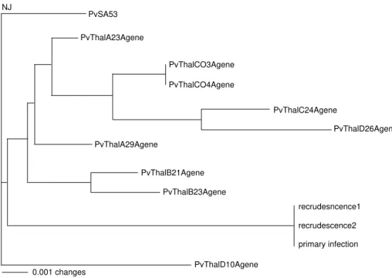
In a panmictic population, diversity should be found in the initial infection and this diversity should represent a subset of variation in the population. Analysis of recrudescent/relapse materials should also represent a subset of population diversity. Departure from random breeding would be indicated by only one type of rRNA being found in the blood. Here we cloned and analyzed three sequences from each the initial infection and two relapse/recrudescence. All nine sequences were identical. We compared these sequences with sequence taken from randomly chosen infections. For the purposes of displaying the extent of diversity in randomly taken sequences, we chose material both from El Salvador and nine sequences from patients in Thailand. The point of the dendogram is to graphically show the d i fferences in degrees of diversity from the diff e r e n t sources (Figure 1).
primary infection
PvThalD10Agene
recrudescence2 recrudesncence1 NJ
0.001 changes
Figure 1 - Dendogram of Plasmodium vivaxfrom different sources. PvSA53
PvThalA23Agene
PvThalCO3Agene
PvThalCO4Agene
PvThalC24Agene
PvThalD26Agene
PvThalA29Agene
PvThalB21Agene
Re vista d a Socie d ad e Brasile ira d e Me d icina Trop ical 32:303-305, m ai-jun, 1999.
305 We routinely find variation in isolates in both
Manaus and villages in Thailand. The identity of sequences shown in our patient indicates a degree of isolation of this sequence from other P. vivax isolates. Genetic isolation is suggested by our finding that the three different genes on three different chromosomes were identical in each sample, much like the situation in clones of P. falc iparu m. This finding suggests a lack of random interbreeding. Furthermore, the source of sporozoites was unusually homogeneous because their relapses/recrudescences were the same as the initial infection. These observations indicate that the original source of parasites (infected h u m a n ) yielded an apparently homogeneous blood feed with regard to parasites. Interpretation of these data seems to suggest that we are following the expansion of a clone.
The changes in P. vivax infection in Manaus are very striking. In part, these changes could reflect an urbanization of malaria that simply favors transmission of P. vivax over P. falcip aru m. H o w e v e r, along with this process of malaria urbanization are changes in infection severity and susceptibilities to chloroquine. Still, even these changes possible relate to extrinsic factors that alter parasite population make-up, e.g., environmental alterations and vector control measures that influence vector population structure or chemotherapeutic drug pressure on parasite populations. Regardless, a method for following the expansion of parasite "clones" and their relationship to malaria burden is important. Parasites like the one described here which carry sequences not seen before represent prime candidates for prospective study.
REFERENCES 1. Alecrim MGC, Alecrim WD, Macêdo V.Plasmodium vivax
resistance to chloroquine (R2) and mefloquine (R3) in Brazilian Amazon region. Revista da Sociedade Brasileira de Medicina Tropical 32:67-68, 1999.
2. Instituto de Medicina Tropical do Amazonas. Boletins informativos do Instituto de Medicina Tropical do Amazonas, 1998-1999
3. Kawamoto F, Miyake H, Haneko O, Kimura, Dung NT, Liu Q, Zhou M, Dao LD, Kawai S, Isomura S, Wataya Y. Sequence variation in the 18S rRNA gene, a target for PCR-based malaria diagnosis, in Plasmodium ovale from Southern Vietnam. Journal of Clinical Microbiology 34:2287-2289, 1996.
4. Li J, Wirtz RA, McConkey GA, Sattabongkot J, Waters AP, Rogers MJ, McCutchan TF. Plasmodium: genus-conserved primers for species identification and quantitation. Experimental Parasitology 81:182-190, 1995.
5. Pan American Health Organization. Status of malaria programs in the Americas. XLII Report. PAHO/WHO. Washington, D.C. 112pp, 1994.
6. Pan American Health Organization. Status of malaria programs in the Americas. XLIII Report. PAHO/WHO. Washington, D.C. 25pp, 1995.
7. Pan American Health Organization. Status of malaria programs in the Americas. XLIV Report. PAHO/WHO. Washington, D.C. 23pp, 1996.
8. Pan American Health Organization. Status of malaria programs in the Americas. XLV Report. PAHO/WHO. Washington, D.C. 25pp, 1997.